

Loading, please wait...
Loading, please wait...
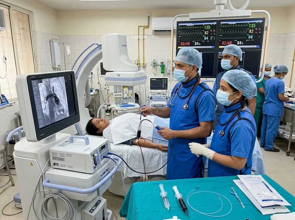
Managing hemodynamic shock caused by massive thromboembolism presents a severe clinical challenge when patients have concurrent severe bleeding. In this life-threatening setting, systemic thrombolytic therapy remains contraindicated due to catastrophic hemorrhage risks. Consequently, interventional clinicians increasingly rely on aspiration thrombectomy in pulmonary embolism as an indispensable emergency reperfusion strategy. A recent case highlighted this delicate balancing act in a patient suffering from obstructive shock alongside profound, symptomatic blood loss.
Clinicians frequently encounter complex medical dilemmas in intensive care units, but few scenarios rival high-risk pulmonary embolism occurring alongside life-threatening anemia. In a recently documented emergency case, a 48-year-old female presented in profound obstructive shock with a blood pressure of 74/47 mm Hg. Simultaneously, laboratory analyses revealed a critical hemoglobin level of merely 41 g/L. The source of this profound anemia was an unremitting three-year history of severe uterine bleeding driven by advanced adenomyosis. Diagnostic imaging swiftly delineated the catastrophic anatomy. Computed tomography pulmonary angiography demonstrated extensive bilateral emboli, including complete occlusion of the right main pulmonary artery trunk. Concurrently, bedside echocardiography identified severe pulmonary hypertension with evident right ventricular strain, while vascular ultrasonography confirmed extensive deep vein thrombosis in her left lower extremity. Standard practice guidelines dictate immediate intravenous thrombolysis or surgical embolectomy for hemodynamically unstable pulmonary embolism. However, systemic thrombolysis in the presence of active uterine hemorrhage and profound anemia carries an unacceptable risk of fatal exsanguination. Therefore, medical teams must navigate an urgent therapeutic tightrope. They must rapidly relieve mechanical pulmonary arterial obstruction while aggressively staunching ongoing hemorrhagic blood loss.
The primary cause of death in massive pulmonary embolism is acute right ventricular failure triggered by a sudden surge in pulmonary vascular resistance. In this patient, severe acute anemia compounded the hemodynamic collapse by drastically diminishing systemic arterial oxygen delivery. Consequently, the combination of hypoxia, right ventricular ischemia, and severe metabolic acidosis demanded prompt afterload reduction. Systemic thrombolysis was clearly precluded, and open surgical pulmonary embolectomy presented prohibitive surgical mortality risks under extreme anemia. Under these circumstances, endovascular therapy provided the most logical and life-saving alternative. Catheter-based interventions allow immediate physical debulking of obstructive thrombi without requiring high doses of systemic fibrinolytic agents. Furthermore, contemporary endovascular platforms enable operators to extract central clot burden swiftly while monitoring real-time hemodynamics. The treating team recognized that reducing pulmonary arterial pressures by even a modest margin could break the vicious cycle of right ventricular failure. Thus, emergency catheter-directed mechanical therapy served as a bridge to survival. By prioritizing physical clot extraction over pharmacological lysis, the clinical team protected the patient from catastrophic uterine bleeding while systematically restoring cardiovascular stability.
The interventional team promptly transferred the patient to the catheterization laboratory to perform emergency mechanical intervention. Specifically, operators utilized large-bore aspiration thrombectomy in pulmonary embolism using sequential 22F, 20F, and 16F aspiration catheters. Large-bore suction devices offer superior mechanical suction force, enabling rapid extraction of dense, organized thromboemboli directly from the main pulmonary arteries. Because excessive procedural blood loss remains a significant hazard with large-bore aspiration devices, meticulous volume management was paramount. The operators carefully filtered the aspirate and successfully quantified total procedural blood loss at approximately 100 mL, preserving essential circulating volume. During the intervention, the aspiration catheters successfully engaged and retrieved extensive central thrombi from the occluded right main pulmonary artery. Following mechanical debulking, invasive pulmonary artery pressures dropped immediately from 51/23 mm Hg to 34/12 mm Hg. This dramatic hemodynamic decompression promptly restored pulmonary perfusion and stabilized systemic arterial pressure. Concurrently, the interventional radiologists placed a retrievable inferior vena cava filter to shield the pulmonary circulation against recurrent thromboemboli from the extensive lower-limb deep vein thrombosis.
Successful resuscitation required extensive cross-specialty collaboration extending far beyond the catheterization laboratory. Initially, critical care intensivists and hematologists prioritized massive transfusion therapy, ultimately delivering 13 units of packed red blood cells to correct tissue hypoxia. Meanwhile, gynecologists intervened urgently to control the source of ongoing pelvic blood loss. The gynecological surgical team performed emergent dilatation and curettage alongside initiation of gonadotropin-releasing hormone agonist therapy to arrest adenomyosis-related uterine hemorrhage. Once local hemostasis was established, clinicians faced the next critical challenge: determining when and how to introduce anticoagulation. Because indwelling inferior vena cava filters and extensive residual deep vein thrombosis create ongoing thrombotic risks, anticoagulation cannot be omitted indefinitely. Therefore, the team instituted carefully titrated parenteral anticoagulation once active vaginal bleeding ceased. Subsequently, clinicians transitioned the patient to oral rivaroxaban as her clinical status improved. Over a comprehensive 24-day hospitalization, her hemoglobin concentration rose steadily to 72 g/L, while bilateral lower extremity edema fully resolved. At discharge, the patient remained completely free of dyspnea, walking comfortably without supplemental oxygen support.
This compelling clinical case illustrates several vital principles for emergency physicians, cardiologists, and critical care specialists managing complex thromboembolic disease. First, large-bore mechanical thrombectomy serves as an invaluable rescue modality when absolute contraindications preclude systemic thrombolytic administration. By physically clearing the central pulmonary trunk, operators achieve rapid hemodynamic decompression with palliative rather than curative intent. Clinicians must recognize that complete angiographic clearance is unnecessary; restoring partial flow suffices to break acute right ventricular failure. Second, meticulous procedural technique and aspiration filtering are essential to avoid worsening pre-existing, life-threatening anemia. Third, temporary inferior vena cava filters provide crucial short-term protection when full-dose anticoagulation must be temporarily delayed. Nevertheless, single case reports cannot establish comparative safety, long-term efficacy, or universal protocols across heterogeneous patient populations. Randomized controlled clinical trials are currently evaluating catheter-directed interventions against conventional therapy to better define standard care algorithms. Above all, this scenario underscores the indispensable value of a dedicated, coordinated Pulmonary Embolism Response Team. Rapid alignment among interventionalists, critical care specialists, and gynecologists remains the cornerstone of survival in high-risk thromboembolic crises.
Systemic thrombolytic agents circulate throughout the entire vascular tree, non-selectively dissolving protective fibrin plugs at all vascular breach points. In a patient with active uterine bleeding and profound anemia, systemic fibrinolysis can induce unstoppable hemorrhagic shock and fatal exsanguination. Therefore, international guidelines consider ongoing or high-risk active bleeding an absolute contraindication, necessitating purely mechanical interventions like catheter aspiration to restore pulmonary perfusion safely.
Large-bore aspiration catheters remove obstructive thrombi directly from the proximal pulmonary arterial branches through mechanical suction. By debulking central vascular occlusions, this intervention immediately reduces elevated pulmonary vascular resistance and pulmonary arterial pressures. Consequently, right ventricular wall tension falls rapidly, improving right coronary artery perfusion and myocardial contractility. This prompt mechanical decompression breaks the cycle of obstructive shock without requiring systemic pharmacological clot dissolution.
An inferior vena cava filter provides temporary mechanical protection against recurrent, fatal emboli originating from lower-extremity deep vein thrombosis. In patients presenting with life-threatening active hemorrhage, immediate therapeutic anticoagulation is unsafe and must be temporarily withheld. Therefore, placing a retrievable filter creates an essential physical barrier that prevents further clot migration to the pulmonary circulation while clinicians aggressively manage active bleeding and correct severe anemia.
Disclaimer: This content is for informational and educational purposes only and should not be considered medical advice or used to guide patient care. Refer to the latest local and national guidelines for clinical practice.
References

Read summarized clinical updates, watch expert medical content, and earn CME certifications right from your smartphone.
Open your smartphone camera to scan and install.





In high-risk pulmonary embolism complicated by severe anemia and active uterine bleeding, systemic thrombolysis is contraindicated. Large-bore aspiration thrombectomy provides rapid hemodynamic rescue without fibrinolysis, supported by inferior vena cava filter placement and multidisciplinary critical care.
Today

A retrospective study evaluates contrast-associated acute kidney injury (CA-AKI) incidence in oncology patients receiving active chemotherapy with preserved renal function, highlighting low overall risk and timing considerations.
Today
A cross-sectional study in Zanzibar reveals that 46.5% of adults with type 2 diabetes experience multidomain autonomic dysfunction on the COMPASS-31 tool, primarily orthostatic intolerance, while 45.9% exhibit cardiovascular autonomic neuropathy, highlighting the urgent need for structured clinical screening.
Today

A secondary analysis of a randomized controlled trial using deep learning models shows that diluted epinephrine irrigation achieves superior visual clarity and requires lower pump pressure compared to intravenous tranexamic acid during arthroscopic rotator cuff repair, with equivalent safety profiles.
Today
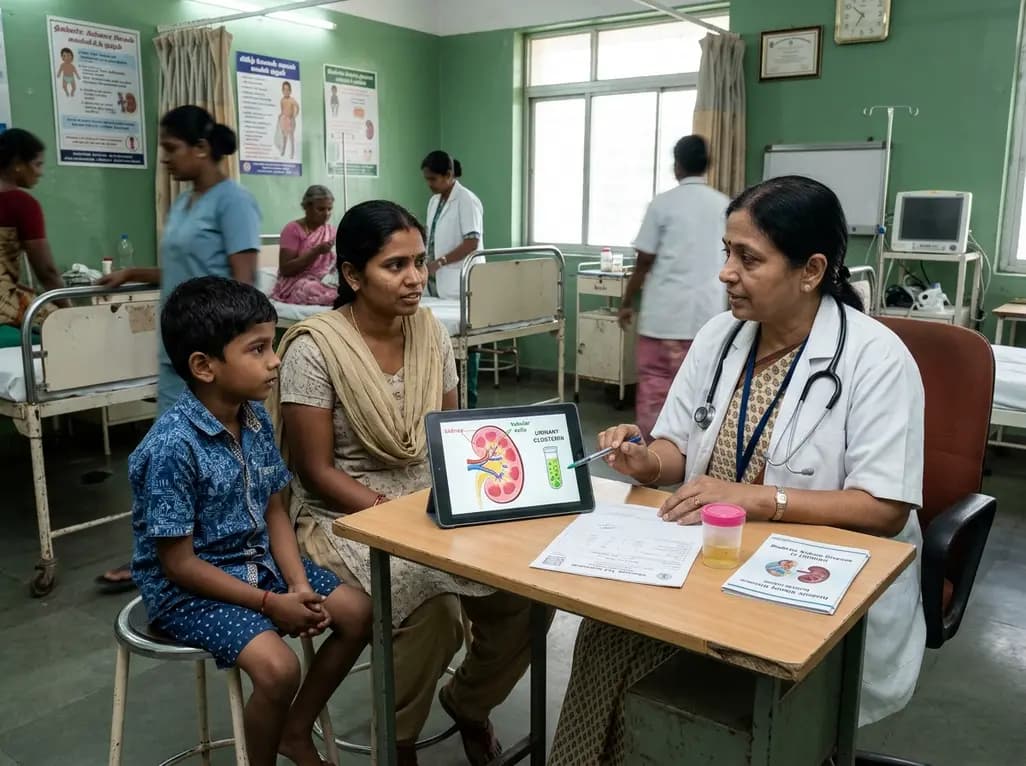
A recent case-control study reveals that urinary clusterin provides superior diagnostic performance for early diabetic kidney disease detection in pediatric type 1 diabetes mellitus, identifying tubular damage before conventional microalbuminuria occurs.
Today
