.webp&w=384&q=75)

Loading, please wait...
Loading, please wait...
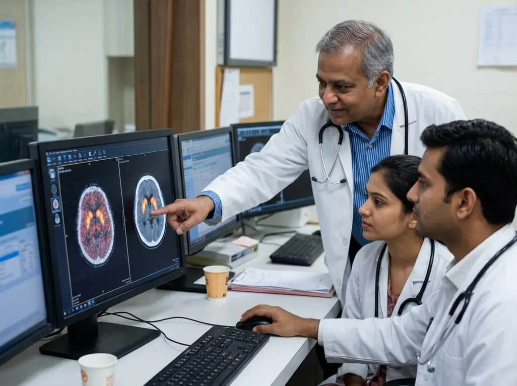
Positron emission tomography has fundamentally reshaped clinical diagnostics across oncology, cardiology, and neurology. However, conventional imaging protocols face persistent physical constraints, including spatial resolution boundaries and radiotracer dose limitations. Recent breakthroughs in computational algorithms have established AI PET reconstruction as a powerful tool to overcome these imaging challenges. Rather than replacing established physical principles, deep learning frameworks now augment the reconstruction workflow from raw coincidence event detection to final clinical visualization. Consequently, nuclear medicine departments can achieve high-fidelity diagnostic images while minimizing scan durations and patient radiotracer exposure.
Historically, nuclear medicine specialists relied on filtered backprojection to invert projection data into diagnostic tomograms. Although computationally rapid, this analytic method frequently amplified high-frequency noise and produced streak artifacts in count-starved scenarios. Subsequently, iterative algorithms, particularly maximum likelihood expectation maximization and ordered subsets expectation maximization, became the routine clinical standard. These iterative approaches successfully integrated statistical Poisson noise models and coarse scanner geometry. Nevertheless, standard iterative reconstructions struggle to balance noise suppression against high-contrast spatial recovery.
To resolve this limitation, computer scientists and medical physicists developed deep learning models that optimize image formation. Modern time-of-flight technology and long-axial field-of-view scanners generate massive datasets that demand immense processing power. Therefore, researchers train deep convolutional neural networks and vision transformers on paired low-count and high-count datasets. As a result, these neural networks learn complex mappings that recover true radioactivity concentrations without degrading structural edges. Furthermore, hybrid methods combine mathematical forward projection with data-driven regularizers. Consequently, the imaging community now embraces algorithms that stabilize low-count scans while maintaining reproducible quantification across diverse clinical indications.
Raw data handling forms the initial foundation of any successful molecular imaging study. During acquisition, modern digital detectors register millions of prompt coincidence events alongside random and scattered coincidences. Furthermore, physical attenuation from patient tissues dramatically attenuates true coincidence photon pairs. Traditionally, system hardware applied uniform mathematical corrections that frequently suffered from patient movement or anatomical mismatch. In contrast, artificial intelligence refines these early corrections directly within the sinogram or list-mode domain.
Specifically, deep neural networks estimate scatter components rapidly by modeling individual patient habitus and gamma attenuation profiles. Additionally, deep learning models synthesize accurate pseudo-attenuation correction maps directly from non-contrast low-dose computed tomography or magnetic resonance sequences. This capability proves exceptionally valuable for hybrid PET/MRI scanners, where bone signal absence historically compromised attenuation accuracy. Moreover, machine learning algorithms track respiratory and cardiac motion vectors throughout list-mode data streams. Consequently, the pipeline corrects physical displacement before the iterative loop begins. By resolving corrupting physical phenomena early, these intelligent pre-processing modules provide clean projection datasets that safeguard subsequent image reconstruction from severe geometric distortions.
Purely data-driven reconstruction models sometimes encounter generalization failures when faced with rare pathology or atypical scanner geometry. Therefore, leading computational researchers favor unrolled iterative networks that embed artificial intelligence within traditional physics-based frameworks. In these hybrid systems, the algorithm preserves trusted system matrix formulations, time-of-flight kernels, and Poisson likelihood models. Simultaneously, neural networks replace simplistic regularization penalties, such as Gaussian smoothing or basic quadratic priors.
Moreover, these unrolled neural architectures execute alternating steps of statistical data consistency and deep feature refinement. For example, deep image priors utilize patient-specific structural priors from co-registered anatomical imaging to guide tracer distribution estimates. Consequently, the reconstruction system preserves subtle photopenic defect boundaries and small hypermetabolic malignant foci with remarkable clarity. Furthermore, physics-informed neural networks restrict intermediate solution spaces to physically plausible tracer concentrations. Thus, the algorithm avoids spurious mathematical artifacts while recovering true quantitative standard uptake values. Because the framework retains strict data fidelity at each iteration, clinicians gain confidence that final pixel intensities reflect genuine tracer accumulation rather than synthetic algorithmic hallucinations.
Image filtering in the spatial domain represents the most commercially widespread implementation of machine intelligence today. After standard reconstruction finishes, residual image noise can obscure tiny sub-centimeter metastatic deposits or subtle cortical hypometabolism. Historically, clinicians applied isotropic Gaussian post-filters, which reduced image noise but severely compromised spatial resolution and lesion conspicuity. In contrast, advanced post-reconstruction deep learning models perform edge-preserving denoising across three-dimensional volumetric images.
Specifically, convolutional networks and generative architectures differentiate random Poisson noise textures from true tracer biodistribution. These deep filters adaptively suppress high-frequency background fluctuations while preserving sharp tumor margins and physiological uptake gradients. Furthermore, multi-center trials confirm that AI-driven denoising allows clinical teams to reduce injected radiopharmaceutical activity by fifty to seventy-five percent without degrading diagnostic confidence. Similarly, high-throughput diagnostic centers can shorten acquisition bed durations, significantly reducing patient motion artifacts and improving comfort. Nevertheless, nuclear medicine physicians must carefully calibrate post-filtering parameters against known baseline acquisitions. Overly aggressive denoising algorithms could smooth out faint hypermetabolic lesions, which might impair accurate restaging in oncological patients.
Translating artificial intelligence from academic engineering laboratories into routine hospital workflows requires rigorous validation protocols. Because clinicians base critical oncological decisions on standardized uptake values and metabolic tumor volumes, reconstructed images must demonstrate rock-solid quantitative fidelity. Consequently, multicenter trials compare AI-enhanced reconstructions against standard full-dose benchmarks across heterogeneous patient cohorts. Notably, these studies show non-inferior diagnostic accuracy and high lesion detectability for solitary pulmonary nodules, neurodegenerative disease patterns, and lymphoma staging.
However, clinical translation introduces complex regulatory and software lifecycle challenges. Health regulatory agencies, including the United States Food and Drug Administration and the European Medicines Agency, mandate clear documentation regarding algorithm training transparency, intended use specifications, and domain generalization. Furthermore, medical physicists must conduct periodic phantom quality assurance to monitor potential drift in quantitative metrics across scanner hardware updates. Additionally, hospital information systems require robust interoperability and audit trails to track algorithmic software versions across Picture Archiving and Communication Systems. Ultimately, establishing standardized validation frameworks ensures that intelligent reconstruction tools enhance diagnostic efficiency while maintaining uncompromising patient safety.
Deep learning models extract authentic tracer distribution signals from sparse, count-starved raw data. Consequently, these algorithms effectively filter Poisson noise without blurring anatomical margins. By leveraging extensive prior training on high-count datasets, the software reconstructs diagnostic-quality scans from scans acquired with up to seventy-five percent lower radiotracer activity. This significant dosage reduction protects pediatric and adult oncology patients undergoing frequent restaging scans from cumulative ionizing radiation exposure while preserving diagnostic accuracy.
Unconstrained generative networks occasionally create hallucinated structures or erroneously smooth out faint lesions. However, modern hybrid pipelines safeguard against this vulnerability by anchoring machine learning within physics-based iterative frameworks. These systems enforce strict mathematical data consistency against original raw projection data at each iteration step. Consequently, the algorithm cannot generate uncorroborated hot spots. Furthermore, rigorous clinical validation across diverse patient populations ensures that reconstructed image features reflect true biological radiotracer uptake.
Regulatory authorities, including the United States FDA, evaluate artificial intelligence reconstruction software as medical devices through rigorous clearance pathways. Manufacturers must submit prospective validation data demonstrating non-inferior diagnostic accuracy, robust lesion detectability, and reproducible standard uptake value quantification across diverse scanner models. Furthermore, agencies require continuous post-market surveillance, strict cybersecurity controls, clear labeling of intended clinical use, and comprehensive software lifecycle management protocols to ensure patient safety and algorithm reliability in routine practice.
Disclaimer: This content is for informational and educational purposes only and does not constitute medical advice, diagnosis, or treatment. Healthcare professionals should exercise their independent clinical judgment when interpreting imaging findings and making management decisions. The application of artificial intelligence in nuclear medicine and diagnostic radiology continues to evolve; clinicians must verify AI-generated reconstruction outputs against raw acquisition data and institutional protocols where appropriate. Refer to the latest local and national guidelines for clinical practice.
References

Read summarized clinical updates, watch expert medical content, and earn CME certifications right from your smartphone.
Open your smartphone camera to scan and install.





Artificial intelligence is transforming positron emission tomography by augmenting the reconstruction pipeline. From sinogram corrections to physics-informed iterative models and denoising, deep learning optimizes image resolution and quantitative accuracy while enabling low-dose scanning.
Today

A comprehensive scoping review synthesizes multilevel determinants influencing mobile health application adoption among older adults with chronic heart disease, emphasizing the essential role of interface design, caregiver engagement, and clinical nursing pathways.
Today
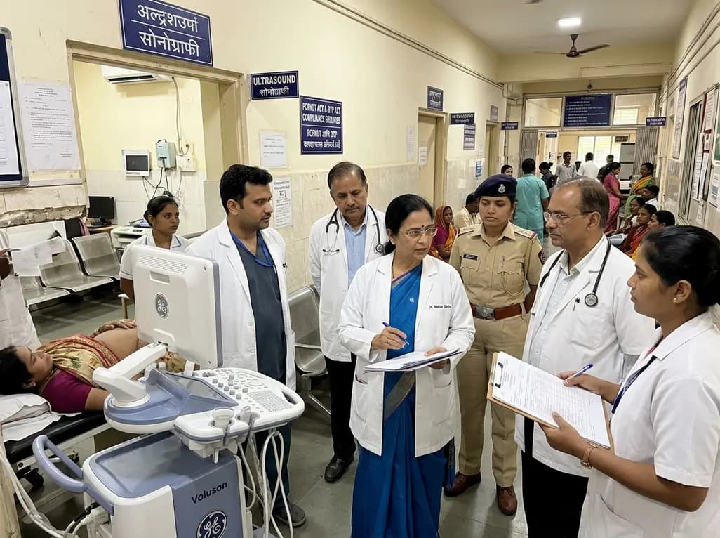
The Maharashtra public health department has established a specialized task force headed by Dr. Neelam Gorhe to eliminate illegal sex determination and unauthorized abortions. The team will execute surprise inspections of ultrasound facilities, audit medical documentation, and enforce strict statutory standards.
Today

A multi-state cohort study of 11 million children reveals that nonsyndromic congenital heart disease significantly increases specific childhood cancer hazards, including lymphoma, hepatoblastoma, and leukemia. These findings underscore the need for targeted clinical vigilance and shared developmental research.
Today
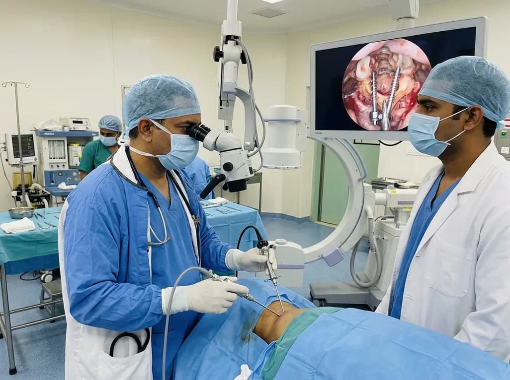
Endoscopic posterior cervical fusion combines minimally invasive decompression, joint preparation, and rigid screw-rod fixation for atlantoaxial pathologies. Early clinical findings demonstrate solid bony union, excellent symptom relief, and minimal soft-tissue morbidity without significant vascular compromise.
Today
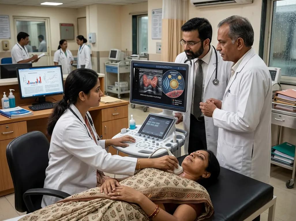
Researchers have developed an innovative ultrasound-responsive nano-contrast agent that co-delivers CIITA-siRNA and rapamycin directly to inflamed thyroid tissue, significantly reducing autoimmune injury and thyroid autoantibodies in preclinical models of Hashimoto thyroiditis.
Today
