.webp&w=384&q=75)

Loading, please wait...
Loading, please wait...
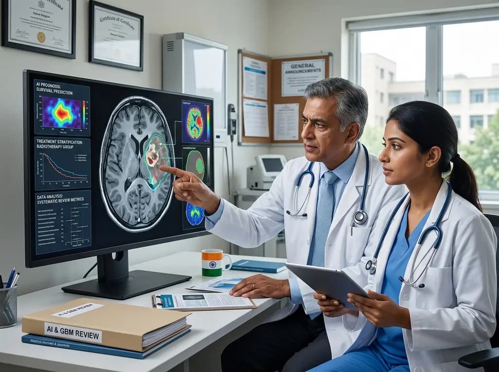
Glioblastoma (GBM) remains the most aggressive and lethal primary brain tumor in adults, posing significant challenges for neurosurgeons worldwide. Modern neurosurgical decision-making increasingly relies on the integration of complex imaging, molecular, and perioperative data. Consequently, AI in glioblastoma neurosurgery has emerged as a revolutionary approach to enhance clinical outcomes and streamline complex workflows. These computational tools aim to assist clinicians by providing data-driven insights into prognosis and treatment stratification. Furthermore, the ability of artificial intelligence to process high-dimensional datasets allows for a level of precision that traditional clinical assessment often lacks. Nevertheless, the transition of these models from research environments to bedside practice requires rigorous evaluation. Recent systematic reviews highlight the growing interest in these technologies, yet they also point toward a landscape marked by significant heterogeneity. As we move toward a more personalized era of neuro-oncology, understanding the nuances of these AI models becomes paramount for every practicing neurosurgeon and oncologist. By leveraging machine learning, clinicians can potentially navigate the biological complexity of GBM with greater confidence and accuracy.
Radiomics currently represents the most common model family used for survival-focused tasks in neuro-oncology. Specifically, this approach involves the extraction of quantitative features from standard-of-care MRI scans that are often imperceptible to the human eye. Researchers have found that radiomics, when paired with conventional machine learning algorithms, can effectively predict patient survival and molecular markers. For instance, approximately 43.3% of current studies utilize this combination to analyze the spatial and textural variations within the tumor. Moreover, these models are particularly adept at identifying subtle imaging signatures that correlate with treatment resistance. Consequently, clinicians can use these insights to tailor surgical resections and adjuvant therapy protocols more effectively. However, the reliability of radiomics depends heavily on the quality of image segmentation and the standardization of feature extraction. Despite these technical challenges, the synergy between radiomics and machine learning continues to drive innovation in preoperative planning. By providing a non-invasive window into the tumor’s molecular landscape, these tools offer a significant advantage over traditional biopsy methods in certain clinical scenarios. Ultimately, the integration of these features into routine practice could refine how we categorize GBM patients into distinct risk groups.
Deep learning has also carved out a significant niche, accounting for roughly 26.7% of the methodologies explored in glioblastoma research. Unlike traditional machine learning, deep learning architectures like convolutional neural networks (CNNs) can automatically learn relevant features directly from raw image data. This eliminates the need for manual feature engineering, which is often time-consuming and subjective. Furthermore, hybrid approaches that combine deep learning with radiomics are gaining traction, representing about 13.3% of the literature. These hybrid models leverage both automated pattern recognition and structured quantitative features to achieve superior predictive performance. In addition, these advanced algorithms are increasingly being applied to predict recurrence and differentiate between true progression and pseudoprogression. This distinction is critical for avoiding unnecessary second-rate surgeries or premature changes in chemotherapy. Notably, deep learning models excel at handling multimodal data, including genomics and proteomics, alongside imaging. By synthesizing these diverse data streams, hybrid models provide a more holistic view of the patient’s disease state. Nevertheless, the "black-box" nature of some deep learning systems remains a hurdle for clinical acceptance. Therefore, ongoing research focuses on developing explainable AI frameworks that provide transparent reasoning for their predictions.
Despite the promise of AI in glioblastoma neurosurgery, several systemic challenges impede its widespread clinical adoption. A primary concern is the extreme heterogeneity found across current studies, ranging from diverse patient populations to varying imaging protocols. This lack of uniformity makes it difficult to compare results and establish standardized benchmarks for performance. Furthermore, external validation remains remarkably uncommon, with only about 16.7% of studies testing their models on independent datasets. Without robust validation across different institutions, there is a high risk that these models will fail to generalize in real-world clinical settings. Most studies currently rely on internal validation, which can lead to overoptimistic performance metrics due to overfitting. Consequently, the clinical reliability of these tools is often questioned by regulatory bodies and healthcare providers alike. In addition, inconsistent methodological reporting further complicates the landscape, making it hard for other researchers to replicate findings. To overcome these barriers, the scientific community must prioritize multi-institutional collaborations and data-sharing initiatives. Only through rigorous, large-scale testing can we ensure that AI-driven decisions are safe and effective for all glioblastoma patients.
Successfully integrating AI tools into the neurosurgical workflow requires more than just high-performing algorithms. It necessitates a shift in how clinicians interact with technology and data. For example, AI should be viewed as an augmented intelligence tool that supports, rather than replaces, human judgment. Furthermore, the implementation of these systems must be accompanied by comprehensive training for neurosurgeons and radiologists. This ensures that the outputs are interpreted correctly within the broader clinical context of each patient. Moreover, regulatory frameworks must evolve to provide clear guidelines for the approval and monitoring of AI-based medical devices. In India, where healthcare resources can vary significantly between centers, AI offers a unique opportunity to standardize high-quality care. By providing decision-support systems in regional centers, AI can help bridge the gap in expertise between specialized oncology hubs and general hospitals. However, this transition must be handled with care to avoid creating new biases or ethical dilemmas. Transparency in how models are trained, especially regarding diverse ethnic and demographic data, is essential for equitable healthcare. Ultimately, the goal is to create a seamless interface where AI provides real-time, actionable insights during both the planning and intraoperative phases of surgery.
The future of neuro-oncology lies in the realization of truly personalized medicine, guided by advanced computational frameworks. As AI models become more sophisticated, they will likely play a central role in real-time intraoperative guidance. For instance, AI-driven augmented reality could help surgeons visualize tumor margins and critical white matter tracts with unprecedented clarity. In addition, the integration of longitudinal data will allow for dynamic risk assessment throughout the patient’s treatment journey. This means that prognosis will no longer be a static calculation made at the time of diagnosis but a continuously updated metric. Furthermore, the move toward biologically informed AI models will bridge the gap between imaging and the underlying tumor microenvironment. By predicting how specific regions of a tumor will respond to targeted therapies, AI can help design more effective, individualized treatment plans. Consequently, the lethal nature of glioblastoma may eventually be managed with greater chronic control. To reach this future, the focus must remain on methodological rigor, external validation, and patient-centered outcomes. As researchers refine these tools, the potential for AI to transform the survival landscape of GBM patients remains a beacon of hope for the medical community.
AI enhances survival prediction by analyzing quantitative imaging features, often called radiomics, that are invisible to the human eye. By combining these signatures with molecular data, AI models provide a more personalized prognosis than clinical variables alone. This allows for better-informed discussions regarding surgical outcomes and postoperative care strategies.
External validation ensures that an AI model performs accurately on data from different institutions and patient populations. Many current models suffer from overfitting to their specific training datasets. Without robust testing across diverse clinical environments, these tools may fail to generalize, making validation a mandatory prerequisite for clinical adoption.
Hybrid models combine the strengths of hand-crafted radiomic features with the automated pattern recognition of deep learning. While traditional machine learning requires manual feature extraction, deep learning identifies complex spatial hierarchies automatically. Merging these approaches often yields higher predictive accuracy for molecular subtypes and survival despite increased model complexity.
Disclaimer: This content is for informational and educational purposes only and does not constitute medical advice, diagnosis, or treatment. Always seek the advice of your physician or other qualified health provider with any questions you may have regarding a medical condition. Refer to the latest local and national guidelines for clinical practice.
References
Reyes JS et al. AI for prognosis and treatment stratification in glioblastoma neurosurgery: a systematic review. J Neurooncol. 2026 Jun 19. doi: 10.1007/s11060-026-05676-3. PMID: 42319645.
Awuah WA et al. Predicting survival in malignant glioma using artificial intelligence. Eur J Med Res. 2025;30:61. doi: 10.1186/s40001-025-01678-x.
Nadeem MW et al. Brain tumor analysis empowered with deep learning: A review, taxonomy, and future challenges. Brain Sci. 2020;10(2):118. doi: 10.3390/brainsci10020118.
"
Read summarized clinical updates, watch expert medical content, and earn CME certifications right from your smartphone.
Open your smartphone camera to scan and install.





A systematic review examines the transformative potential of artificial intelligence in glioblastoma neurosurgery. While AI models show promise in predicting survival and treatment response, significant hurdles like study heterogeneity and limited external validation remain for clinical implementation.
3 months ago

As monsoon seasons elevate arboviral disease threats, Tamil Nadu has initiated an ICMR-monitored evaluation of the tetravalent dengue vaccine in Tenkasi district. Clinicians emphasize clinical monitoring, public prevention, and high-quality early pediatric care to reduce disease morbidity across endemic regions.
Today

A comparative cohort study evaluates vNOTES vs TLH hysterectomy for benign conditions, demonstrating shorter operative duration, faster return to daily activities, and equivalent female sexual function scores without increased dyspareunia.
Today

Review how tailored exercise programs benefit frail older patients with heart failure. Explore pathophysiological mechanisms, individualized training prescriptions, and strategies to overcome adherence barriers in clinical practice.
Today

This comparative study evaluates posterior labral tears with and without glenohumeral instability, highlighting distinct demographic patterns, MRI underestimation in stable shoulders, and successful arthroscopic repair outcomes.
Today

A clinical study demonstrates that early intervention with tocilizumab, either as monotherapy or bridged with corticosteroids, achieves significant proptosis reduction and resolves active inflammation in treatment-naive thyroid eye disease.
Today
