

Loading, please wait...
Loading, please wait...
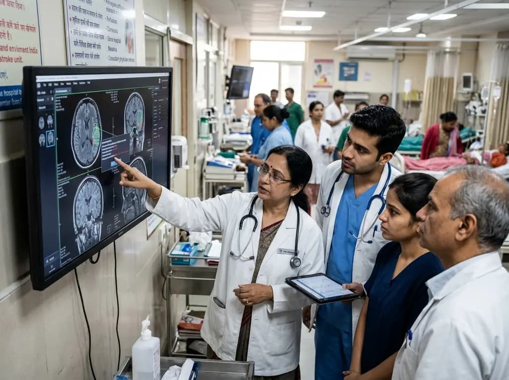
Acute ischemic stroke remains a primary driver of morbidity and mortality across the globe, demanding hyper-acute clinical coordination. Consequently, modern health systems are transitioning toward automated digital pathways to eliminate avoidable prehospital and hospital delays. The recent clinical integration of AI-assisted stroke care exemplifies this necessary paradigm shift in emergency neurovascular medicine. Coordinated clinical pathways now link emergency medical services directly with acute neurology specialists, advanced imaging suites, and dedicated neuro-intensive care units. Furthermore, this comprehensive approach emphasizes prompt diagnosis, early intravenous recanalization, and emergent endovascular intervention. Ultimately, timely intervention preserves ischemic brain tissue and significantly improves functional independence for patients.
Prehospital delay frequently represents the largest barrier to successful revascularization in acute stroke care. Therefore, modern emergency response networks deploy advanced ambulances equipped with high-speed 5G communication telemetry. Paramedics assess stroke severity rapidly using standardized clinical scales before arriving at the tertiary medical center. In addition, onboard digital equipment transmits vital signs, continuous electrocardiograms, and point-of-care laboratory data directly to hospital stroke teams. Consequently, the receiving emergency department activates specialized stroke codes and prepares neuroimaging suites well before ambulance arrival. This pre-arrival coordination eliminates avoidable door-to-imaging delays that otherwise compromise precious neuronal viability. Furthermore, mobile telemedicine platforms enable direct visual consultations between emergency physicians, paramedical staff, and on-call neurovascular specialists. Clinicians can immediately review baseline medical records, check treatment eligibility, and rule out acute contraindications while transit continues. Additionally, early prehospital activation alerts the interventional neuroradiology suite if large vessel occlusion appears clinically probable. Dedicated emergency hotlines, such as specialized 24-hour telephone helplines, urge families to seek rapid professional emergency transport rather than private conveyance. Ultimately, this seamless coordination between prehospital ambulances and emergency departments secures rapid patient triage. Patients move seamlessly from arrival bays directly into diagnostic imaging suites without unnecessary administrative triage holdups.
Rapid neuroimaging interpretation forms the cornerstone of contemporary ischemic stroke management. Consequently, modern emergency facilities incorporate AI-assisted stroke care algorithms directly into routine radiographic diagnostic workflows. As soon as a patient completes initial non-contrast computed tomography, deep learning algorithms analyze brain parenchymal data. For instance, automated software calculates the Alberta Stroke Program Early CT Score within seconds, reducing human inter-observer variability. Furthermore, deep learning platforms evaluate CT angiograms to detect subtle large-vessel occlusions across both anterior and posterior cerebral circulations. Automated alerts notify the on-call neurologist and interventional radiologist directly on secure mobile devices, bypassing administrative logging bottlenecks. In addition, automated CT perfusion software accurately maps core ischemic tissue against hypoperfused salvageable penumbra. This rapid calculation eliminates clinical guesswork and establishes tissue viability parameters with remarkable precision. However, clinicians recognize that artificial intelligence operates as a powerful clinical decision-support tool rather than an autonomous decision-maker. Radiologists and neurologists review every algorithmic overlay to prevent false-positive activations driven by chronic vascular artifacts or motion degradation. Moreover, machine learning platforms continuously refine their diagnostic accuracy across diverse patient populations. Ultimately, automated imaging analysis accelerates decision-making and ensures timely reperfusion for eligible patients facing acute neurological deterioration.
Historically, therapeutic windows for acute ischemic stroke restricted endovascular interventions strictly to within six hours of onset. However, pivotal clinical trials completely redefined acute stroke paradigms through perfusion-guided patient selection. Clinicians now safely perform mechanical thrombectomy up to 24 hours from symptom onset in carefully selected individuals. Advanced perfusion imaging software identifies a favorable clinical-core mismatch, demonstrating viable brain tissue despite prolonged arterial occlusion. Consequently, wake-up strokes and unwitnessed presentations now receive life-saving mechanical recanalization rather than purely conservative medical management. For patients presenting within four and a half hours, intravenous thrombolysis remains the primary pharmacological standard of care. Therefore, emergency teams rapidly administer intravenous thrombolytics while simultaneously preparing eligible thrombectomy candidates for arterial groin puncture. When large vessel occlusion occurs, neurointerventional radiologists advance microcatheters to retrieve or aspirate obstructing thromboemboli. In addition, sophisticated biplane angiography suites allow precise visualization of cerebral vasculature during complex endovascular maneuvers. Successful recanalization promptly restores microvascular perfusion and halts the expansion of irreversible core necrosis. Furthermore, extended-window thrombectomy significantly reduces long-term physical disability and bed-bound dependency. Ultimately, protocolized intervention pathways ensure that late-presenting patients receive prompt, evidence-based revascularization when salvageable penumbra persists.
Revascularization represents only the initial phase of comprehensive acute neurovascular management. Following successful recanalization, patients require intensive physiological monitoring in dedicated neuro-intensive care units. Consequently, multidisciplinary teams closely control hemodynamics to prevent secondary reperfusion injury and cerebral hyperperfusion syndrome. Strict blood pressure titration protects fragile blood-brain barrier architecture and minimizes catastrophic hemorrhagic transformation risks. Furthermore, neuro-intensivists continuously track core body temperature, oxygen saturation, and serum glycemic levels to avert secondary neuronal metabolic stress. Dedicated neuro-ICUs utilize multimodal intracranial pressure monitoring when malignant edema threatens midline tissue structures. In severe cases of large hemispheric infarction, neurosurgeons perform emergent decompressive hemicraniectomy to reduce intractable intracranial pressure. Moreover, advanced surgical suites equipped with intraoperative MRI capabilities allow neurosurgeons to assess intracranial anatomy without risky patient transport. Specialized neuro-nursing teams monitor neurological status using frequent standardized assessments to detect subtle neurological declines immediately. Additionally, early dysphagia screening prevents aspiration pneumonia, which represents a common and lethal post-stroke complication. Therefore, seamless integration between neurointerventional suites, specialized neuro-ICUs, and neurosurgical operating rooms maximizes long-term survival. Patients benefit from continuous hemodynamic optimization during the critical 72-hour window following acute cerebral ischemia.
Complete neurovascular recovery extends far beyond the acute emergency department and intensive care setting. Consequently, structured stroke programmes initiate multidisciplinary rehabilitation within the first twenty-four to forty-eight hours of clinical stability. Physiotherapists collaborate with occupational therapists to encourage early mobilization and combat post-stroke muscle spasticity. Furthermore, speech and language pathologists address aphasia and swallowing deficits through targeted, evidence-based neuroplasticity exercises. These coordinated inpatient therapies significantly accelerate motor retraining and promote cortical reorganization around infarcted zones. In addition, medical teams conduct rigorous diagnostic workups to identify underlying etiologies, such as paroxysmal atrial fibrillation or carotid stenosis. Clinicians initiate targeted secondary prevention therapies, including antiplatelet agents, direct oral anticoagulants, and high-intensity statins. Moreover, comprehensive care models incorporate connected home-based telerehabilitation systems following hospital discharge. Digital monitoring platforms allow recovery specialists to evaluate gait stability, fine motor progress, and cognitive recovery remotely. Families receive structured education regarding medication adherence, lifestyle modification, and recurrent stroke recognition. Therefore, holistic recovery pathways bridge acute inpatient management with sustained outpatient rehabilitation. Ultimately, connected post-stroke follow-up reduces hospital readmission rates and restores meaningful functional independence for stroke survivors.
Q1: What defines an extended intervention window up to 24 hours?
Historically, endovascular interventions were limited to six hours after symptom onset. However, modern clinical guidelines permit mechanical thrombectomy up to twenty-four hours in select individuals. Specialized perfusion neuroimaging identifies patients who possess a favorable clinical-radiological mismatch with significant salvageable penumbra. Consequently, even late-arriving patients or individuals with unwitnessed wake-up strokes can safely undergo catheter-based clot retrieval to restore cerebral perfusion and prevent severe, permanent functional disability.
Q2: How does automated triage improve door-to-needle time in emergency centers?
Artificial intelligence algorithms analyze brain computed tomography scans instantly, identifying intracranial hemorrhages and large vessel occlusions within seconds. Furthermore, automated systems immediately notify stroke neurologists, radiologists, and catheterization teams on secure mobile platforms. This automated parallel notification completely bypasses traditional sequential communication bottlenecks within busy hospitals. Consequently, medical teams administer thrombolytic medications and prepare neurointerventional suites far more rapidly, preserving millions of critical neurons through expedited vascular recanalization.
Q3: Why is specialized neuro-intensive care vital following successful revascularization?
Endovascular clot retrieval successfully reopens occluded vessels, but vulnerable brain parenchyma remains susceptible to reperfusion injury. Therefore, dedicated neuro-intensive care teams meticulously titrate systemic blood pressure to prevent secondary hemorrhagic transformation. Additionally, specialists monitor intracranial pressure, optimize cerebral perfusion pressure, and strictly manage body temperature and blood glucose. This vigilant post-procedural monitoring prevents secondary ischemic insult, manages malignant cerebral edema promptly, and creates the ideal physiologic environment for neurological recovery.
Disclaimer: This content is for informational and educational purposes only. It does not constitute medical advice or replace professional judgment. Refer to the latest local and national guidelines for clinical practice.
References

Read summarized clinical updates, watch expert medical content, and earn CME certifications right from your smartphone.
Open your smartphone camera to scan and install.





Modern acute neurovascular management demands rapid triage and seamless intervention. Integrating artificial intelligence into prehospital transit, neuroimaging analysis, and endovascular thrombectomy pathways up to 24 hours dramatically reduces door-to-needle times and preserves viable brain tissue for better outcomes.
Today

With over 10,000 cases reported across Africa, health agencies warn of global transmission risks from the Bundibugyo ebolavirus strain. Indian physicians must master emergency triage protocols, clinical differential diagnoses, infection prevention measures, and airport screening guidelines to ensure readiness.
Today

A nationwide cohort study published in JAMA Pediatrics reveals that maternal exposure to wildfire fine particulate matter, especially in late pregnancy, is linked to an elevated risk of childhood autism. Clinicians should evaluate ambient air quality risks and provide proactive protective counseling to mothers.
Today
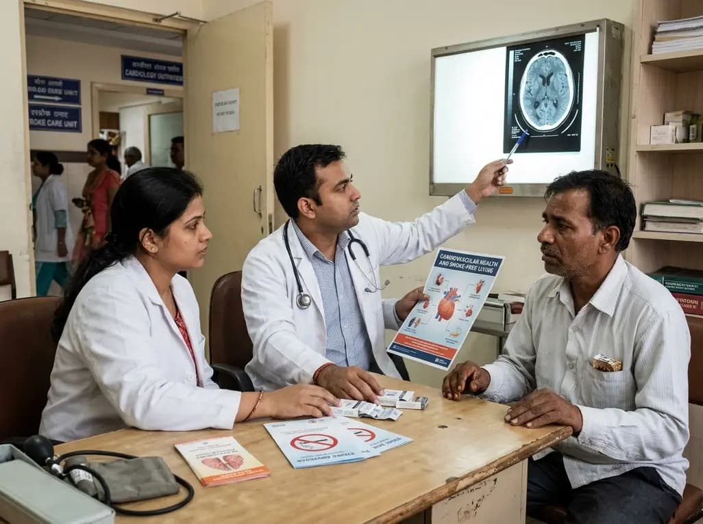
India faces an escalating challenge with vascular diseases, where tobacco use and hypertension together drive substantial cerebrovascular mortality. Addressing nicotine addiction as a clinical neurochemical disorder rather than a lack of willpower is vital for nationwide prevention and sustainable cessation support.
Today
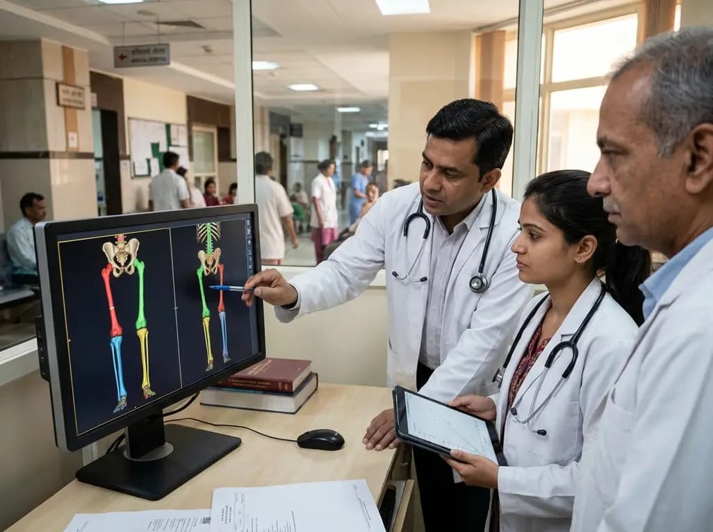
Modern 3D anthropometry reveals that pediatric leg growth ceases years before spinal elongation, exposing the clinical illusion of the classical Anderson-Green model. Learn how segmental growth dissociation improves surgical timing for epiphysiodesis in adolescent leg length discrepancy.
2 days back

With packaged goods giant HUL backing FSSAI's proposed front-of-pack red warning labels for foods high in fat, sugar, and salt, India takes a major step toward curbing non-communicable diseases. Learn how clearer nutrition disclosures empower clinicians, inform consumer choices, and drive dietary reform.
Today
