.webp&w=384&q=75)

Loading, please wait...
Loading, please wait...
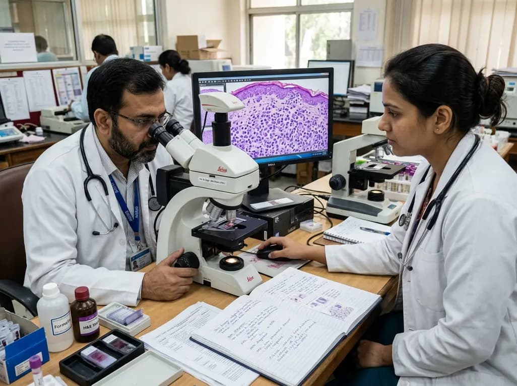
The clinical emergence of leukemia cutis in AML represents a well-documented extramedullary manifestation of myeloid malignancy. Although skin involvement occurs in a minority of acute myeloid leukemia cases, its presence carries profound diagnostic and clinical implications. Patients typically present with non-tender, violaceous, indurated plaques, firm papules, or discrete nodules distributed across the trunk, extremities, or facial regions. Furthermore, cutaneous infiltrates can emerge at varied chronological points during the patient's disease course. In some individuals, skin findings precede detectable peripheral blood or marrow blasts, an uncommon entity termed aleukemic leukemia cutis. In other patients, cutaneous lesions manifest synchronously with active bone marrow disease or signify an early extramedullary relapse following chemotherapy. Consequently, oncologists and dermatologists must maintain clinical vigilance when examining refractory or unexplained cutaneous eruptions. Because early lesions frequently mimic benign reactive inflammatory dermatoses or drug eruptions, prompt punch biopsy remains essential. Performing early histological evaluation accelerates systemic staging and prevents significant treatment delays. Therefore, recognizing these cutaneous signals empowers clinical teams to initiate timely systemic intervention and optimize outcomes.
Microscopic evaluation of cutaneous tissue sections demonstrates characteristic architectural patterns that guide definitive identification. Typically, malignant myeloid blasts assemble within the reticular dermis and frequently extend into deeper subcutaneous adipose tissue. In addition, these neoplastic cells often track in a single-file arrangement between preexisting dermal collagen bundles. Notably, a distinct subepidermal grenz zone separates the dense dermal aggregate from the overlying epidermis. Dermatopathologists rarely observe epidermotropism, which helps distinguish myeloid infiltration from epidermotropic cutaneous T-cell lymphomas. Cytologically, the infiltrating blasts exhibit moderate cytoplasm, finely dispersed chromatin, and irregular or indented nuclear membranes with prominent nucleoli. Mitotic figures and apoptotic bodies appear regularly throughout dense aggregates. However, architecture varies depending on whether granulocytic or monocytic differentiation predominates. Monocytic variants frequently display sheet-like expansions that efface normal dermal architecture, whereas immature myeloblastic lesions show accentuated perivascular and periadnexal distributions. Additionally, crush artifacts in superficial biopsies can obscure these diagnostic features. Pathologists must therefore examine adequate full-thickness biopsies to evaluate the complete dermal and subcutaneous compartments accurately.
Accurate diagnostic confirmation requires immunohistochemical validation of myeloid lineage. Historically, pathologists anticipated that cutaneous blasts would mirror the precise immunophenotype observed in bone marrow aspirates. However, contemporary cohort studies reveal substantial immunophenotypic discordance between cutaneous infiltrates and corresponding marrow populations. For example, traditional immature precursor markers, particularly CD34 and CD117, frequently show downregulation or total absence in skin specimens. Conversely, mature myelomonocytic markers, such as CD68, CD43, and lysozyme, demonstrate reliable and prominent immunoreactivity in dermal tissue. Furthermore, myeloperoxidase yields inconsistent expression, staining negative in a large percentage of monocytic cases. Diagnosticians also frequently detect CD56 in cutaneous infiltrates, where it promotes tissue homing and extravasation. Consequently, clinicians who rely exclusively on marrow flow cytometry profiles risk missing cutaneous disease. Dermatopathologists must therefore apply an extensive antibody battery comprising CD33, CD163, CD4, and CD123 alongside conventional myeloid markers. This balanced panel ensures diagnostic accuracy and clarifies confusing atypical presentations. In addition, recognizing phenotypic shift prevents misclassification of myeloid blasts as non-hematopoietic tumors.
Molecular investigations provide deep insight into the biology of cutaneous infiltration in acute myeloid leukemia. Specifically, clinical studies document an enrichment of NPM1 mutations and FLT3 internal tandem duplications among patients with skin manifestations. In addition, cytogenetic analyses frequently reveal recurrent anomalies, including trisomy 8 and complex karyotypic alterations. These genetic mutations stimulate cellular proliferation and alter cell-matrix adhesion, encouraging malignant blasts to exit the vascular tree and settle in dermis. Moreover, researchers identify activating mutations within the RAS pathway, such as NRAS and KRAS, which further drive extranodal migration. Historically, extramedullary involvement heralded an exceptionally adverse prognosis and shortened survival. Nonetheless, the development of targeted therapies has revolutionized treatment paradigms for these complex presentations. Clinicians now deploy selective FLT3 inhibitors and targeted epigenetic regulators to achieve improved systemic disease control. Therefore, routine next-generation sequencing on bone marrow and cutaneous biopsy specimens remains vital for contemporary patient stratification. Combining molecular data with morphology allows clinicians to tailor precision therapies and track minimal residual disease effectively.
Differentiating cutaneous myeloid disease from histopathologic mimickers requires vigilant clinicopathologic correlation. In daily clinical workflows, leukemic infiltrates often mimic inflammatory dermatoses, such as Sweet syndrome or leukemia cutis-like drug eruptions. However, while Sweet syndrome features prominent dermal edema and mature neutrophils with leukocytoclasia, leukemic infiltrates contain immature blasts displaying irregular nuclei and mitotic activity. In addition, diagnosticians must distinguish cutaneous AML from blastic plasmacytoid dendritic cell neoplasm. Both entities often express CD4 and CD56; therefore, pathologists rely on CD123, TCF4, and TCL1 to confirm dendritic lineage. Furthermore, non-Hodgkin lymphomas, poorly differentiated carcinomas, and malignant melanoma represent critical diagnostic pitfalls that require exclusion using lineage-specific panels. Once pathologists confirm cutaneous involvement, medical teams must initiate comprehensive systemic staging. Staging evaluations include repeat bone marrow biopsy, cytogenetics, and cerebrospinal fluid examination to assess central nervous system sanctuary sites. Prompt initiation of systemic induction chemotherapy remains the cornerstone of treatment, while allogeneic hematopoietic stem cell transplantation offers durable remission for eligible patients. Thus, coordinated multidisciplinary care ensures accurate diagnosis, rapid treatment deployment, and optimized patient survival.
Yes, leukemia cutis can present prior to detectable peripheral blood or bone marrow involvement, a rare phenomenon termed aleukemic leukemia cutis. In these challenging cases, neoplastic myeloid blasts home preferentially to cutaneous tissue before expanding systemically. However, virtually all untreated patients develop overt bone marrow disease within weeks to months. Consequently, prompt skin biopsy, immunohistochemistry, and immediate initiation of systemic chemotherapy remain vital to arrest systemic progression and improve survival.
Cutaneous leukemic blasts frequently exhibit immunophenotypic discordance due to microenvironmental adaptation and selective cellular differentiation during tissue migration. As leukemic cells enter the dermal microenvironment, they often downregulate immature progenitor markers like CD34 and CD117 while upregulating mature monocyte-associated antigens such as CD68 and CD163. Additionally, blasts acquire homing molecules like CD56 that facilitate tissue extravasation. Therefore, pathologists must utilize expansive immunohistochemical panels rather than relying solely on initial marrow flow cytometry results.
Patients with leukemia cutis show notable enrichment in specific genetic mutations, most frequently involving NPM1, FLT3, and RAS signaling pathway genes like NRAS and KRAS. Cytogenetic studies also reveal recurrent chromosomal abnormalities, particularly trisomy 8. These molecular alterations drive aggressive cellular growth, alter cell-matrix interactions, and promote blast invasion through endothelial barriers. Furthermore, identifying these specific mutations assists clinicians in selecting targeted molecular inhibitors, which refines prognostication and personalizes therapeutic management.
Disclaimer: This content is for informational and educational purposes only and does not constitute medical advice, diagnosis, or treatment recommendations. Clinical decisions should be made by qualified healthcare professionals based on individual patient evaluation and current medical standards. Refer to the latest local and national guidelines for clinical practice.
References
1. Erem AS et al. Acute Myeloid Leukemia Involving the Skin. J Cutan Pathol. 2026 Sep 20. doi: 10.1111/cup.70219. PMID: 42764255.
2. Cho-Vega JH, Medeiros LJ, Prieto VG, Vega F. Leukemia cutis. Am J Clin Pathol. 2008;129(1):130-142.
3. Cronin DMP, George TI, Sundram UN. An updated approach to the diagnosis of myeloid leukemia cutis. Am J Clin Pathol. 2009;132(1):101-110.
4. Wagner G, Fenchel K, Back W, Schulz A, Sachse MM. Leukemia cutis - epidemiology, clinical presentation, and differential diagnoses. J Dtsch Dermatol Ges. 2012;10(1):27-36.

Read summarized clinical updates, watch expert medical content, and earn CME certifications right from your smartphone.
Open your smartphone camera to scan and install.





A comprehensive clinical and histopathologic guide to leukemia cutis in acute myeloid leukemia, highlighting diagnostic criteria, immunophenotypic discordance, and molecular drivers.
Today

The Central Council for Research in Ayurvedic Sciences emphasizes that integrating traditional medicine with mainstream healthcare requires scientific rigor. Enhanced pharmacovigilance networks, adverse drug reaction monitoring, and validated clinical trials remain pivotal to elevating clinical credibility.
Today
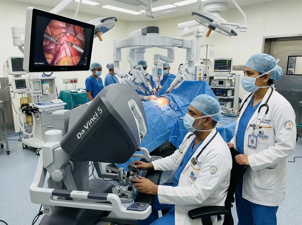
India expands its minimally invasive surgical landscape as Intuitive unveils its fifth-generation robotic platform. Equipped with real-time force feedback and immense computing performance, this innovation assists clinical specialists across urology, gynecology, and general surgery in optimizing operative care.
Today

Large-scale biobank research demonstrates that low-grade systemic inflammation quietly remodels ventricular architecture years before cardiovascular symptoms appear. Learn how emerging biomarkers like GlycA and targeted anti-inflammatory pathways could reshape preventive cardiovascular screening and clinical risk evaluation.
Today

Systematic review comparing labral repair and reconstruction in primary hip arthroscopy shows comparable short- to mid-term patient-reported outcomes, with lower THA conversion after repair but superior outcomes for reconstruction in patients aged 40 and older.
Today

A preclinical study reveals that alpinetin significantly alters glimepiride pharmacokinetics by inhibiting CYP2C9. This drug-herb interaction enhances hypoglycemic potency and underscores critical safety concerns regarding severe hypoglycemia in diabetic management.
Today
