.webp&w=384&q=75)

Loading, please wait...
Loading, please wait...

Femoroacetabular impingement and labral tears cause persistent hip pain and mechanical dysfunction in active individuals. Over the past two decades, primary hip arthroscopy has transformed surgical preservation of the native hip joint. When addressing labral pathology, orthopedic surgeons historically debated between debridement and anatomical refixation. Modern practice, however, focuses on restoring the acetabular fluid seal through either labral repair or reconstruction. Both procedures aim to alleviate symptoms, restore hip stability, and delay secondary degenerative joint disease.
Recent systematic evidence evaluates clinical efficacy following labral preservation versus replacement techniques. Researchers analyzed six comparative Level III investigations encompassing more than two thousand operative hips. Across follow-up periods ranging from two to nearly six years, both surgical approaches produced significant symptomatic improvements. Patients in both cohorts consistently achieved validated success metrics. Notably, achievement rates for the minimal clinically important difference and patient acceptable symptomatic state exceeded 70 percent in both groups. Direct comparisons demonstrated largely equivalent functional scores across standardized metrics, including the modified Harris Hip Score. Two included studies noted comparable postoperative gains between repair and reconstruction cohorts. Conversely, two investigations reported greater functional improvements favoring reconstruction, while one study observed higher adjusted scores for labral repair. Consequently, surgeons can reassure patients that both surgical approaches reliably improve hip joint function, decrease disability, and enhance overall quality of life.
Although labral repair remains the preferred primary treatment, specific intraoperative conditions warrant alternative solutions. Labral reconstruction replaces absent, ossified, or severely degenerated tissue with an autograft or allograft substitute. During primary hip arthroscopy, surgeons often encounter hypoplastic labra measuring less than four millimeters or complex irreparable tears. Under such circumstances, simple repair cannot adequately restore the critical acetabular fluid suction seal. Therefore, surgeons utilize graft tissues such as the iliotibial band or semitendinosus tendon to reconstruct anatomical labral volume. Restoring circumferential height stabilizes the femoroacetabular articulation and redistributes intra-articular contact pressures. In addition, surgeons favor reconstruction when encountering extensive chondrolabral disruption or severe intrasubstance degeneration. While technically demanding, arthroscopic reconstruction provides a robust biomechanical solution when native tissue cannot support anchor fixation. Thus, patient-specific anatomy and intraoperative tissue viability dictate the optimal operative strategy.
Patient age represents a decisive variable influencing surgical durability in hip preservation. In primary hip arthroscopy, clinical trajectories often diverge when evaluating patients chronologically. Systematic evidence indicates that patients aged 40 years and older derive distinct advantages from labral reconstruction. In one comparative cohort, patients aged 40 or older undergoing reconstruction achieved greater functional improvements than repair counterparts. Furthermore, this mature cohort experienced significantly lower overall surgical failure rates when treated with reconstruction. Older patients frequently present with chronic, degenerative tissue changes rather than clean, acute traumatic separations. In these individuals, native labral tissue often lacks sufficient cellularity and vascularity to support robust biological healing. Consequently, providing a pristine structural graft via reconstruction may enhance mechanical longevity. Conversely, younger patients with healthy tissue typically thrive following anatomical repair. Clinicians must therefore weigh chronological age alongside tissue quality during preoperative planning.
Joint survivorship represents an essential benchmark when comparing labral repair against reconstruction. Across published comparative trials, revision arthroscopy rates showed no significant differences between the two procedures. However, the risk of conversion to total hip arthroplasty revealed noteworthy distinctions between treatment groups. Systematic data demonstrate that labral repair yields lower overall rates of conversion to joint arthroplasty compared to reconstruction. This observed disparity likely reflects baseline selection bias rather than an inherent structural flaw of reconstruction. Patients selected for reconstruction typically present with more severe pre-existing articular cartilage loss and advanced chondrolabral disease. As a result, underlying joint osteoarthritis predisposes these hips to eventual prosthetic replacement. Additionally, graft remodeling requires time, which may accelerate degeneration if cartilage wear already exists. Surgeons must counsel patients that pre-existing chondral damage substantially elevates the mid-term likelihood of total hip replacement.
Achieving optimal surgical outcomes requires meticulous preoperative planning and precise arthroscopic execution. Surgeons must evaluate bony morphology on advanced imaging to identify underlying cam, pincer, or mixed femoroacetabular impingement. Failure to adequately resect osseous deformities increases mechanical stress on repaired or reconstructed labra, causing early failure. Intraoperatively, the arthroscopist must critically inspect tissue vascularity, width, and structural integrity before choosing fixation or replacement. Furthermore, surgeons must manage concurrent pathologies, including acetabular chondral defects and capsular laxity, during the index procedure. Routine capsular closure preserves hip stability and prevents iatrogenic microinstability. In the Indian orthopedic setting, where patients often present late with advanced joint degeneration, accurate risk stratification proves especially vital. Consequently, surgeons must avoid a rigid, standardized approach. Instead, they should individualize treatment based on patient age, baseline cartilage status, mechanical alignment, and tissue quality to optimize preservation.
Surgeons generally choose labral reconstruction when the native acetabular labrum is structurally deficient, hypoplastic, calcified, or severely degenerated. If native tissue cannot hold suture anchors or fails to restore the acetabular fluid seal, reconstruction provides necessary structural volume. Additionally, in patients aged 40 years and older with chronic degenerative tears, evidence suggests reconstruction may yield superior functional scores and lower failure rates compared to primary labral repair.
The lower rate of total hip arthroplasty conversion following labral repair largely stems from baseline patient selection rather than technical superiority. Surgeons typically reserve labral reconstruction for hips exhibiting more extensive labral damage and significant concomitant articular cartilage degeneration. Consequently, these joints already carry an elevated biological risk for progressive osteoarthritis. While repair preserves native tissue in healthier joints, severe pre-existing chondral breakdown in reconstruction cohorts predisposes patients to eventual arthroplasty.
Surgeons commonly utilize either autografts or allografts to reconstruct the acetabular labrum during arthroscopic surgery. Autograft options frequently include the iliotibial band and the gracilis or semitendinosus tendons, which offer excellent biological compatibility and eliminate disease transmission risks. Alternatively, anterior tibialis, semitendinosus, or fascia lata allografts are popular choices because they avoid donor-site morbidity and reduce operative time. Both graft sources demonstrate comparable biomechanical integrity and clinical success in restoring joint stability.
Disclaimer: This content is for informational and educational purposes only and does not constitute medical advice, diagnosis, or treatment. Healthcare professionals should rely on their clinical judgment, institutional protocols, and current evidence-based literature when making treatment decisions. Patient presentations and clinical circumstances vary; therefore, management strategies must be tailored to individual needs. The authors and publishers assume no liability for any injury, loss, or adverse outcomes resulting from the application of the information presented herein. Refer to the latest local and national guidelines for clinical practice.
References
Messer KP et al. Labral Repair and Reconstruction Yield Comparable Patient-Reported Outcomes at Short- to Mid-Term Follow-Up During Primary Hip Arthroscopy: A Systematic Review. Arthroscopy. 2026 Sep 20. doi: 10.1002/arj.70625. PMID: 42763809.
Bert T, Kraeutler MJ, Mei-Dan O. Clinical outcomes of arthroscopic hip labral reconstruction versus repair in the primary setting: A systematic review and meta-analysis. Arthroscopy. 2025;41(12):3450-3461.
Okoroha KR, Patel BH, Jildeh TR, et al. Two-Year Outcomes of Primary Arthroscopic Surgery in Patients with Femoroacetabular Impingement: A Comparative Study of Labral Repair and Labral Reconstruction. Am J Sports Med. 2024;52(11):2815-2824.

Read summarized clinical updates, watch expert medical content, and earn CME certifications right from your smartphone.
Open your smartphone camera to scan and install.





Systematic review comparing labral repair and reconstruction in primary hip arthroscopy shows comparable short- to mid-term patient-reported outcomes, with lower THA conversion after repair but superior outcomes for reconstruction in patients aged 40 and older.
Today

A systematic review evaluated molecular testing for UTIs across 12 clinical and microbiological studies. While PCR provides rapid results and symptom reduction, low specificity and industry sponsorship leave clinical utility uncertain.
Today
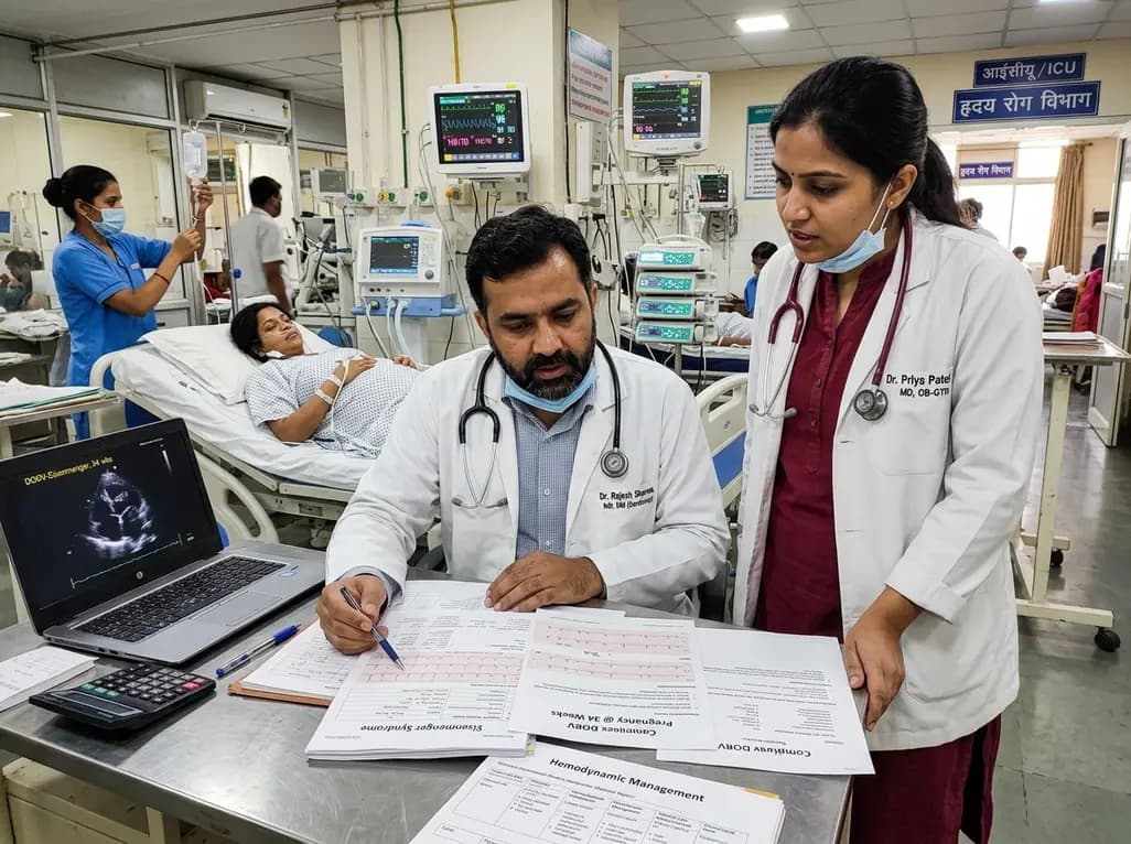
A recent case highlights favorable maternal and neonatal survival in a patient presenting at 34 weeks with probable Eisenmenger physiology and suspected double outlet right ventricle (DORV). Here is a critical review of hemodynamic challenges, delivery strategies, and peripartum management in complex cardiac disease.
Today

An updated systematic review and meta-analysis of 7,875 patients reveals that routine microaxial left ventricular support during nonemergent high-risk PCI does not reduce mortality or ischemic complications, while significantly increasing major bleeding events.
Yesterday
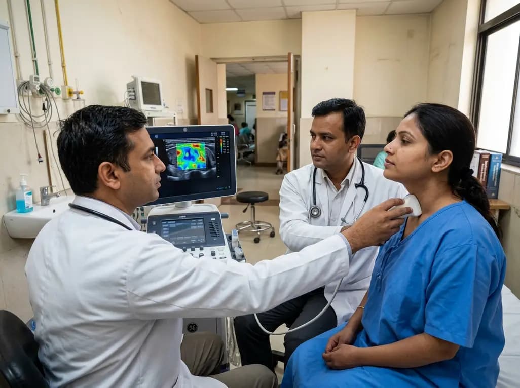
Subacute thyroiditis presents diagnostic dilemmas when differentiating it from malignancy or chronic thyroiditis. Discover how 2D shear wave elastography provides quantitative tissue stiffness metrics that enhance diagnostic accuracy and assist in tracking disease resolution during therapy.
Today
