.webp&w=384&q=75)

Loading, please wait...
Loading, please wait...

Anterior cervical discectomy and fusion remains the standard surgical intervention for degenerative cervical disc disease. Traditionally, spine surgeons combine anterior plates and screws with structural allograft bone to establish mechanical stability. However, the advent of the 3D-printed porous titanium cage introduces an innovative stand-alone alternative that eliminates plate-related instrumentation. Recent comparative evidence demonstrates that this porous additive technology yields equivalent radiologic fusion alongside meaningful clinical benefits, prompting clinicians to modernize their cervical reconstruction strategies.
Cervical degenerative disc disease frequently precipitates debilitating radiculopathy or progressive myelopathy, necessitating surgical decompression when medical therapy fails. For decades, conventional anterior reconstruction has paired structural allograft bone with metallic anterior plate-and-screw stabilization. This time-tested construct provides reliable rigid immobilization, which fosters robust osseous fusion across diverse patient populations. Nevertheless, anterior plate fixation introduces notable technical and anatomical concerns. Specifically, the anterior physical profile of the plate often impinges upon the adjacent esophagus, resulting in prolonged postoperative dysphagia. Furthermore, the extensive soft tissue retraction required for plate positioning elevates localized inflammation and prevertebral swelling. Surgeons also encounter potential risks of screw loosening, plate hardware migration, and adjacent segment ossification during extended follow-up. Because of these recognized complications, spine specialists have long pursued stand-alone interbody cages that remove the need for anterior plating. Early smooth polyetheretherketone or dense metallic cages occasionally displayed elevated risks of graft dislodgement or endplate subsidence. Consequently, modern biomedical engineering turned toward additive manufacturing to overcome these structural limitations. By engineering interconnected micro-porous architectures, contemporary 3D-printed cages provide primary frictional stability, ensuring dependable structural support without supplemental anterior fixation plates.
Additive manufacturing has revolutionized orthopedic implant design through laser powder bed fusion technology. Specifically, the novel 3D-printed porous titanium cage features an interconnected open-pore architecture that closely mimics native trabecular bone geometry. This biomimetic porosity promotes rapid osteoblast attachment, osteogenic differentiation, and extensive capillary infiltration directly throughout the scaffold core. In addition, the porous micro-structure effectively lowers the modulus of elasticity, aligning the cage much closer to natural vertebral bone stiffness. Consequently, this structural alignment markedly reduces pathological stress-shielding, which historically hampered solid metallic interbody spacers. Furthermore, the micro-textured exterior generates immediate frictional purchase against the cartilaginous vertebral endplates upon insertion. This primary stability maintains disc space distraction and lordotic alignment without requiring supplemental anchor screws. Laboratory biomechanical assessments confirm that porous titanium constructs distribute axial loads evenly across adjacent vertebral surfaces. Therefore, osseointegration occurs continuously through the central matrix rather than solely around the implant periphery. By combining physiological load transmission with active bone ingrowth properties, additively manufactured porous titanium delivers exceptional structural integrity while accelerating long-term biologic fixation. These osteoconductive features make the device an ideal platform for modern stand-alone cervical reconstruction.
A recent propensity-matched cohort study in Neurology India evaluated stand-alone 3D-printed porous cages against traditional plated allograft constructs over two years. The researchers reviewed sixty propensity-matched patients undergoing single-level cervical procedures to compare longitudinal clinical and radiologic metrics. Interestingly, the investigation demonstrated superior neck pain relief among patients who received the stand-alone porous titanium device. Visual analog scale scores for axial neck pain improved significantly more in the porous cage group across all follow-up time points. Although both surgical cohorts experienced comparable improvements in upper limb radicular pain and Neck Disability Index scores, axial neck symptoms resolved faster without plates. Spine specialists attribute this distinctive advantage to reduced surgical dissection and less traumatic soft tissue retraction. Because surgeons do not need to denude anterior vertebral surfaces for plate application, anterior cervical muscular structures endure less intraoperative traction injury. Additionally, intraoperative blood loss and postoperative wound drainage remained minimal in both treatment arms. Patients receiving the stand-alone device mobilized comfortably during early postoperative recovery. Thus, these findings highlight that avoiding anterior plates substantially enhances early patient comfort while preserving two-year functional outcomes.
Postoperative dysphagia represents one of the most common and distressing complications following anterior cervical surgery. Standard anterior plate systems project anteriorly beyond the vertebral boundary, where they continuously contact and irritate mobile retro-esophageal soft tissues. Conversely, the zero-profile stand-alone porous titanium spacer remains confined entirely within the decompressed intervertebral disc space. Consequently, the propensity-matched study reported a dramatic decrease in dysphagia incidence among patients managed with stand-alone cages. At postoperative day three, only ten patients in the porous cage group experienced swallowing difficulties, compared with nineteen patients in the plated cohort. By one month postoperatively, dysphagia persisted in just three stand-alone patients versus ten individuals with plates. Quantitative radiographic measurements confirmed that prevertebral soft tissue swelling was significantly smaller in the stand-alone group at day three, one month, and three months. By eliminating chronic mechanical friction against the pharyngoesophageal complex, the stand-alone construct preserves natural swallowing biomechanics. Furthermore, patients experience less pharyngeal discomfort and require fewer anti-inflammatory interventions during convalescence. Therefore, eliminating anterior hardware profiles effectively neutralizes a primary driver of anterior cervical surgical morbidity.
Concerns regarding pseudoarthrosis and endplate subsidence have historically surrounded stand-alone cervical implants. However, two-year radiologic follow-up conclusively demonstrated a one hundred percent solid bony fusion rate in both cohorts. Thin-cut computed tomography and dynamic flexion-extension radiographs confirmed continuous trabecular bridging through the porous titanium matrix without motion. Furthermore, cage subsidence occurred in 33.3% of porous cage patients compared to 30.0% in plated allograft patients, showing no statistical difference. Importantly, these radiographic subsidence events remained entirely asymptomatic and did not produce recurrent radicular symptoms. The study also revealed superior surgical efficiency with stand-alone implants, evidenced by significantly lower intraoperative fluoroscopy counts. Surgeons required an average of only 5.7 fluoroscopic images for porous cage seating, compared to 7.9 images for plate contouring and screw placement. This reduction lowers cumulative radiation exposure for operative staff and shortens anesthesia times. In high-volume Indian spine centers, streamlined procedures and reduced hardware requirements offer meaningful clinical and economic advantages. Consequently, stand-alone porous titanium cages deliver dependable fusion and exceptional safety, establishing an evidence-based alternative to conventional plated cervical fusion.
The 3D-printed porous titanium cage features a biomimetic trabecular architecture that closely resembles cancellous bone. This open microscopic network encourages immediate cellular adherence, capillary ingrowth, and osteointegration throughout the construct. Furthermore, high surface friction and physiological load distribution provide primary stability, facilitating solid fusion without anterior plating hardware.
Conventional anterior plates sit directly on the anterior vertebral body, protruding into retro-esophageal soft tissues and causing localized inflammation or friction. In contrast, stand-alone cages remain entirely within the intervertebral disc space. This low-profile positioning avoids esophageal irritation, significantly reducing prevertebral soft tissue swelling and accelerating the recovery of normal swallowing function.
Stand-alone porous cages are primarily indicated for single-level degenerative disc disease with preserved bone mineral density. In cases involving multi-level cervical spondylotic myelopathy, gross segmental instability, or severe osteoporosis, supplemental anterior plating or posterior instrumentation is still recommended to ensure construct rigidity and prevent pathological endplate subsidence or cage migration.
Disclaimer: This content is for informational and educational purposes only and does not constitute medical advice, diagnosis, or treatment. Healthcare professionals should exercise their independent clinical judgment when making treatment decisions. Patient details and clinical presentations may vary, and medical knowledge is continually evolving. Refer to the latest local and national guidelines for clinical practice.
References

Read summarized clinical updates, watch expert medical content, and earn CME certifications right from your smartphone.
Open your smartphone camera to scan and install.





A propensity-matched cohort study in Neurology India shows that stand-alone 3D-printed porous titanium cages in single-level ACDF achieve 100% fusion with significantly less dysphagia, smaller prevertebral swelling, and superior neck pain relief compared to traditional allograft and plating constructs.
Today
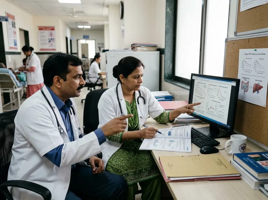
Recent research reveals that subclinical cholestasis, marked by elevated serum gamma-glutamyl transferase, reflects gut dysbiosis and predicts resistance to immune checkpoint blockade. Bile acid remodeling and mucosal MAdCAM-1 loss impair antitumor immunity, highlighting novel therapeutic targets.
Today
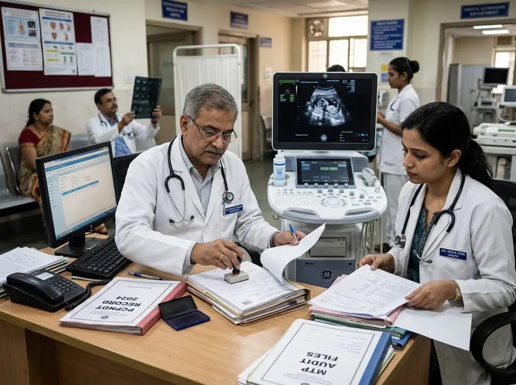
Maharashtra has initiated the appointment of two dedicated special public prosecutors in every district alongside a joint police-health SOP to fast-track PCPNDT and MTP cases. Medical imaging centers, radiologists, and obstetricians must strengthen statutory documentation and audit preparedness.
Today
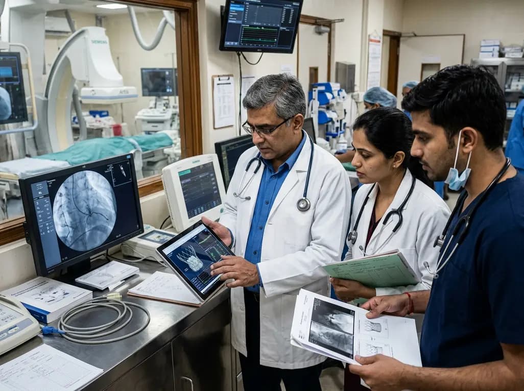
The US FDA has released an early safety alert regarding Edwards Lifesciences Commander M delivery catheters used in transcatheter mitral valve replacement. Reports of balloon bursting during deployment led to one death and one injury, prompting a recall of specific lots and urgent clinical inventory reviews.
Today
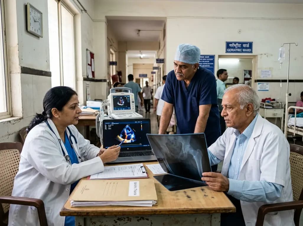
A meta-analysis evaluates the impact of preoperative transthoracic echocardiography on surgical delay and hospital stay in geriatric hip fracture patients. Evidence emphasizes balancing cardiac workups against operative urgency and adhering to clinical guidelines.
Today
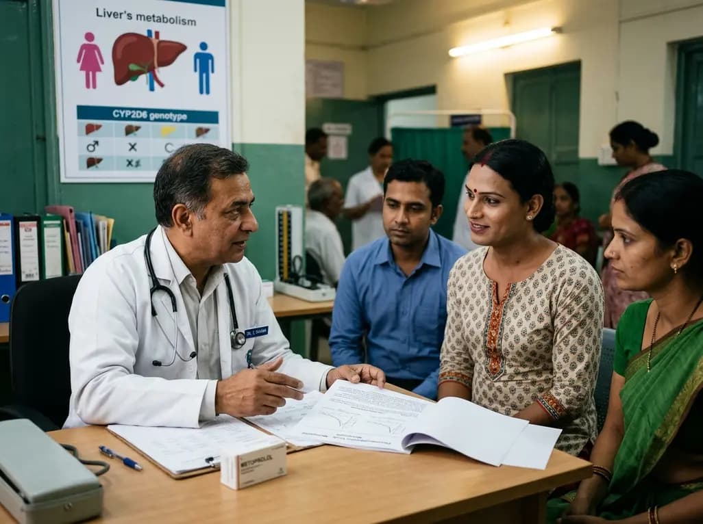
An exploratory pilot study evaluates metoprolol pharmacokinetics across cisgender males, cisgender females, and transgender women on gender-affirming hormone therapy. Findings reveal distinct drug disposition trends, emphasizing the critical need for genotype-stratified research in transgender cardiovascular care.
Today
