.webp&w=384&q=75)

Loading, please wait...
Loading, please wait...
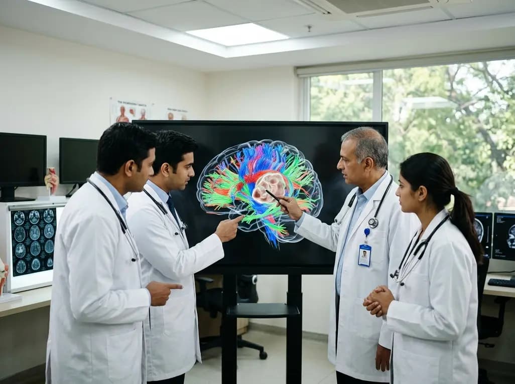
The complex landscape of neuro-oncology continues to evolve as clinicians and researchers seek better ways to preserve neurological function during aggressive tumor resections. Central to this challenge is the white matter tumor interaction, which defines how intracranial lesions affect the structural and functional integrity of associative fasciculi. Historically, surgeons relied primarily on anatomical landmarks and basic imaging to navigate the brain. However, modern neuroimaging, particularly diffusion tensor imaging (DTI), now allows for a more detailed visualization of subcortical pathways. Understanding how tumors displace, infiltrate, or destroy these tracts is paramount for optimizing surgical strategies. Specifically, the relationship between microstructural integrity, measured by fractional anisotropy (FA), and the physical morphometry of these tracts remains a subject of intense investigation. This knowledge directly impacts the preservation of critical functions like language and visual-spatial orientation. Consequently, medical educators emphasize the need for standardized DTI protocols to characterize these interactions across various tumor histotypes. By analyzing these relationships, neurosurgeons can better predict postoperative outcomes and tailor their approaches to individual patient anatomy.
To deepen the understanding of tract alterations, researchers conducted a multicenter retrospective study involving 156 patients. Each participant underwent surgery for brain tumors located near the inferior fronto-occipital fasciculus (IFOF) or the arcuate fasciculus (AF). The research team utilized standardized DTI protocols to perform preoperative deterministic tractography, ensuring consistency across different clinical sites. Moreover, they employed linear mixed-effects models to assess the relationship between fractional anisotropy and various morphometric parameters, such as tract volume and length. These models included adjustments for relevant covariates, including tumor type and hemispheric location. Therefore, the study provided a robust framework for evaluating how different lesions influence white matter architecture. By focusing on both the microstructural level through FA and the macrostructural level through volume, the investigators could capture a comprehensive picture of tumor impact. This methodological rigor allows for more reliable comparisons between low-grade gliomas, high-grade gliomas, and brain metastases. Furthermore, the use of deterministic tractography highlights the practical utility of these tools in everyday clinical environments in India and globally.
The study results revealed a significant and strong association between higher fractional anisotropy and increased tract volume in both the AF and IFOF. Specifically, the data showed that microstructural integrity, as represented by FA, reflects the overall physical presence and health of the white matter bundles. Additionally, researchers observed that FA positively correlated with tract length, suggesting that higher anisotropy often accompanies longer, better-preserved fibers. This finding is particularly important because it suggests that FA serves as a reliable surrogate marker for the degree of tract preservation. In contrast, lower FA values typically indicated significant tract disruption or infiltration by the tumoral process. Furthermore, the study noted that tumor type significantly influenced these associations. For example, the relationship between FA and morphometry varied between primary brain tumors and metastatic lesions. Consequently, these findings underscore the necessity of integrating both quantitative DTI metrics and volumetric data when planning surgical interventions. This integrated approach provides a more nuanced understanding of the structural challenges posed by different intracranial masses, ultimately guiding safer resections.
Understanding the white matter tumor interaction has direct implications for neurosurgical planning and patient counseling. Notably, high-grade gliomas (HGG) often exhibit more aggressive infiltration compared to low-grade gliomas (LGG) or metastases. This infiltration typically results in a more pronounced reduction in FA and a subsequent decrease in tract volume. Therefore, recognizing these patterns preoperatively helps surgeons decide between radical resection and more conservative, function-preserving strategies. Additionally, the study highlighted that the arcuate fasciculus, which is vital for language processing, often shows unique patterns of displacement versus destruction depending on the tumor's location. Similarly, the IFOF, essential for semantic processing and visual integration, requires careful monitoring to avoid permanent deficits. Furthermore, the researchers found that metastases often cause displacement rather than direct axonal destruction, which may explain the potential for better functional recovery in some cases. Consequently, the preoperative assessment of FA and tract morphometry provides a roadmap for navigating these eloquent areas. By using this data, surgical teams can maximize the extent of resection while minimizing the risk of devastating postoperative neurological impairments.
As tractography becomes more prevalent in clinical practice, the importance of standardized imaging protocols cannot be overstated. The study demonstrated that consistent DTI acquisition allows for more accurate longitudinal monitoring of white matter changes. Moreover, standardized metrics like FA and tract volume facilitate better communication between multidisciplinary teams, including radiologists, oncologists, and neurosurgeons. Therefore, medical institutions in India should strive to implement these advanced imaging techniques as part of their routine preoperative workup for brain tumors. Additionally, the integration of these findings into neuronavigation systems allows for real-time visualization of tracts during surgery. This technological synergy enhances the surgeon's ability to identify critical boundaries between the lesion and functional pathways. Furthermore, ongoing research into probabilistic tractography may offer even greater precision in areas of high edema or complex fiber crossing. Ultimately, the goal remains the same: to leverage every available piece of microstructural information to improve patient safety. Consequently, continued education on tractography interpretation is essential for the next generation of neuro-oncology specialists.
The field of neuroimaging is rapidly advancing, with new techniques offering even deeper insights into white matter health. While FA remains a cornerstone of DTI, other metrics like mean diffusivity and axial anisotropy are gaining traction as complementary markers. Additionally, researchers are exploring the role of neuroplasticity in the context of white matter tumor interaction. This includes investigating how the brain might reorganize its pathways in response to slow-growing lesions like low-grade gliomas. Furthermore, the development of artificial intelligence algorithms to automate tract segmentation could significantly reduce the time required for preoperative analysis. Therefore, the future of neuro-oncology lies in a more personalized, data-driven approach to brain tumor management. By combining quantitative imaging with functional mapping, clinicians can offer patients more accurate prognoses and better-tailored treatment plans. Notably, the integration of advanced DTI into clinical trials will help validate these metrics as primary endpoints for evaluating treatment efficacy. Consequently, the ongoing evolution of tractography will continue to reshape our understanding of brain connectivity and its resilience in the face of disease.
Fractional anisotropy (FA) measures the directionality of water diffusion in white matter. In neuro-oncology, higher FA values usually indicate intact, healthy nerve fibers, while lower values suggest tract infiltration or destruction. This metric helps surgeons identify functional pathways that must be avoided during tumor resection to preserve neurological capabilities.
High-grade gliomas often infiltrate and destroy white matter tracts, significantly reducing FA and tract volume. In contrast, metastases frequently cause mass effect, displacing tracts without necessarily destroying the axonal structure initially. Understanding these differences allows neurosurgeons to choose the most appropriate surgical approach for each specific tumor histotype.
The arcuate fasciculus is critical for language, while the inferior fronto-occipital fasciculus (IFOF) supports semantic processing and visual-spatial functions. Because these tracts are often located near common tumor sites, mapping them with tractography is essential to prevent permanent deficits in speech, comprehension, and vision following surgical intervention.
Disclaimer: This content is for informational and educational purposes only. It is not intended to be a substitute for professional medical advice, diagnosis, or treatment. Always seek the advice of your physician or other qualified health provider with any questions you may have regarding a medical condition. Refer to the latest local and national guidelines for clinical practice.
References
Altieri R et al. Rethinking white matter-tumor interaction: a tractography based analysis of associations between fractional anisotropy and morphometry in the IFOF and arcuate fasciculus. J Neurooncol. 2026 Jul 07. doi: 10.1007/s11060-026-05691-4. PMID: 42412124.
Li, Z. et al. (2013). Diffusion tensor tractography of the arcuate fasciculus in patients with brain tumors: Comparison between deterministic and probabilistic models. Journal of Biomedical Science and Engineering, 6.
Sarubbo S. et al. (2025). Tractography in brain tumor surgery: current clinical impact and future challenges. PubMed Central.

Read summarized clinical updates, watch expert medical content, and earn CME certifications right from your smartphone.
Open your smartphone camera to scan and install.





A multicenter study highlights the strong association between fractional anisotropy and white matter tract morphometry in patients with brain tumors, offering new insights for surgical planning and understanding tumor-related microstructural changes in critical pathways like the IFOF and arcuate fasciculus.
3 months ago
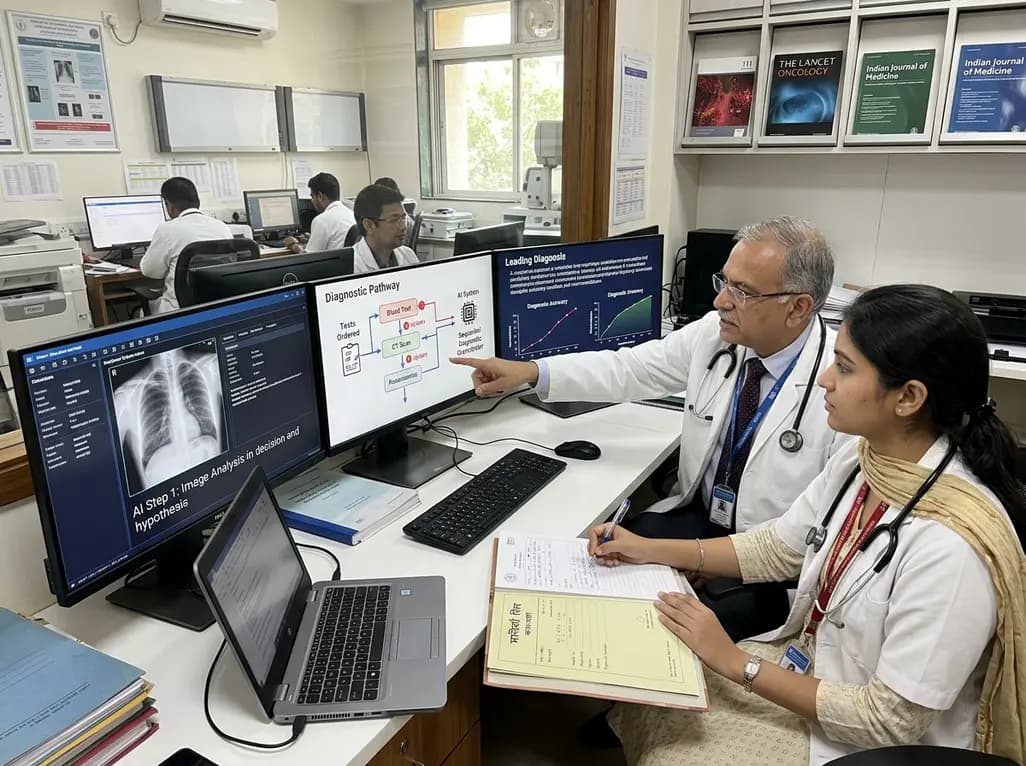
A breakthrough peer-reviewed study demonstrates that a sequential AI diagnosis system can mirror real-world clinical reasoning. By ordering stepwise tests and updating hypotheses dynamically, this multi-agent orchestrator achieved superior diagnostic accuracy while reducing investigative costs.
Today

A prenatal case highlights severe glutaric acidemia type II presenting as bilateral hyperechoic cystic kidneys at 18 weeks. Whole-exome sequencing revealed a novel homozygous ETFDH frameshift variant, demonstrating the power of rapid genetic testing in fetal renal anomalies.
Today

Review how tailored exercise programs benefit frail older patients with heart failure. Explore pathophysiological mechanisms, individualized training prescriptions, and strategies to overcome adherence barriers in clinical practice.
Yesterday

This comparative study evaluates posterior labral tears with and without glenohumeral instability, highlighting distinct demographic patterns, MRI underestimation in stable shoulders, and successful arthroscopic repair outcomes.
Yesterday

A clinical study demonstrates that early intervention with tocilizumab, either as monotherapy or bridged with corticosteroids, achieves significant proptosis reduction and resolves active inflammation in treatment-naive thyroid eye disease.
Yesterday
