.webp&w=384&q=75)

Loading, please wait...
Loading, please wait...
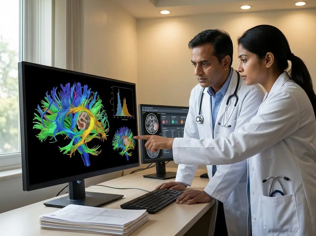
Neuro-oncological surgery has recently witnessed a major paradigm shift toward maximizing functional outcomes rather than focusing solely on tumor volume reduction. Consequently, white matter tractography stands at the forefront of this clinical transformation, offering a non-invasive window into the brain\'s complex architectural network. Historically, surgeons relied almost entirely on anatomical landmarks for guidance during resection. However, these landmarks often shift significantly due to the mass effect exerted by growing tumors. Today, advanced diffusion tensor imaging (DTI) provides critical insights into the microstructural health of associative fasciculi. Furthermore, understanding the intimate interaction between neoplasms and white matter is essential for preserving eloquent functions like language, vision, and movement. Recent research emphasizes that the relationship between fractional anisotropy (FA) and tract morphometry remains highly complex and varies across pathologies. Specifically, the Inferior Fronto-Occipital Fasciculus (IFOF) and the Arcuate Fasciculus (AF) represent two of the most critical pathways involved in high-level human cognition. As tumor cells infiltrate or displace these fibers, their physical dimensions and integrity undergo measurable changes. This study highlights how multicenter retrospective analyses help clarify these interactions across various tumor histotypes. By utilizing advanced white matter tractography, clinicians can better predict how different tumors, from glioblastomas to metastases, affect the brain\'s connectivity. Ultimately, this leads to more personalized surgical strategies and improved patient quality of life.
To appreciate the clinical utility of white matter tractography, one must first understand the significance of fractional anisotropy. FA serves as a scalar value that describes the degree of anisotropy of a diffusion process. In the context of the brain, it reflects the directionality of water diffusion along myelinated axons. Specifically, high FA values typically indicate healthy, well-organized white matter where diffusion occurs primarily along the axis of the fibers. Conversely, lower FA values often signal a loss of microstructural integrity, which may result from edema, infiltration, or physical destruction by a tumor. Moreover, the relationship between FA and tract volume is not always linear. Notably, in the presence of a lesion, the FA value provides a snapshot of the remaining functional capacity of a fasciculus. Researchers have found that higher FA is strongly associated with both AF and IFOF tract volume. This association suggests that as the microstructure remains intact, the overall macroscopic dimensions of the tract are preserved. Additionally, this study demonstrated that FA also correlates with tract length, emphasizing that integrity affects the entire span of the fasciculus. Therefore, monitoring FA throughout the preoperative phase allows clinicians to gauge the severity of tract involvement. This microstructural assessment proves invaluable for differentiating between tracts that are merely displaced and those that have undergone irreversible degradation.
Morphometric analysis involves measuring the physical characteristics of white matter tracts, such as volume and length. In the presence of intracranial tumors, these parameters often change in predictable and unpredictable ways. For instance, the Inferior Fronto-Occipital Fasciculus (IFOF), which connects the frontal and occipital lobes, is frequently displaced by tumors in the temporal or insular regions. Similarly, the Arcuate Fasciculus (AF), a key component of the language network, is highly susceptible to displacement and infiltration. The study utilized linear mixed-effects models to assess how FA relates to these morphometric changes. Interestingly, the researchers adjusted for relevant covariates to ensure the accuracy of their findings. They discovered that higher FA values consistently mapped to greater tract volumes and lengths across the cohort. Furthermore, this relationship was particularly evident in the IFOF, which appeared more sensitive to microstructural changes than the AF in certain tumor types. These morphometric alterations do not occur in isolation but are part of a broader reorganize process within the peritumoral environment. As a result, surgeons must consider both the physical location and the microstructural health of these tracts. Failing to account for these changes could lead to the accidental disruption of critical pathways during surgery. Consequently, preoperative mapping through white matter tractography has become an indispensable tool for protecting these vital neural highways.
One of the most compelling findings of recent research is how different tumor histotypes impact white matter tracts uniquely. Specifically, the study compared glioblastomas, metastases, and meningiomas to see how they altered the relationship between FA and morphometry. Glioblastomas are notoriously infiltrative, often invading white matter tracts long before physical changes are visible on standard MRI. In contrast, metastases tend to be more circumscribed, typically displacing tracts rather than infiltrating them directly. However, the study revealed that the interaction between tumor type and FA had a significant effect on tract volume. For example, the association between FA and IFOF volume was significantly stronger in patients with metastases compared to those with glioblastomas. This suggests that in metastatic disease, the physical volume of the tract remains more closely tied to its microstructural health. Conversely, the infiltrative nature of glioblastomas may decouple this relationship, as the tract might retain some volume despite significant microstructural damage. Furthermore, the analysis showed that tumor location within a specific hemisphere also played a role in these dynamics. These findings underscore the importance of histotype-specific surgical planning. By recognizing that a glioblastoma might require a different tractographic interpretation than a metastasis, neurosurgeons can better navigate the delicate balance between resection and preservation. Therefore, histotype-driven analysis of white matter tractography represents a significant step forward in personalized neuro-oncology.
The practical application of white matter tractography in the operating room cannot be overstated. By integrating DTI data into neuronavigation systems, surgeons can visualize critical tracts in real-time. This visualization allows for a more aggressive resection of the tumor while maintaining a safe distance from eloquent white matter. Specifically, the preservation of the Arcuate Fasciculus is vital for maintaining language fluency and comprehension. If a surgeon knows that the AF is displaced medially, they can adjust their surgical approach to enter the tumor from a lateral corridor. Moreover, the quantitative data provided by FA values can help determine the "resection limit." If the FA value drops significantly at the tumor-tract interface, it may indicate a zone of infiltration where the risk of permanent deficit is high. Consequently, the surgeon might choose a subtotal resection in that specific area to preserve function. Additionally, intraoperative mapping with subcortical stimulation often correlates well with preoperative tractography findings. This dual approach provides a "safety net" for the patient, ensuring that the visual map produced by DTI matches the functional reality observed during surgery. Ultimately, the goal is to achieve a maximal safe resection, which is the cornerstone of modern neuro-oncology. The insights gained from analyzing 156 patients in this study confirm that white matter tractography is a robust and reliable tool for achieving this objective.
Looking ahead, the integration of advanced white matter tractography into the multidisciplinary care of brain tumor patients will only deepen. Beyond surgical planning, these metrics offer potential for monitoring treatment response and predicting long-term outcomes. For instance, serial DTI scans could track the recovery of white matter integrity following chemotherapy or radiation. Furthermore, as artificial intelligence and machine learning algorithms improve, the automated segmentation of the IFOF and AF will become faster and more accurate. Notably, this will allow for the widespread adoption of tractography even in centers without specialized neuroradiologists. Additionally, understanding the baseline microstructural health of the brain may help identify patients who are at higher risk for postoperative cognitive decline. By using FA and morphometry as biomarkers, clinicians can tailor rehabilitation programs to the specific needs of the individual. In conclusion, this study provides a foundational understanding of how tumor type and microstructural integrity interact to shape the brain\'s architecture. It highlights the need for a nuanced approach to imaging that goes beyond simple tumor localization. As we continue to rethink white matter-tumor interactions, the use of white matter tractography will remain central to our efforts to improve the lives of patients facing these challenging diagnoses. Indeed, the future of neuro-surgery lies in our ability to see not just the tumor, but the intricate web of connections that define the human experience.
Fractional anisotropy measures the directionality of water diffusion, which is often high in healthy white matter and low in disrupted tissue. By mapping FA values near a tumor, clinicians can identify areas where the white matter is being infiltrated or destroyed, helping to define the physiological boundaries of the lesion beyond what is visible on standard scans.
The IFOF and arcuate fasciculus are long, associative tracts that traverse multiple brain regions, making them highly likely to be encountered by growing intracranial tumors. Because they support essential cognitive functions like language and semantic processing, any physical displacement or microstructural damage caused by a tumor can lead to significant clinical deficits for the patient.
While tractography shows the physical path of fibers, combining it with fractional anisotropy (FA) and mean diffusivity (MD) helps distinguish between these states. Displaced tracts usually maintain relatively high FA, whereas infiltrated tracts show a significant drop in FA as tumor cells disrupt the organized bundle of axons, indicating a higher risk during resection.
Disclaimer: This content is for informational and educational purposes only and does not constitute professional medical advice, diagnosis, or treatment. Always seek the advice of your physician or other qualified health provider with any questions you may have regarding a medical condition. Refer to the latest local and national guidelines for clinical practice.
References
Altieri R et al. Rethinking white matter-tumor interaction: a tractography based analysis of associations between fractional anisotropy and morphometry in the IFOF and arcuate fasciculus. J Neurooncol. 2026 Jul 07. doi: 10.1007/s11060-026-05691-4. PMID: 42412124.
Rueckriegel SM, Bruhn H, Thomale UW, Driever PH. Cerebral white matter fractional anisotropy and tract volume as measured by MR imaging are associated with impaired cognitive and motor function in pediatric posterior fossa tumor survivors. Pediatr Blood Cancer. 2015;62(7):1232-1240.
Abhinav K, Yeh FC, Mansouri A, Zadeh G, Fernandez-Miranda JC. High-definition fiber tractography for the evaluation of perilesional white matter tracts in high-grade glioma surgery. Neuro Oncol. 2015;17(9):1199-1209.

Read summarized clinical updates, watch expert medical content, and earn CME certifications right from your smartphone.
Open your smartphone camera to scan and install.





This multicenter study explores the association between fractional anisotropy (FA) and morphometry in the IFOF and arcuate fasciculus across various brain tumor types, highlighting how histotypes like glioblastoma and metastases uniquely impact white matter integrity.
3 months ago

A breakthrough study evaluated H5N1 transmission efficiency across mammalian isolates. Researchers discovered that human-derived D1.1 harboring the PB2 D701N mutation spread efficiently in ferrets, unlike strains carrying PB2 E627K. This finding highlights critical genetic markers for zoonotic surveillance.
Today

A comparative cohort study evaluates vNOTES vs TLH hysterectomy for benign conditions, demonstrating shorter operative duration, faster return to daily activities, and equivalent female sexual function scores without increased dyspareunia.
Today

Review how tailored exercise programs benefit frail older patients with heart failure. Explore pathophysiological mechanisms, individualized training prescriptions, and strategies to overcome adherence barriers in clinical practice.
Yesterday

This comparative study evaluates posterior labral tears with and without glenohumeral instability, highlighting distinct demographic patterns, MRI underestimation in stable shoulders, and successful arthroscopic repair outcomes.
Yesterday

A clinical study demonstrates that early intervention with tocilizumab, either as monotherapy or bridged with corticosteroids, achieves significant proptosis reduction and resolves active inflammation in treatment-naive thyroid eye disease.
Today
