.webp&w=384&q=75)

Loading, please wait...
Loading, please wait...
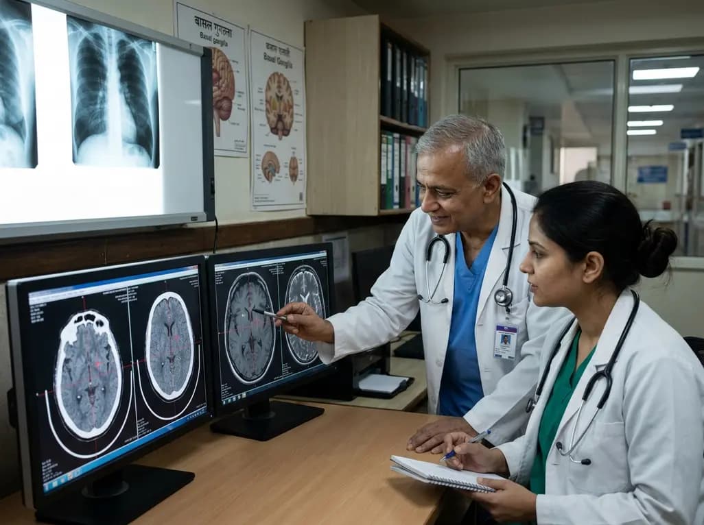
Deep brain stimulation (DBS) has revolutionized the management of medically refractory essential tremor and Parkinsonian tremor. Specifically, targeting the ventral intermediate thalamic nucleus (Vim) serves as the gold standard for achieving significant tremor suppression. In many clinical settings, surgeons must choose between different imaging modalities to guide the electrode placement. While magnetic resonance imaging (MRI) offers superior soft-tissue contrast, computed tomography (CT) remains a robust tool in frame-based stereotaxy. Consequently, the debate persists regarding which modality yields better functional outcomes for the patient. Understanding the nuances of Vim DBS targeting techniques is essential for neurosurgeons aiming to optimize surgical precision. This is particularly relevant in high-volume centers where both resources might be available but workflow efficiency is paramount. Ultimately, the primary goal of any targeting strategy is to ensure accurate electrode positioning within the motor portion of the thalamus while minimizing side effects. Therefore, comparative studies play a vital role in validating existing protocols. By examining long-term outcomes, clinicians can determine if one imaging modality offers a statistically significant advantage over the other in real-world practice.
Historically, the localization of the Vim nucleus relied heavily on indirect targeting methods. These techniques utilize the anterior commissure-posterior commissure (AC-PC) line and stereotactic atlases to estimate the target's coordinates. However, individual anatomical variability often challenges the precision of these atlas-based estimates. In contrast, modern Vim DBS targeting techniques have evolved to include direct visualization through advanced MRI sequences. Specifically, proton density-weighted imaging or susceptibility-weighted imaging can sometimes delineate the thalamic substructures. Despite these advancements, many centers still rely on CT-guided frame-based registration due to its high geometric accuracy and compatibility with stereotactic frames. Furthermore, intraoperative microelectrode recording (MER) and macrostimulation are frequently employed to refine the final lead position regardless of the initial imaging choice. This physiological mapping acts as a crucial safety net, compensating for potential discrepancies between imaging and functional anatomy. Moreover, the choice of modality often depends on institutional expertise and the specific hardware available at the facility. For instance, some surgeons prefer the speed and reliability of CT when using a traditional stereotactic frame. Meanwhile, others advocate for the anatomical detail provided by MRI to reduce reliance on intraoperative testing. Consequently, establishing the equivalence of these methods is necessary for standardized neurosurgical care.
A recent retrospective analysis investigated whether a statistically significant difference exists between CT-guided and MRI-guided targeting for Vim DBS. Researchers evaluated a series of patients who underwent surgery over a seven-year period at a specialized neurosurgical department. Notably, the study focused on the Fahn-Tolosa-Marin Tremor Scale (FTM TS) to measure clinical efficacy. The results indicated that both groups experienced substantial improvements in tremor severity following the procedure. Specifically, the pre-surgery mean scores were comparable between the CT and MRI cohorts. Following the intervention, the 1-year assessment revealed that the reduction in tremor was not significantly different between the two imaging groups. This suggests that the precision achieved with CT-guided targeting is effectively on par with MRI-guided methods when integrated into a comprehensive surgical workflow. Furthermore, statistical tests confirmed that neither group held a significant advantage in terms of raw score reduction. Therefore, clinicians can remain confident in the use of CT when MRI is contraindicated or logistically difficult to obtain. Additionally, these findings underscore the importance of the surgeon's expertise and the use of supplementary physiological markers during lead placement. As a result, the choice between CT and MRI may be driven more by practical considerations than by differences in therapeutic efficacy.
Beyond the objective measurement of tremor amplitude, the impact of Vim DBS targeting techniques on a patient's quality of life is a critical consideration. The EQ-5D score, a standardized instrument for measuring health-related quality of life, was utilized to compare the two groups. Interestingly, both the CT and MRI patient groups showed marked improvements in their EQ-5D scores at the one-year follow-up. Specifically, the median difference between pre- and post-surgery scores was identical for both groups in some parameters. This parity indicates that the subjective benefit perceived by the patient is not influenced by the primary imaging modality used for planning. Furthermore, functional improvements in activities of daily living, such as writing and eating, were consistently observed across the entire cohort. Consequently, the focus remains on the accuracy of the final electrode position within the thalamic circuit rather than the specific pixels used to find it. Moreover, the lack of significant difference in quality of life scores suggests that both methods successfully target the relevant neural networks. In addition, the stability of these outcomes over a year points to the durability of the tremor suppression achieved through either planning route. Therefore, the decision-making process for surgical planning can safely incorporate institutional preference without compromising patient-centered outcomes.
When implementing Vim DBS targeting techniques, several technical factors must be addressed to ensure patient safety. CT-guided planning typically involves a fusion process where the pre-operative MRI is registered to an intraoperative frame-based CT scan. While this method is highly accurate, it introduces the risk of registration errors or image fusion artifacts. On the other hand, direct MRI-based planning avoids the fusion step but may be susceptible to magnetic field distortions. Specifically, these distortions can affect the geometric fidelity of the images, potentially leading to targeting inaccuracies if not properly calibrated. However, the study results suggest that these theoretical risks do not translate into clinical differences for the majority of patients. Furthermore, the integration of microelectrode recording provides a functional validation that transcends the limitations of static imaging. Specifically, the characteristic firing patterns of the Vim neurons help the surgical team confirm the lead's location in real-time. Additionally, intraoperative testing allows for the immediate assessment of side effects, such as paresthesia or dysarthria. Consequently, the surgical workflow effectively mitigates the minor technical variances between imaging modalities. As a result, the high success rate of Vim DBS remains consistent across different technological approaches. Therefore, the robustness of the procedure is a testament to the complementary nature of imaging and neurophysiology.
The findings regarding Vim DBS targeting techniques have significant implications for functional neurosurgery in India. Given the diverse infrastructure across various medical centers, the validation of CT-guided targeting is particularly valuable. Specifically, in many regions, access to high-field 3T MRI units for stereotactic planning may be limited or cost-prohibitive. Consequently, the ability to achieve excellent clinical results using CT-guided frames ensures that DBS remains a viable option for more patients. Furthermore, the equivalence in outcomes allows neurosurgeons to adopt more flexible workflows based on available resources. For instance, a center may choose to use MRI for initial target identification and CT for the day-of-surgery frame registration. This hybrid approach leverages the strengths of both modalities while managing costs and scheduling constraints. Moreover, training programs for neurosurgical residents can continue to emphasize the importance of traditional stereotactic principles alongside modern imaging. Ultimately, the objective is to broaden the accessibility of life-changing therapies for essential tremor. Therefore, these results support the continued use of diverse targeting methodologies in a variety of clinical settings. By prioritizing functional accuracy and patient safety, Indian neurosurgeons can maintain world-class standards in movement disorder surgery.
Yes, research indicates that CT-guided targeting for Vim DBS provides clinical outcomes comparable to MRI-guided methods. While MRI offers better visualization of brain tissue, CT remains the gold standard for geometric accuracy in frame-based stereotaxy. When combined with microelectrode recording, both modalities ensure high precision in lead placement for tremor control.
Vim DBS significantly reduces tremor amplitude, which directly enhances the patient's ability to perform essential daily tasks like writing, drinking, and self-care. Standardized quality of life assessments, such as the EQ-5D, consistently show marked improvement post-surgery, reflecting the profound positive impact on the patient's independence and emotional well-being.
The Fahn-Tolosa-Marin Tremor Scale (FTM TS) is the most frequently used tool for objectively measuring tremor severity in clinical studies. Additionally, the Essential Tremor Rating Assessment Scale (TETRAS) is often used due to its validated sensitivity. These scales allow clinicians to quantify the degree of improvement and compare different surgical techniques accurately.
Disclaimer: This content is for informational and educational purposes only and does not constitute medical advice, diagnosis, or treatment. Always seek the advice of a qualified healthcare provider regarding a medical condition. Refer to the latest local and national guidelines for clinical practice.
References
Gough M et al. Locating the ventral intermediate thalamic nucleus for deep brain stimulation surgery: analysis of a case series comparing CT and MR targeting. Br J Neurosurg. 2025 Oct. doi: 10.1080/02688697.2024.2313674. PMID: 38372013.
Benabid AL et al. Long-term suppression of tremor by chronic stimulation of the ventral intermediate thalamic nucleus of the thalamus. Lancet. 1991;337(8738):403-406. doi: 10.1016/0140-6736(91)91179-8.
Gross RE et al. Advances in neuroimaging for deep brain stimulation. Neurosurgery. 2006;58(suppl_4):ONS-260. doi: 10.1227/01.NEU.0000205545.91896.7E.

Read summarized clinical updates, watch expert medical content, and earn CME certifications right from your smartphone.
Open your smartphone camera to scan and install.





A comparative study of CT and MRI-guided targeting for Vim-DBS in tremor patients reveals no significant difference in clinical outcomes or quality of life at one year, supporting the reliability of both imaging modalities in functional neurosurgery.
3 months ago

Discover the crucial role of glymphatic dysfunction in stroke, where perivascular clearance failure and neuroinflammation exacerbate cerebral edema and post-stroke brain injury.
Today

A prenatal case highlights severe glutaric acidemia type II presenting as bilateral hyperechoic cystic kidneys at 18 weeks. Whole-exome sequencing revealed a novel homozygous ETFDH frameshift variant, demonstrating the power of rapid genetic testing in fetal renal anomalies.
Today

Review how tailored exercise programs benefit frail older patients with heart failure. Explore pathophysiological mechanisms, individualized training prescriptions, and strategies to overcome adherence barriers in clinical practice.
Yesterday

This comparative study evaluates posterior labral tears with and without glenohumeral instability, highlighting distinct demographic patterns, MRI underestimation in stable shoulders, and successful arthroscopic repair outcomes.
Yesterday

A clinical study demonstrates that early intervention with tocilizumab, either as monotherapy or bridged with corticosteroids, achieves significant proptosis reduction and resolves active inflammation in treatment-naive thyroid eye disease.
Yesterday
