.webp&w=384&q=75)

Loading, please wait...
Loading, please wait...

Evaluating hemorrhage risk in brain arteriovenous malformations (bAVMs) remains a significant neurovascular challenge. Clinicians frequently encounter unruptured lesions where determining treatment urgency proves exceptionally complex. Recently, magnetic resonance vessel wall imaging (VWI) has emerged as an innovative diagnostic technique. Specifically, vessel wall enhancement on high-resolution MRI shows immense promise in identifying unstable vascular beds. While conventional catheter angiography outlines the vascular lumen, it cannot evaluate intramural biological activity. In contrast, vessel wall enhancement directly highlights pathological alterations occurring within the blood vessel tunic. Researchers have successfully applied this modality to intracranial aneurysms and vasculopathies. Consequently, neurosurgeons are now investigating its role in brain arteriovenous shunts. Recent evidence indicates that contrast hyperintensity correlates closely with local tissue vulnerability and impending mechanical failure. Therefore, identifying abnormal enhancement patterns may clarify why certain unruptured niduses bleed unpredictably. In this context, clinicians seek reliable, noninvasive biomarkers to guide proactive surveillance and timely interventional decisions. By visualizing microstructural stress before catastrophic parenchymal hemorrhage occurs, high-resolution VWI provides an indispensable anatomical perspective. This article explores recent clinical research linking intramural enhancement to radiological and histopathological instability markers.
A pioneering study evaluated twenty-six consecutive patients with brain arteriovenous malformations to characterize mural biology. All participants underwent high-resolution magnetic resonance vessel wall imaging to examine detailed nidal architecture. The imaging protocol utilized post-contrast T1-weighted 3D turbo spin-echo sequences to achieve superior black-blood luminal suppression. Furthermore, two independent neuroradiologists evaluated each scan to establish diagnostic reproducibility. The reviewers categorized vessel wall enhancement patterns into focal, circumferential, or diffuse morphology. Additionally, the medical team acquired multiparametric sequences to assess surrounding parenchymal changes comprehensively. They identified perifocal edema on fluid-attenuated inversion recovery (FLAIR) images. Similarly, the radiologists detected hemosiderin deposition using T2*-weighted gradient-echo scans. Precontrast T1-weighted sequences allowed researchers to identify intraluminal thrombosis without contrast enhancement interference. The authors then conducted univariate statistical analyses to assess correlations with clinical presentations. Among the twenty-six patients, nineteen individuals demonstrated unambiguous vessel wall enhancement. This substantial prevalence indicates that intramural tissue remodeling occurs extensively within arteriovenous malformation beds. However, enhancement intensity and distribution varied substantially between stable and unstable cases. By systematically pairing advanced magnetic resonance protocols with clinical datasets, researchers created a reliable paradigm for appraising vascular vulnerability.
The quantitative findings revealed compelling connections between mural contrast uptake and clinical instability. Most notably, all ten patients presenting with acute intracranial hemorrhage demonstrated positive vessel wall enhancement. This produced a statistically significant correlation with hemorrhagic presentation. Furthermore, in eight of these ten ruptured cases, the enhancing vessel segment lay immediately adjacent to the parenchymal hematoma. This precise spatial alignment strongly suggests that contrast uptake marks the rupture site. Across the overall cohort, enhancement also showed a powerful association with perifocal brain edema. Moreover, researchers documented a statistically significant relationship between enhancement and parenchymal hemosiderin deposition. In contrast, the team identified no meaningful association with intraluminal thrombosis. To exclude post-hemorrhagic artifacts, the authors evaluated a dedicated subgroup of sixteen unruptured patients. Remarkably, vessel wall enhancement remained significantly associated with perifocal edema in this non-hemorrhagic cohort. Therefore, mural contrast hyperintensity does not merely represent a reactive response to prior extravasation. Instead, it captures an active, pre-existing intramural pathological disturbance. Consequently, neuroradiologists can utilize these imaging markers to identify heightened biological stress before hemorrhagic events occur.
To substantiate these imaging findings, investigators examined resected surgical specimens from four operative patients. Specifically, the neurosurgical team correlated enhancing intranidal varices with targeted histopathological sections. They also compared these specimens against a control non-enhancing varix from an operative case. The microscopic evaluation demonstrated striking morphological distinctions between the groups. Enhancing vascular walls consistently exhibited extensive inflammatory cell infiltration. Furthermore, pathologists identified pronounced wall thinning alongside severe extracellular matrix degradation. These enhancing specimens also contained organized mural thrombi and substantial perivascular hemosiderin deposits. These features highlight ongoing micro-hemorrhages and chronic endothelial dysfunction. In sharp contrast, the non-enhancing varix demonstrated preserved structural integrity with only minimal intimal inflammation. Thus, radiological contrast uptake directly reflects intramural inflammatory destruction and leukocyte extravasation. This histopathological validation confirms the biological relevance of high-resolution vessel wall imaging. Rather than showing simple passive hyperemia, contrast hyperintensity signals active enzymatic degradation and mechanical weakening. Consequently, these microscopic insights connect noninvasive neuroimaging directly to the pathophysiological processes that destabilize arteriovenous shunts.
These discoveries provide crucial clinical guidance for neurosurgeons and interventional specialists managing complex malformations. Currently, practitioners rely on anatomical grading scales to evaluate surgical risk. While these classification systems predict operative outcomes well, they cannot measure dynamic biological volatility. Here, vessel wall imaging bridges an essential diagnostic void. Identifying active inflammation helps clinicians differentiate indolent lesions from unstable vascular beds. For example, an unruptured malformation showing focal enhancement and adjacent edema warrants heightened therapeutic consideration. In such scenarios, clinicians may recommend timely endovascular embolization, microsurgical excision, or stereotactic radiosurgery. Conversely, the absence of enhancement may support safe conservative surveillance. Nevertheless, clinicians must interpret these initial retrospective findings carefully. Extravasated blood induces secondary inflammation, meaning enhancement in ruptured cases might partly reflect post-hemorrhagic reparative changes. Therefore, prospective longitudinal studies must evaluate whether vessel wall enhancement independently predicts future rupture in unruptured cohorts. Standardizing imaging parameters across magnetic resonance platforms will also remain critical for clinical translation. Ultimately, combining vessel wall imaging with clinical risk factors promises to refine patient stratification and optimize neurovascular treatment timing.
Intramural contrast enhancement primarily stems from chronic inflammation, localized endothelial breakdown, and neovascularization within the vessel wall. Pathological studies confirm that enhancing segments contain leukocyte infiltration, elastic lamina thinning, organized micro-thrombi, and hemosiderin deposits. These degenerative processes increase vascular permeability, allowing gadolinium contrast agents to accumulate directly within the dysfunctional vessel wall.
While vessel wall enhancement correlates strongly with acute hemorrhage, perifocal edema, and histological instability, it cannot yet definitively predict future rupture. Contrast uptake in ruptured malformations may partially reflect secondary post-hemorrhagic inflammation. Consequently, neurovascular specialists require prospective, longitudinal studies to establish whether enhancement independently predicts bleeding in previously unruptured patients.
Standard luminal techniques, such as digital subtraction angiography and time-of-flight magnetic resonance angiography, outline only intravascular blood flow and luminal geometry. Conversely, black-blood vessel wall imaging suppresses intraluminal blood signal to visualize the actual vascular tunic. This advanced capability enables clinicians to identify intramural inflammation, wall thickening, and structural degeneration invisible on conventional luminograms.
Disclaimer: This content is for informational and educational purposes only and does not constitute medical advice, diagnosis, or treatment. Healthcare professionals should exercise their independent clinical judgment when interpreting clinical research and diagnostic imaging findings. Refer to the latest local and national guidelines for clinical practice.
References

Read summarized clinical updates, watch expert medical content, and earn CME certifications right from your smartphone.
Open your smartphone camera to scan and install.





A retrospective study reveals that vessel wall enhancement on MRI strongly associates with hemorrhage, perifocal edema, and histopathological inflammatory changes in brain arteriovenous malformations, highlighting its promise as a biomarker for lesion instability.
Today
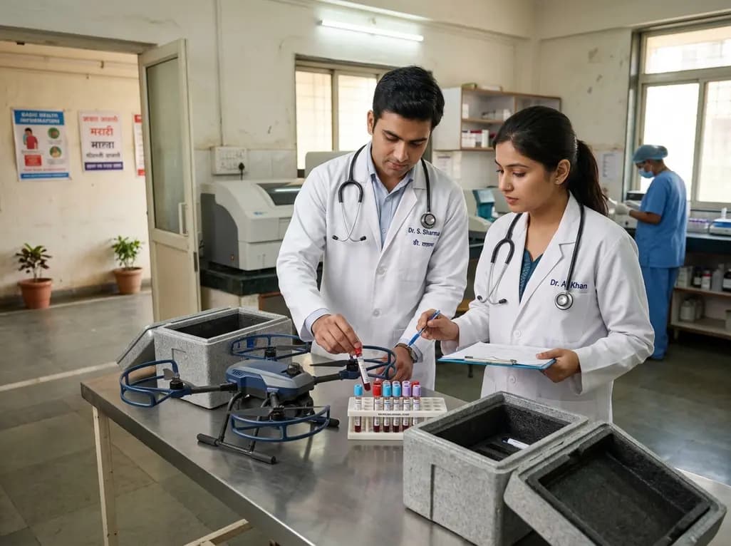
A study evaluating drone transport of blood samples across winter and summer found 31 of 32 routine biochemical analytes remained stable compared to ground transport. While plasma LDH showed minor increases, separation gel caused greater preanalytical alterations, showing aviation rules are the main barrier.
Today
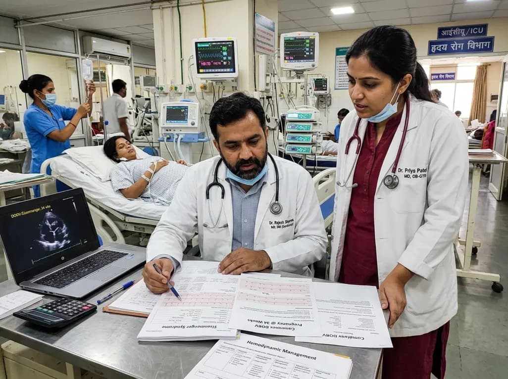
A recent case highlights favorable maternal and neonatal survival in a patient presenting at 34 weeks with probable Eisenmenger physiology and suspected double outlet right ventricle (DORV). Here is a critical review of hemodynamic challenges, delivery strategies, and peripartum management in complex cardiac disease.
Today
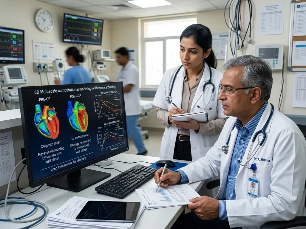
A novel multiscale computational model calibrated with Bayesian algorithms demonstrates that full reverse remodeling after mitral valve regurgitation demands both mechanical correction of volume overload and neurohormonal restoration, shedding light on persistent postoperative left ventricular dysfunction.
Today
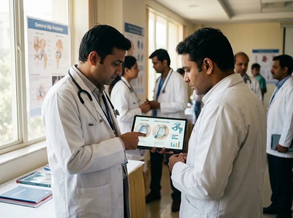
Systematic review comparing labral repair and reconstruction in primary hip arthroscopy shows comparable short- to mid-term patient-reported outcomes, with lower THA conversion after repair but superior outcomes for reconstruction in patients aged 40 and older.
Today

A preclinical study reveals that alpinetin significantly alters glimepiride pharmacokinetics by inhibiting CYP2C9. This drug-herb interaction enhances hypoglycemic potency and underscores critical safety concerns regarding severe hypoglycemia in diabetic management.
Today
