

Loading, please wait...
Loading, please wait...

Reconstructing significant full-thickness eyelid margins represents a challenging surgical endeavor in facial plastic and reconstructive surgery. Historically, reconstructive surgeons restricted primary direct closure to defects involving less than thirty-three percent of the horizontal margin. Defects exceeding this dimension typically mandate complex tissue mobilization or staged procedures to prevent severe eyelid distortion. However, innovative surgical refinements now provide alternative reconstructive pathways for larger periorbital wounds. The vertical tarsotomy technique offers an elegant solution that expands residual lid tissues to bridge substantial defects without regional flap transfer. By performing serial vertical incisions through the residual tarsal plate, surgeons can overcome structural tissue resistance. Consequently, this surgical innovation permits tension-free primary margin closure across moderate-to-large defects. This surgical evolution significantly transforms periorbital oncologic management, offering superior aesthetic preservation and functional protection.
The biomechanical architecture of the eyelid relies heavily on the rigid fibrous tarsus for horizontal stability and globe apposition. In traditional oculoplastic protocols, attempting primary closure under excessive tension produces severe complications, including eyelid retraction, notch formation, and cicatricial ectropion. Consequently, reconstructive algorithms have historically shifted toward local flaps whenever defects exceed one-third of the lid length. The vertical tarsotomy technique fundamentally alters this paradigm by directly modifying posterior lamellar mechanics. Specifically, surgeons place sequential vertical full-thickness incisions across the residual tarsal plate away from the defect margin. These precise cuts interrupt the rigid internal architecture of the tarsal collagen framework without severing the overlying marginal arcade. As a direct result, the remaining tarsus expands like an accordion under gentle horizontal traction. Furthermore, this structural relaxation lengthens the usable posterior lamella while preserving essential vascularity. Thus, surgeons achieve significant horizontal elongation without sacrificing structural integrity or compromising ocular support.
Surgeons managing moderate-to-large periocular defects traditionally utilize rotational flaps or two-stage procedures such as the Hughes tarsoconjunctival flap. While these conventional methods provide reliable coverage, they carry notable morbidities. For instance, the Hughes flap occludes the visual axis for several weeks, creating substantial distress in elderly individuals. Moreover, extensive local flap mobilization often produces skin texture mismatches, donor site scarring, and persistent lower eyelid edema. In contrast, the vertical tarsotomy approach sidesteps these procedural burdens by maximizing the native eyelid tissue. Patients retain continuous binocular vision throughout the entire postoperative period. Additionally, direct closure preserves the native mucocutaneous junction and cilia alignment far better than composite tissue transfers. Therefore, eliminating complex flap harvest shortens operative duration, minimizes donor site complications, and substantially accelerates postoperative functional recovery.
Executing this reconstructive strategy requires meticulous adherence to micro-surgical tissue handling and anatomical planes. First, the surgeon ensures complete margin clearance following oncologic resection, typically involving basal cell carcinoma excision. Next, the surgeon assesses residual eyelid mobility and measures horizontal tissue deficiency. To facilitate mobilization, clinicians frequently execute an internal lateral cantholysis, releasing the inferior crus of the lateral canthal tendon. Subsequently, the surgeon performs serial vertical incisions through the residual tarsal plate and conjunctiva, spacing them several millimeters apart. Clinicians must carefully avoid cutting through the eyelid margin itself to protect the marginal arterial arcade. Furthermore, gentle lateral traction demonstrates progressive tissue expansion after each successive tarsal incision. Once the eyelid reaches adequate horizontal laxity, the surgical team approximates the defect margins in precise anatomical layers. Specifically, surgeons place buried absorbable sutures within the tarsal plate and close the skin with fine non-absorbable interrupted sutures.
Recent clinical investigations substantiate the high reliability and low complication rate associated with this innovative reconstructive approach. In published clinical cohorts, the vast majority of patients presented with defects exceeding fifty percent of the horizontal lid length. Despite these substantial tissue voids, surgeons achieved primary direct closure in all treated patients. Furthermore, eighty percent of patients successfully underwent concurrent internal lateral cantholysis to optimize wound apposition without structural compromise. Follow-up evaluations extending over several months demonstrated outstanding eyelid margin stability, symmetrical palpebral fissures, and intact tear film distribution. Postoperative complications remained minimal and self-limiting. For example, occasional mild lower lid ectropion resolved without requiring secondary surgical revision. In addition, isolated corneal epitheliopathy responded promptly to temporary bandage contact lens placement. Crucially, no patients experienced complete wound breakdown, flap ischemia, or sight-threatening visual compromise.
Integrating serial tarsotomies into standard surgical algorithms provides oculoplastic, dermatologic, and head and neck surgeons with a valuable reconstructive asset. However, proper case selection remains fundamental to achieving optimal outcomes. This technique proves particularly advantageous in elderly patients who demonstrate baseline eyelid laxity, as senescent elastosis facilitates greater mechanical stretching. Conversely, surgeons should exercise caution in young patients possessing exceedingly stiff tarsal plates with minimal tissue laxity. In such instances, combining tarsotomies with a lateral canthal release ensures adequate tension relief. Additionally, clinicians must handle the palpebral conjunctiva with extreme gentleness to avoid postoperative granuloma formation or foreign-body sensation. Regular postoperative slit-lamp evaluation ensures early detection of suture irritation or epithelial breakdown. Ultimately, mastering this versatile technique enables reconstructive specialists to manage significant eyelid defects efficiently while minimizing surgical morbidity.
The vertical tarsotomy technique prevents vertical lid retraction by preserving the normal anatomical vector of the eyelid. By making controlled, full-thickness vertical cuts strictly within the remaining tarsal plate, the surgeon selectively releases horizontal stiffness. Consequently, this controlled mechanical relaxation allows the lid margin to stretch horizontally across the defect. Because surgeons avoid vertical traction, the lower eyelid maintains stable apposition to the ocular surface without provoking cicatricial ectropion.
Surgeons frequently perform an internal lateral cantholysis when residual eyelid tension remains high after primary defect assessment. In clinical series, approximately eighty percent of cases required cantholysis alongside serial tarsotomies to achieve direct apposition. Combining lateral canthal tendon release with vertical tarsotomies provides supplemental tissue advancement. Therefore, this dual approach mobilizes adequate tissue safely. Consequently, surgeons can successfully bridge full-thickness margin defects exceeding half the total horizontal eyelid length.
Clinicians must monitor patients closely for transient ocular surface irritation and subtle eyelid malposition. In published cohorts, mild lower lid ectropion developed in a minority of patients, resolving with conservative management or minimal intervention. Furthermore, temporary corneal epitheliopathy may arise from altered tear film dynamics or suture exposure. Fortunately, this condition responds rapidly to bandage contact lens wear. Crucially, studies report no instances of wound dehiscence, flap necrosis, or sight-threatening functional complications during postoperative recovery.
Disclaimer: This content is for informational and educational purposes only. It is not intended to substitute for professional medical advice, diagnosis, or treatment. Always seek the advice of a qualified healthcare provider with any questions you may have regarding a medical condition. Refer to the latest local and national guidelines for clinical practice.
References

Read summarized clinical updates, watch expert medical content, and earn CME certifications right from your smartphone.
Open your smartphone camera to scan and install.





Serial vertical tarsotomies allow surgeons to reconstruct moderate-to-large full-thickness eyelid defects via primary closure, minimizing reliance on complex flaps while maintaining ocular stability.
Today
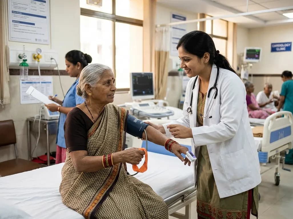
Review how tailored exercise programs benefit frail older patients with heart failure. Explore pathophysiological mechanisms, individualized training prescriptions, and strategies to overcome adherence barriers in clinical practice.
Today
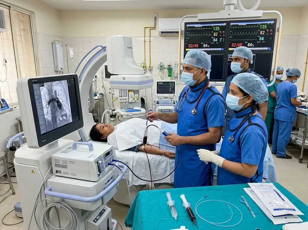
In high-risk pulmonary embolism complicated by severe anemia and active uterine bleeding, systemic thrombolysis is contraindicated. Large-bore aspiration thrombectomy provides rapid hemodynamic rescue without fibrinolysis, supported by inferior vena cava filter placement and multidisciplinary critical care.
Today

A cross-sectional study in Zanzibar reveals that 46.5% of adults with type 2 diabetes experience multidomain autonomic dysfunction on the COMPASS-31 tool, primarily orthostatic intolerance, while 45.9% exhibit cardiovascular autonomic neuropathy, highlighting the urgent need for structured clinical screening.
Today

This comparative study evaluates posterior labral tears with and without glenohumeral instability, highlighting distinct demographic patterns, MRI underestimation in stable shoulders, and successful arthroscopic repair outcomes.
Today
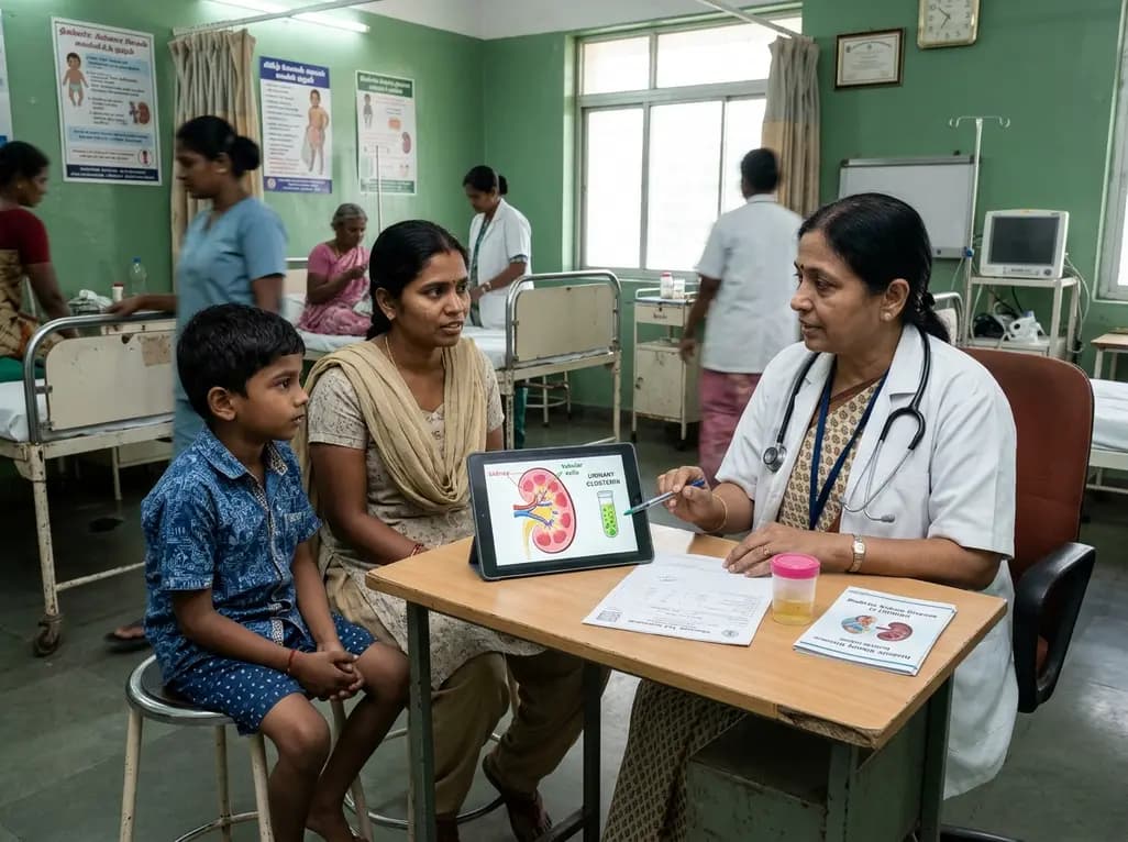
A recent case-control study reveals that urinary clusterin provides superior diagnostic performance for early diabetic kidney disease detection in pediatric type 1 diabetes mellitus, identifying tubular damage before conventional microalbuminuria occurs.
Today
