.webp&w=384&q=75)

Loading, please wait...
Loading, please wait...

Routine pediatric immunization protects children from severe infectious morbidity, yet rare ocular adverse reactions occasionally present diagnostic challenges. A recently published clinical report details a rare occurrence of vaccine-associated retinitis presenting as bilateral maculopathy in a thirteen-month-old infant following measles, mumps, rubella, and varicella (MMRV) vaccination. Recognizing these atypical inflammatory patterns allows clinicians to intervene promptly and preserve visual function. Furthermore, understanding the benign long-term prognosis provides reassurance to families and physicians alike.
The thirteen-month-old infant presented with acute bilateral visual deterioration two weeks after receiving the combined live MMRV vaccine. Consequently, the parents observed uncoordinated wandering eye movements and an inability to fixate on familiar objects. An urgent ophthalmic evaluation revealed marked bilateral retinitis accompanied by retinal vasculitis. In addition, dilated fundus examination demonstrated serous exudative retinal detachments involving both posterior poles. Bilateral non-necrotizing retinitis of this severity in early childhood represents an exceptional clinical scenario. Therefore, clinicians immediately suspected an acute immune-mediated or infectious event temporally linked to the recent immunization. Although live attenuated viral vaccines exhibit outstanding safety profiles worldwide, viral antigens can occasionally trigger profound ocular inflammation in susceptible hosts. The latency interval of two weeks aligned closely with systemic antibody synthesis and immune complex activation. As a result, differentiating between active viral replication and immune-mediated parainfectious inflammation became the most pressing diagnostic objective. Clinicians quickly assembled a multidisciplinary team to evaluate the infant thoroughly.
Clinicians promptly organized an exhaustive diagnostic investigation to identify the underlying etiology and exclude alternative infections. Serological testing demonstrated positive measles IgM and IgG antibody titers, reflecting active immunological stimulation following vaccination. However, quantitative polymerase chain reaction (PCR) tests of the aqueous humor and urine yielded completely negative results for measles virus ribonucleic acid. In contrast, comprehensive testing for herpes simplex virus, cytomegalovirus, and varicella-zoster virus proved entirely negative. Furthermore, detailed pediatric neurologic evaluations showed no focal deficits or altered mental status. Neuroimaging with brain magnetic resonance imaging confirmed normal intracranial architecture, effectively ruling out central nervous system involvement such as encephalitis. Lumbar puncture analysis also yielded normal cerebrospinal fluid cell counts and chemistry. Thus, negative intraocular viral PCR in the presence of positive systemic serology strongly supported a sterile immune-mediated hypersensitivity reaction. Consequently, these definitive findings confirmed presumed vaccine-associated retinitis rather than active herpetic necrotizing retinopathy.
High-resolution optical coherence tomography (OCT) played an essential role in defining the sequential structural evolution of the macular lesions. Initially, baseline scans revealed severe neurosensory retinal detachment accompanied by prominent hyperreflective inflammatory deposits within the outer retinal layers. Furthermore, OCT demonstrated diffuse disruption of the foveal photoreceptor ellipsoid zone and external limiting membrane in both eyes. Over sequential follow-up examinations, these inflammatory subretinal exudates gradually consolidated into dense hyperreflective deposits. In addition, the subretinal fluid progressively resorbed as the outer blood-retinal barrier regained integrity. Eventually, the retinitis resolved completely, leaving bilateral, well-demarcated macular scars with focal retinal pigment epithelium alterations. Notably, this clinical report offers the first sequential OCT documentation of presumed measles vaccine-associated retinitis in an infant. These structural insights demonstrated that outer neurosensory retinal layers can stabilize despite marked early damage. Consequently, multimodal imaging proved invaluable for objectively monitoring therapeutic response over time.
Given the initial diagnostic ambiguity between viral retinitis and immune-mediated uveitis, clinicians initiated empirical dual antimicrobial and anti-inflammatory therapy. Specifically, they administered intravenous acyclovir alongside high-dose systemic corticosteroids. This regimen simultaneously covered potential herpesvirus necrotizing retinitis while blunting intense intraocular tissue destruction. Once molecular tests excluded intraocular viral replication, clinicians tapered the systemic corticosteroids gradually over several months. As a result, the posterior uveitis and retinal vasculitis subsided steadily without progressing to catastrophic retinal necrosis. During an extensive six-year longitudinal follow-up, the child maintained stable ocular architecture without recurrent intraocular inflammation. Furthermore, no systemic autoimmune diseases or secondary ocular complications, such as cataracts or glaucoma, appeared. Most importantly, the patient achieved a final visual acuity of 6/9 in each eye at six years of age. This encouraging visual recovery proves that rapid, targeted medical intervention can preserve central visual potential in bilateral infantile maculopathy.
In India, pediatric healthcare providers widely administer the measles-rubella (MR) vaccine through the Universal Immunization Programme, alongside MMR and MMRV vaccines in private clinical settings. While adverse events following immunization remain exceedingly rare, clinicians must maintain clinical vigilance for unusual neuro-ophthalmic presentations. Pediatricians and family physicians should recognize visual fixation loss, acute nystagmus, or wandering eye movements as urgent indicators for ophthalmic evaluation. Furthermore, prompt referral to a pediatric vitreoretinal specialist ensures timely intervention and prevents irreversible amblyopia. Clinicians should also recognize that systemic corticosteroids effectively control inflammatory damage once viral pathogens are excluded. However, healthcare workers must proactively reassure parents that such adverse reactions occur with extraordinary rarity. Therefore, these isolated occurrences must never compromise confidence in vital national immunization campaigns. Ultimately, preventing the devastating consequences of wild measles infection remains paramount for pediatric public health across India.
Clinicians differentiate vaccine-associated retinitis by combining detailed serology, intraocular fluid PCR testing, and careful funduscopic observation. True viral retinitis typically exhibits rapid progression, progressive retinal necrosis, and positive PCR findings for viral DNA in aqueous humor. In contrast, vaccine-associated retinitis frequently demonstrates negative intraocular PCR results alongside positive serology. Furthermore, the clinical course responds favorably to systemic corticosteroid therapy, demonstrating an immune-mediated parainfectious pathophysiology rather than direct microbial destruction.
The recommended management involves immediate collaboration between ophthalmologists and pediatric infectious disease specialists. Clinicians usually initiate empirical systemic antiviral therapy with intravenous acyclovir to cover potential viral etiologies like herpes simplex or varicella. Concurrently, high-dose systemic corticosteroids are administered to arrest severe intraocular inflammation. Once viral PCR testing returns negative, clinicians gradually taper corticosteroids over weeks or months. Regular monitoring with optical coherence tomography ensures complete resolution of exudates.
Subsequent immunizations require individualized risk assessment by an expert pediatric allergy and infectious disease committee. Generally, clinicians avoid revaccinating the child with the specific suspected live viral vaccine to prevent recurrent intraocular inflammation. However, other essential childhood vaccines that do not share cross-reacting antigens can often proceed according to national schedules under supervised observation. Public health specialists strongly emphasize completing unrelated vaccinations to protect vulnerable pediatric patients against life-threatening infections.
Disclaimer: This content is for informational and educational purposes only, and should not be taken as medical advice. It is not intended to be used as a substitute for professional medical advice, diagnosis, or treatment. Always seek the advice of your physician or other qualified health provider with any questions you may have regarding a medical condition. Refer to the latest local and national guidelines for clinical practice.
References

Read summarized clinical updates, watch expert medical content, and earn CME certifications right from your smartphone.
Open your smartphone camera to scan and install.





A 13-month-old infant developed bilateral retinitis and maculopathy following MMRV vaccination. Diagnostic workup ruled out active infection, and prompt antiviral and corticosteroid therapy led to resolution with 6/9 vision at 6-year follow-up, offering key insights into vaccine-associated ocular complications.
Today
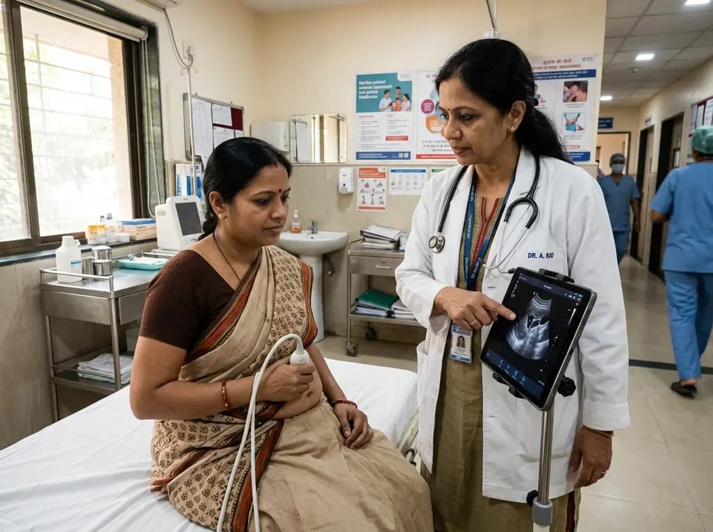
Self-performed ultrasound empowers patients to capture diagnostic-quality scans for remote clinical review. This review evaluates its accuracy, technological challenges, and integration hurdles.
Today
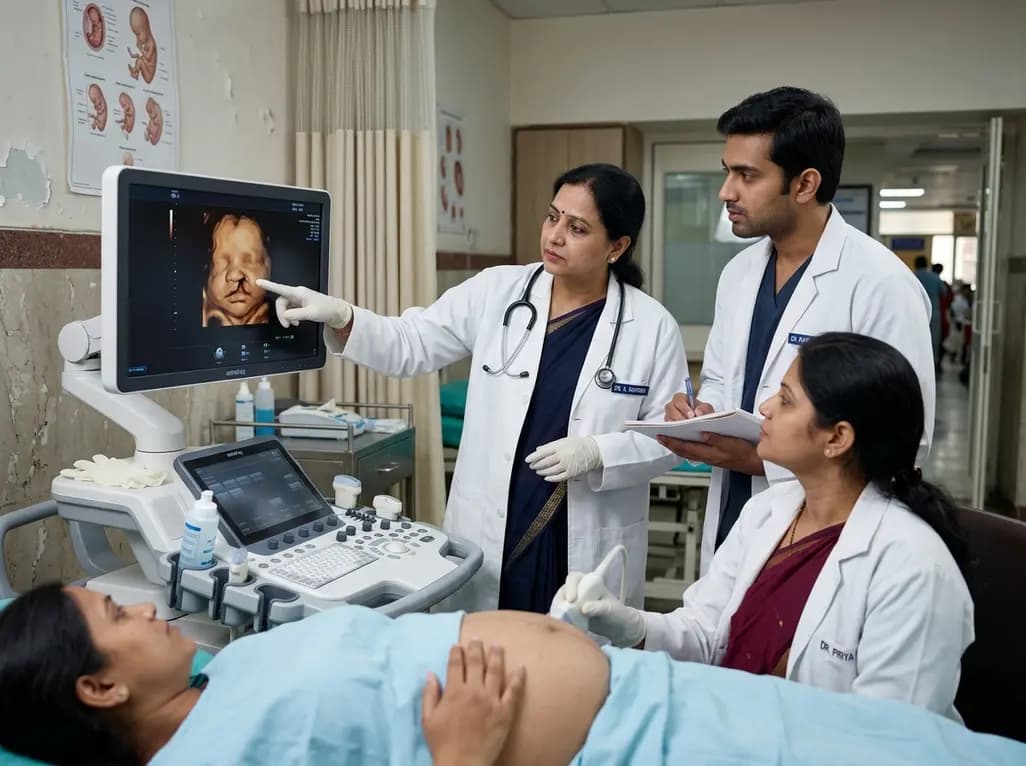
A 5-year retrospective study evaluates prenatal ultrasound detection of fetal cleft lip and palate. While overall detection reached 87%, accuracy varied from 99.3% in combined clefts to 50% in isolated cases. Combined defects showed elevated risks of concurrent structural anomalies and chromosomal abnormalities.
Today

An updated systematic review and meta-analysis of 7,875 patients reveals that routine microaxial left ventricular support during nonemergent high-risk PCI does not reduce mortality or ischemic complications, while significantly increasing major bleeding events.
Today
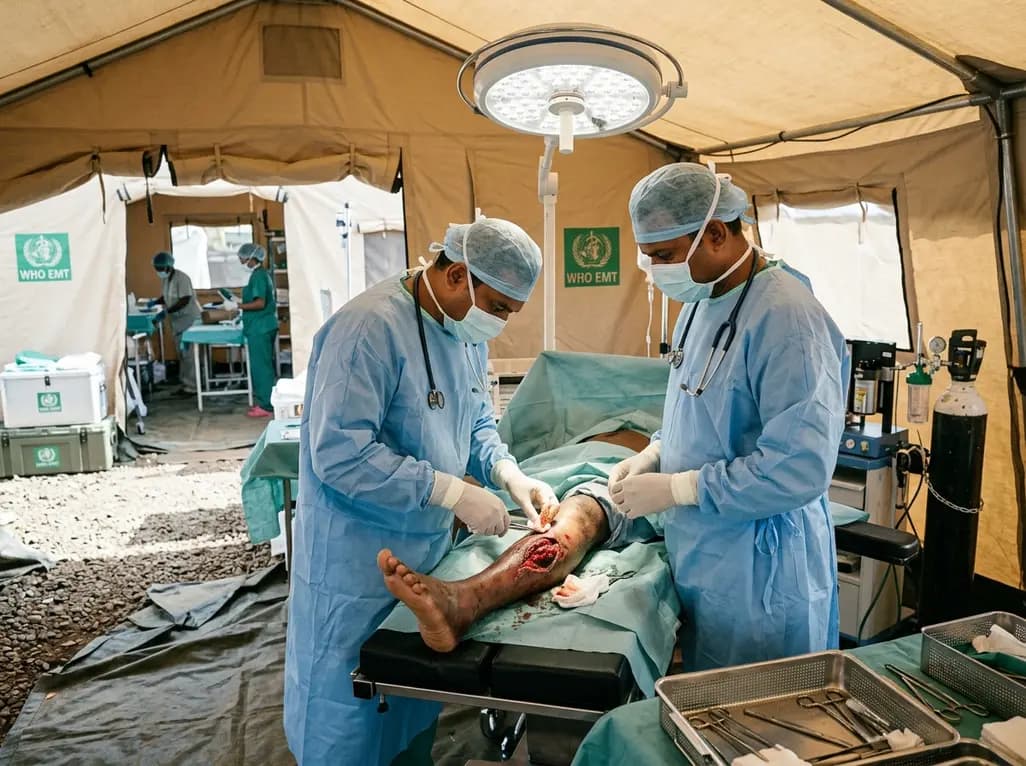
Following Cyclone Chido, a WHO EMT-2 field hospital performed 268 surgeries in Mayotte. While acute trauma is expected initially, 98% of cases were septic wounds of the extremities requiring secondary intention healing, revealing crucial operational insights for disaster surgical preparedness.
Today

The SED-SEEN-SEEP consensus outlines an Integrated Care Pathway for preclinical type 1 diabetes, structuring target screening, autoantibody confirmation, metabolic staging, and proactive follow-up to prevent diabetic ketoacidosis and facilitate early disease-modifying intervention.
Today
