.webp&w=384&q=75)

Loading, please wait...
Loading, please wait...

Congenital facial clefts represent one of the most frequent structural anomalies encountered during pregnancy, requiring precise evaluation for optimal obstetric counseling. Recent clinical research confirms that fetal cleft lip ultrasound serves as an indispensable modality for identifying orofacial defects during prenatal surveillance. However, diagnostic accuracy varies significantly across distinct anatomical subtypes and gestational trimesters. Clinicians must therefore understand these diagnostic nuances to guide diagnostic testing, parental counseling, and neonatal surgical planning effectively.
Recent clinical investigations provide valuable insight into sonographic performance across diverse cleft presentations. Overall, antenatal sonography achieves an encouraging aggregate detection proportion of eighty-seven percent throughout gestation. Nevertheless, diagnostic sensitivity depends heavily on whether the cleft involves only the lip, the secondary palate, or both anatomical regions. For combined cleft lip with cleft palate, ultrasound demonstrates remarkable efficacy, achieving a detection rate of nearly ninety-nine percent. In contrast, sonographers detect isolated cleft lip in only fifty percent of cases, while isolated cleft palate detection reaches approximately fifty-six percent. Consequently, clinicians must recognize that a normal appearance of the alveolar ridge does not entirely rule out isolated palatal clefting. Technical factors such as fetal skull shadowing, acoustic attenuation, and inadequate fetal positioning frequently obscure clear visualization of the secondary palate. Furthermore, standard two-dimensional acoustic planes struggle to evaluate the soft palate consistently. Because missed diagnoses alter perinatal planning, centers increasingly implement advanced multiplanar three-dimensional sweeps and specialized coronal views. Additionally, color Doppler imaging can demonstrate abnormal transpalatal fluid movement during fetal respiration. Therefore, sonographers must exercise thoroughness during targeted mid-trimester structural surveys to avoid diagnostic oversights.
Timing plays a foundational role in the prenatal detection of craniofacial malformations. During the first trimester, between eleven and fourteen weeks, routine screening identifies approximately forty-one percent of all cleft cases. Combined cleft lip and palate accounts for the vast majority of these early discoveries, primarily because severe disruptions in the retronasal triangle create conspicuous sonographic gaps. However, early sonography exhibits major limitations when evaluating milder cleft forms. Incomplete facial ossification and tiny embryonic dimensions make isolated lip defects or solitary palatal gaps exceedingly difficult to discern. Consequently, obstetricians should not interpret an unremarkable first-trimester scan as a definitive exclusion of facial clefting. Instead, high-risk patients require dedicated follow-up examinations during the standard mid-trimester anatomy evaluation between eighteen and twenty-two weeks. During this secondary window, fetal structures demonstrate larger dimensions and superior acoustic contrast. Practitioners can then interrogate the alveolar arch, upper lip continuity, and secondary palate with greater precision. Moreover, longitudinal scanning tracks dynamic amniotic fluid dynamics, which occasionally unmask subtle palatal defects during fetal swallowing. When diagnostic ambiguity persists, fetal magnetic resonance imaging serves as an invaluable complementary modality. Thus, two-stage screening protocols provide the highest sensitivity for complex facial malformations.
The clinical presentation of facial clefts ranges from mild isolated defects to complex syndromic malformations. Evidence demonstrates that the anatomical subtype directly correlates with the likelihood of concurrent systemic anomalies. Fetuses presenting with combined cleft lip and cleft palate exhibit additional structural malformations in forty-seven percent of cases. Common coexisting anomalies involve congenital heart disease, central nervous system malformations, skeletal dysplasias, and renal tract anomalies. In comparison, isolated cleft lip and isolated cleft palate demonstrate significantly lower rates of associated major structural defects. Furthermore, ultrasound specialists frequently identify non-specific sonographic soft markers in affected fetuses, including nuchal translucency enlargement and abnormal extremity posturing. Because multiple malformations sharply increase neonatal morbidity, discovering any facial defect warrants an immediate, rigorous anatomical survey. Clinicians should specifically assess the four-chamber cardiac view, the outflow tracts, and intracranial anatomy. In addition, fetal echocardiography provides essential secondary evaluation when facial clefting occurs. Detailed neurosonography also helps rule out subtle holoprosencephaly spectrum anomalies and corpus callosum dysgenesis. Ultimately, distinguishing an isolated cosmetic defect from a complex multisystem syndrome changes the clinical trajectory completely, enabling teams to offer nuanced guidance to prospective parents.
Genetic evaluation serves as a cornerstone of prenatal management whenever sonographers detect orofacial clefting. Historically, clinicians relied on conventional karyotyping to identify aneuploidies such as trisomy 13 and trisomy 18. Today, chromosomal microarray analysis provides vastly superior resolution, detecting pathogenic submicroscopic copy number variations. Retrospective findings indicate that nearly thirty-three percent of fetuses with combined cleft lip and palate carry significant genetic abnormalities. In contrast, isolated cleft lip shows a substantially lower genetic burden. Pathogenic copy number variations frequently correspond to recognized syndromes, including 22q11.2 deletion syndrome, Van der Woude syndrome, and complex microdeletion syndromes. Therefore, obstetricians must routinely recommend diagnostic invasive testing via amniocentesis or chorionic villus sampling when identifying cleft lip and palate. Geneticists can perform chromosomal microarray analysis alongside next-generation gene panels to identify specific monogenic craniofacial conditions. Furthermore, whole exome sequencing progressively identifies elusive single-gene disorders when chromosomal microarrays yield non-diagnostic results. This comprehensive diagnostic workup empowers clinical geneticists to calculate precise family inheritance patterns. Moreover, knowing the definitive genetic diagnosis clarifies long-term neurodevelopmental expectations and recurrence risks for subsequent pregnancies. Consequently, genetic investigation transforms prenatal counseling from uncertain speculation into individualized, evidence-based clinical guidance.
Prenatal diagnosis profoundly influences parental decision-making and overall pregnancy outcomes. Retrospective data show striking differences in pregnancy termination rates across anatomical groups. Families facing a diagnosis of combined cleft lip and palate choose termination in nearly eighty-nine percent of cases. Conversely, termination rates remain markedly lower for isolated cleft lip at fourteen percent and isolated cleft palate at eighteen percent. These dramatic discrepancies reflect parental concerns regarding associated severe malformations and genetic diagnoses. Therefore, modern maternal-fetal medicine emphasizes proactive, empathetic multidisciplinary counseling. Obstetricians, medical geneticists, pediatric plastic surgeons, and neonatologists should collaboratively engage the parents soon after diagnosis. Surgeons can show pre- and post-repair photographs, outline feeding strategies, and explain staged reconstructive protocols. In addition, neonatal specialists can demystify airway concerns and reassure families regarding successful surgical outcomes for isolated defects. Dedicated speech and language therapists also explain future swallowing and articulation milestones to expectant parents. Furthermore, comprehensive counseling alleviates acute parental anxiety, corrects misconceptions, and supports informed reproductive choices. Ultimately, a structured multidisciplinary framework ensures that families receive balanced medical facts and compassionate psychological support throughout the perinatal journey.
Sonographers frequently struggle to detect isolated cleft palate because the surrounding bony structures of the fetal skull create significant acoustic shadowing. Additionally, clinicians usually examine the upper lip and primary palate in standard transverse and coronal planes, which fail to visualize the secondary hard and soft palates effectively. Consequently, without dedicated multiplanar reconstruction or dynamic fluid-swallowing assessment, subtle defects in the secondary palate remain obscured during routine obstetrical screening scans.
Chromosomal microarray analysis detects microdeletions and microduplications that conventional karyotyping misses entirely. Fetuses with combined cleft lip and palate carry a high risk of genetic abnormalities, reaching approximately thirty-three percent. Furthermore, many of these copy number variations indicate complex syndromic conditions such as 22q11.2 deletion syndrome. Therefore, microarray testing provides indispensable diagnostic precision that clarifies syndromic risks, guides antenatal management, and informs prospective parents about expected long-term developmental outcomes.
Antenatal diagnosis enables multidisciplinary teams to coordinate specialized postnatal care well before delivery occurs. Parents receive education regarding customized feeding devices, which prevents neonatal malnutrition and aspiration complications. Additionally, pediatric craniofacial surgeons can formulate a structured timeline for primary lip repair at three to six months and palatal reconstruction near one year of age. Consequently, prenatal detection minimizes parental shock, optimizes neonatal airway stabilization, and ensures seamless transition into specialized surgical care.
Disclaimer: This content is for informational and educational purposes only and should not be considered a substitute for professional medical judgment. Clinicians must evaluate each patient independently. Refer to the latest local and national guidelines for clinical practice.
References

Read summarized clinical updates, watch expert medical content, and earn CME certifications right from your smartphone.
Open your smartphone camera to scan and install.





A 5-year retrospective study evaluates prenatal ultrasound detection of fetal cleft lip and palate. While overall detection reached 87%, accuracy varied from 99.3% in combined clefts to 50% in isolated cases. Combined defects showed elevated risks of concurrent structural anomalies and chromosomal abnormalities.
Today
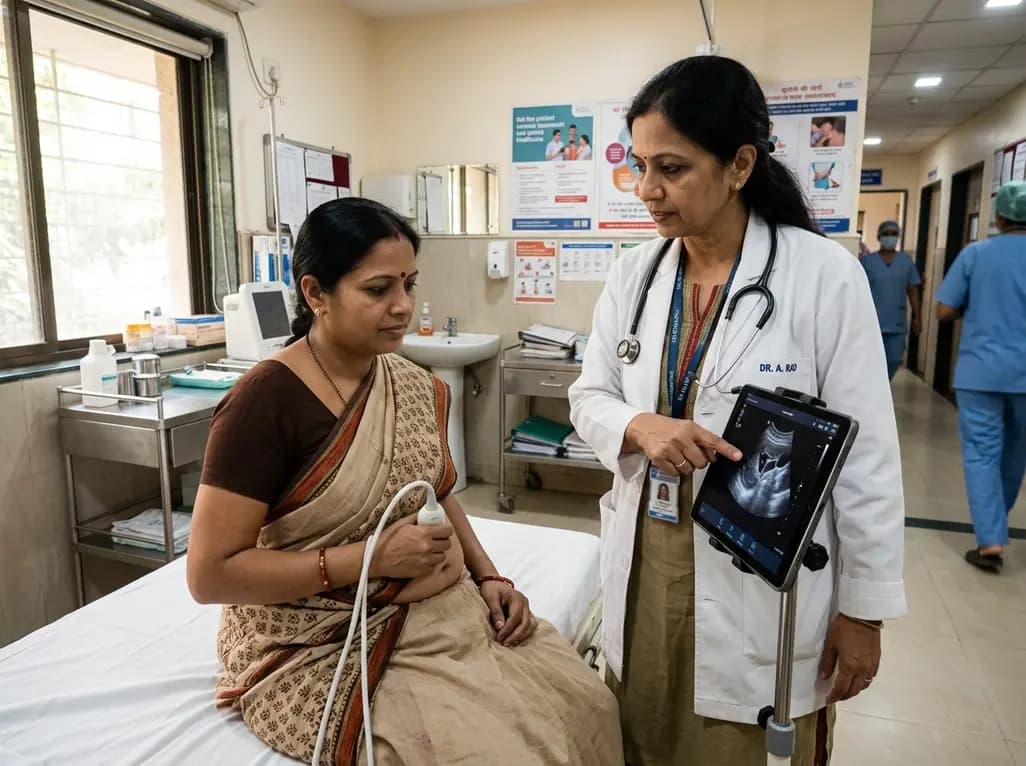
Self-performed ultrasound empowers patients to capture diagnostic-quality scans for remote clinical review. This review evaluates its accuracy, technological challenges, and integration hurdles.
Today

An updated systematic review and meta-analysis of 7,875 patients reveals that routine microaxial left ventricular support during nonemergent high-risk PCI does not reduce mortality or ischemic complications, while significantly increasing major bleeding events.
Today
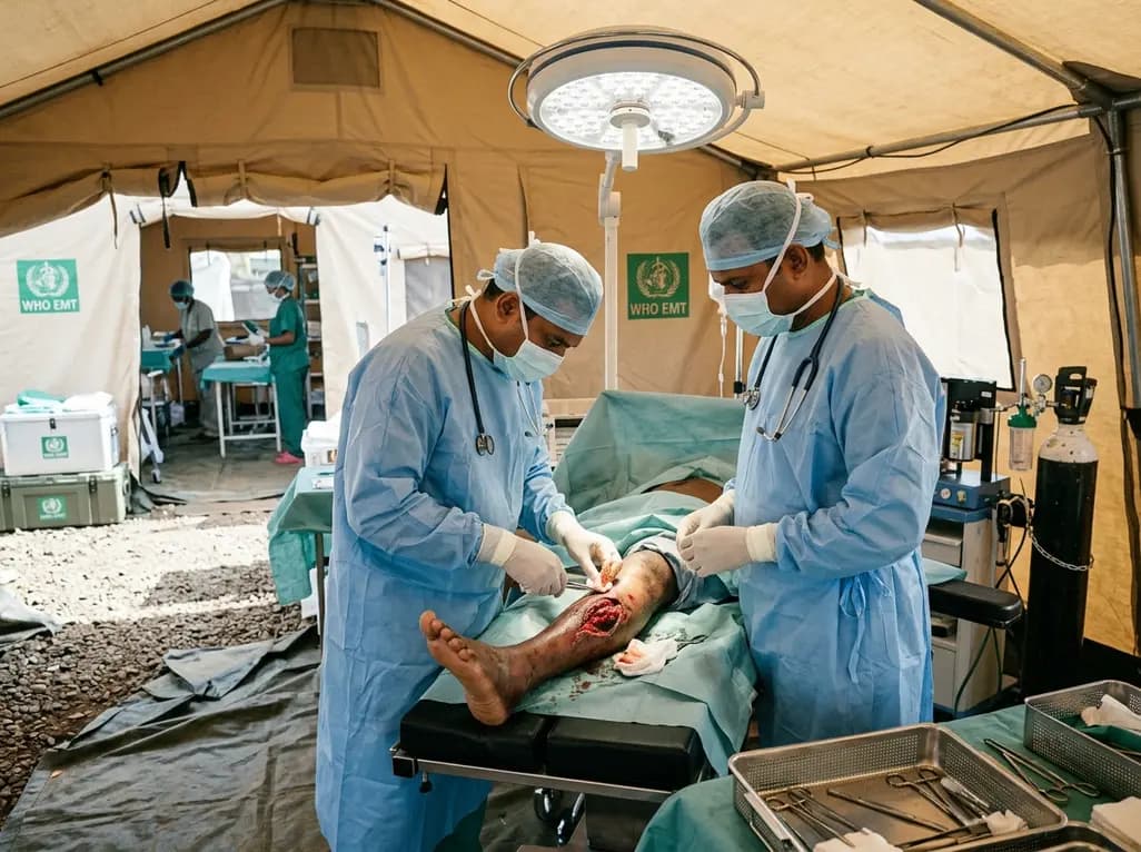
Following Cyclone Chido, a WHO EMT-2 field hospital performed 268 surgeries in Mayotte. While acute trauma is expected initially, 98% of cases were septic wounds of the extremities requiring secondary intention healing, revealing crucial operational insights for disaster surgical preparedness.
Today

The SED-SEEN-SEEP consensus outlines an Integrated Care Pathway for preclinical type 1 diabetes, structuring target screening, autoantibody confirmation, metabolic staging, and proactive follow-up to prevent diabetic ketoacidosis and facilitate early disease-modifying intervention.
Today
