

Loading, please wait...
Loading, please wait...

Clinicians frequently face diagnostic dilemmas when managing chronic, non-resolving intraocular inflammation. Among these complex presentations, uveitis masquerade syndrome represents an essential differential diagnosis in patients presenting with steroid-refractory posterior uveitis. Primary vitreoretinal lymphoma remains the most frequent malignant cause of this deceptive condition. Therefore, ophthalmologists must maintain a high index of suspicion whenever elderly patients demonstrate persistent vitritis or unresponsive retinal lesions. In many clinical scenarios, physicians initially categorize these manifestations as benign idiopathic uveitis. Consequently, patients undergo prolonged empirical immunosuppressive therapy that fails to halt underlying disease progression. In addition, corticosteroid therapy often induces transient, misleading improvements because malignant lymphocytes temporarily lyse under hormonal exposure. However, inflammation invariably recurs once clinicians taper systemic or topical medications. Recognizing this cyclical pattern helps specialists avoid severe delays in definitive therapy. Moreover, malignant cells can silently proliferate within the vitreous and neurosensory retina during these therapeutic pauses. Because timely diagnosis directly influences systemic survival, clinicians must actively look for masquerade features in every refractory case. Establishing early clinical suspicion enables prompt diagnostic intervention before extraocular dissemination occurs.
Diagnosing intraocular lymphoma presents significant clinical pitfalls because malignant presentations frequently mimic common non-neoplastic pathologies. For instance, sub-retinal pigment epithelium infiltrates often resemble drusen or age-related macular degeneration. Consequently, clinicians may misattribute visual deterioration in older adults to routine senile degeneration rather than aggressive malignancy. Furthermore, vitritis often mimics chronic intermediate uveitis, toxoplasmosis, or fungal endophthalmitis. In clinical audits, patients frequently receive corticosteroids or anti-vascular endothelial growth factor injections for several months without achieving genuine disease control. In addition, steroid-induced symptom fluctuation often creates a false sense of security for both the physician and the patient. Nevertheless, careful funduscopic observation reveals subtle, atypical features that distinguish neoplastic processes from standard inflammatory conditions. For example, solid creamy-yellow lesions beneath the retinal pigment epithelium typically signal lymphoma cell infiltration rather than inflammatory drusenoid deposits. In addition, the presence of sheets or clumps of vitreous cells with minimal anterior chamber reaction warrants immediate concern. Thus, practitioners must re-evaluate presumptive uveitis diagnoses whenever expected therapeutic milestones remain unmet. Recognizing these deceptive presentations prevents prolonged misdirection and preserves visual function.
Modern multimodal imaging provides invaluable diagnostic clues that help unmask neoplastic intraocular infiltration. Spectral-domain optical coherence tomography serves as a primary non-invasive tool to distinguish lymphomatous deposits from inflammatory exudates. Specifically, high-resolution scans frequently reveal wavy, nodular hyperreflective elevations situated beneath the retinal pigment epithelium. Furthermore, clinicians often identify hyperreflective vertical infiltrates traversing the neurosensory layers, accompanied by subretinal fluid accumulation. In contrast, standard macular degeneration produces diffuse lipid exudation or classic drusenoid material without distinct cellular vitreal clumps. Fundus autofluorescence offers complementary diagnostic value by displaying characteristic leopard-spot patterns with alternating areas of hyperautofluorescence and hypoautofluorescence. Therefore, these imaging hallmarks guide targeted surgical intervention and pinpoint ideal biopsy locations. In addition, fluorescein angiography typically demonstrates areas of early hypofluorescence with late staining or punctate hyperfluorescence, rather than severe primary retinal vasculitis. Indocyanine green angiography also confirms hypofluorescent lesions that remain distinct throughout late phases. Consequently, integrating these non-invasive imaging modalities allows retina specialists to detect early lymphomatous infiltrates before extensive tissue disruption emerges.
Although multimodal imaging raises strong clinical suspicion, definitive diagnosis requires cytological and molecular confirmation. Therefore, diagnostic pars plana vitrectomy remains the gold standard for securing intraocular specimens. However, vitreous biopsy demands meticulous coordination because malignant lymphoid cells deteriorate rapidly outside their native microenvironment. Vitreoretinal surgeons must avoid corticosteroid administration for several weeks prior to biopsy, as steroids rapidly lyse lymphoma cells and yield false-negative cytological results. Furthermore, the operating team must transfer undiluted vitreous samples immediately to the pathology suite for rapid fixation and analysis. Cytopathology typically reveals large, atypical lymphocytes displaying high nuclear-to-cytoplasmic ratios, prominent nucleoli, and irregular nuclear contours. In addition to conventional cytology, contemporary laboratories utilize flow cytometry to identify monoclonal B-cell populations exhibiting kappa or lambda light chain restriction. Moreover, molecular testing for MYD88 L265P gene mutations provides robust diagnostic specificity, identifying malignant clones even in specimens with low cellular yields. Elevated interleukin-10 levels relative to interleukin-6 in vitreous humor further substantiate lymphoma over inflammatory uveitis. Thus, combining advanced molecular tests with precise cytology drastically elevates diagnostic yield and eliminates diagnostic delay.
Intraocular lymphoma carries a grave systemic prognosis because up to eighty percent of patients eventually develop central nervous system involvement. Consequently, confirming ocular lymphoma necessitates immediate neurological staging and systemic restaging. Clinicians must perform contrast-enhanced brain magnetic resonance imaging alongside lumbar puncture for cerebrospinal fluid cytology and flow cytometry. Even if initial neuroimaging appears completely normal, multidisciplinary surveillance remains vital throughout the disease trajectory. In addition, hematologists, ocular oncologists, neurologists, and pathologists must collaborate closely to design personalized therapeutic regimens. Local ocular treatment frequently incorporates intravitreal methotrexate or rituximab injections to clear vitreoretinal infiltration and restore vision. Meanwhile, systemic management requires high-dose methotrexate-based systemic chemotherapy to eradicate occult central nervous system disease and prolong overall survival. Regular surveillance neuroimaging and repeat funduscopic evaluations ensure early detection of relapse. Furthermore, genetic counseling and supportive care assist patients in navigating prolonged multi-agent therapeutic regimens. Through coordinated interdisciplinary communication, clinicians can transform a historically dismal prognosis into prolonged clinical remission. Therefore, shared vigilance among specialties ensures prompt intervention and substantially improves patient survival.
Clinicians should strongly suspect vitreoretinal lymphoma when an older adult develops persistent, steroid-refractory vitritis or unexplained visual deterioration. Furthermore, the presence of creamy-yellow sub-retinal pigment epithelium infiltrates, atypical vitreous cell clumping, and lack of systemic inflammatory signs should prompt diagnostic investigation. When posterior uveitis improves transiently with systemic corticosteroids but promptly recurs following drug tapering, specialists must immediately pursue masquerade syndrome protocols rather than escalating conventional immunosuppressive therapies.
Corticosteroids exert a potent cytotoxic effect on malignant lymphoid cells, leading to temporary clinical remission and clearing of vitreous haze. However, this transient response masks the underlying neoplastic disease and gives clinicians a false impression of successful anti-inflammatory treatment. Furthermore, corticosteroid exposure causes widespread apoptosis of lymphoma cells, significantly degrading subsequent biopsy specimens. Consequently, diagnostic vitrectomy performed during or immediately after steroid therapy frequently yields non-diagnostic or false-negative results.
Molecular testing dramatically enhances diagnostic accuracy when vitreous cytology contains limited viable cells. Specifically, polymerase chain reaction assays detect the MYD88 L265P somatic mutation, which occurs in over seventy percent of primary vitreoretinal lymphomas. Additionally, clinicians evaluate immunoglobulin heavy chain gene rearrangements through molecular clonality testing. Combined with elevated interleukin-10 to interleukin-6 ratios, these sensitive molecular analyses confirm monoclonal lymphoproliferation and expedite life-saving treatment before central nervous system spread occurs.
Disclaimer: This content is for informational and educational purposes only... Refer to the latest local and national guidelines for clinical practice.
References

Read summarized clinical updates, watch expert medical content, and earn CME certifications right from your smartphone.
Open your smartphone camera to scan and install.





Uveitis masquerade syndrome is a vital differential in steroid-refractory posterior uveitis. Primary vitreoretinal lymphoma often mimics inflammatory disease, requiring multimodal imaging, biopsy, and multidisciplinary care to detect central nervous system involvement and improve patient survival.
Today

A recent cross-sectional study reveals that prolonged residence at high altitude significantly escalates foot frostbite risk among lowland migrants, with chronic mountain sickness acting as a significant physiological mediator through hyperviscosity and microcirculatory stasis.
Today

A litigation-derived series of 252 women highlights the risks of repeat revisions after midurethral sling surgery. Recurrent incontinence occurred in up to 60% of patients, and repeat revision rates reached 70%, emphasizing cautious patient selection and skilled multidisciplinary management.
Today
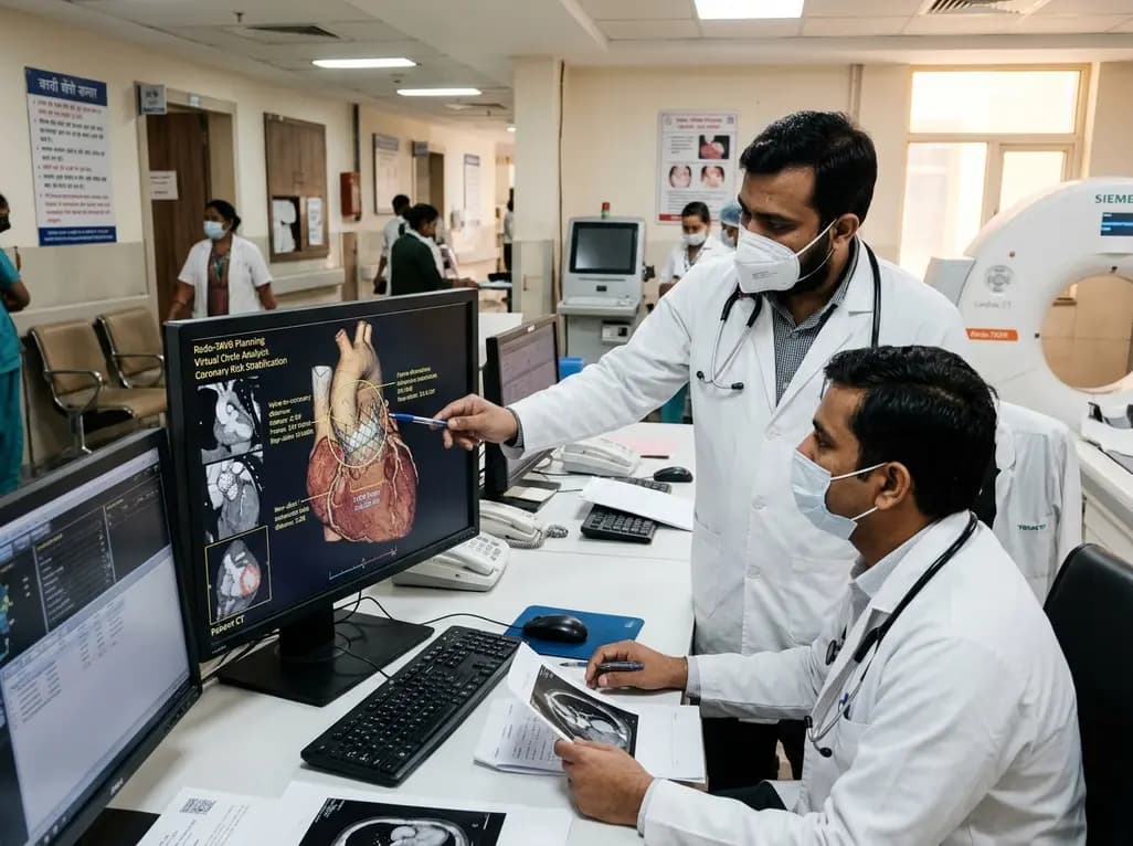
A multicenter study evaluating paired cardiac CT scans reveals that the virtual circle method reliably predicts index transcatheter valve frame expansion and changes in valve-to-aorta distance during redo-TAVR, offering accurate or conservative coronary risk stratification in 85% of cases.
Today

Explore joint-preserving correction of malunited anterior and posterior malleolar fractures. Discover how 3D CT imaging, anatomic reduction of the incisura, and restorative osteotomies prevent posttraumatic ankle osteoarthritis and restore biomechanical stability.
Today

India is poised to transform chronic disease management through wearable health technology. Discover how continuous remote monitoring and predictive algorithms empower clinicians to detect early cardiovascular signals, personalize metabolic interventions, and deliver equitable preventive care across populations.
Today
