

Loading, please wait...
Loading, please wait...
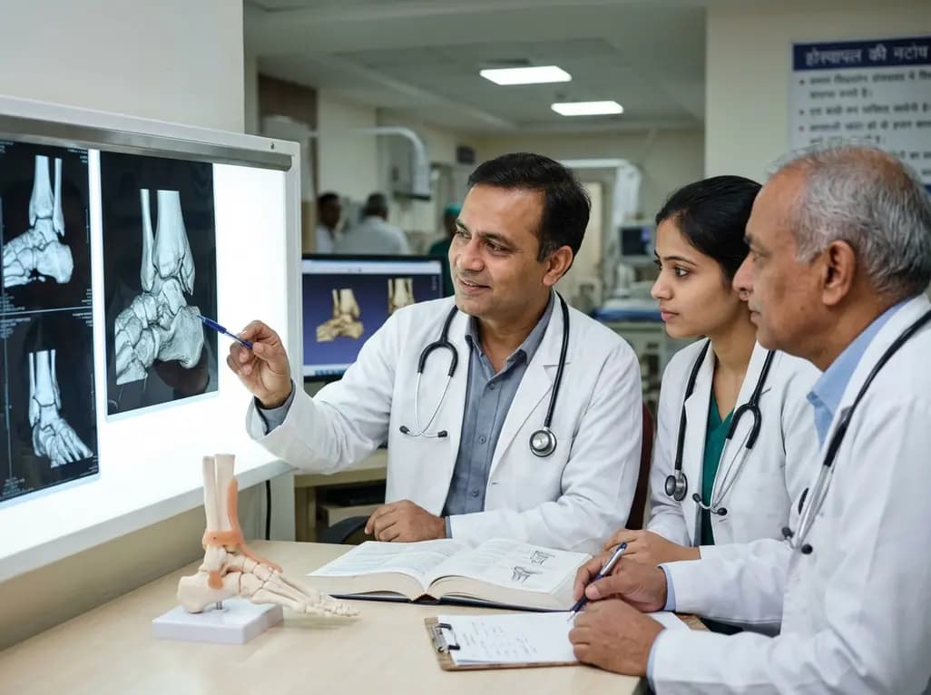
Ankle fractures represent some of the most frequent skeletal injuries encountered in daily orthopedic trauma practice. However, unreduced intra-articular fragments often lead to severe functional impairment and progressive degenerative changes. Timely malleolar malunion correction offers a valuable opportunity to restore native joint mechanics before end-stage arthritis develops. Surgeons must recognize that subtle step-offs or rotational displacements alter load distribution across the tibial plafond. Consequently, secondary joint-preserving reconstruction provides viable salvage pathways for active individuals who wish to avoid early arthrodesis.
The posterior and anterior tibial margins serve vital biomechanical roles in preserving overall ankle integrity. Clinicians historically prioritized the medial and lateral malleoli, often overlooking marginal plafond rims. Orthopedic trauma surgeons now classify the posterior distal tibial margin as the third malleolus. Similarly, they recognize the anterior Chaput tubercle as the fourth malleolus. Both structures provide critical anchor points for the inferior tibiofibular syndesmotic ligaments. In addition, these bony rims define the boundaries of the fibular incisura.
When an ankle fracture heals with residual displacement, profound structural consequences emerge. First, malunion of the posterior rim alters sagittal talar containment and promotes posterior talonavicular destabilization. Furthermore, displacement disrupts the concave morphology of the tibial notch. As a result, the distal fibula shifts out of its physiological groove, causing chronic syndesmotic instability. Persistent step-offs also concentrate contact pressures onto small areas of articular cartilage. Over time, this abnormal mechanical loading triggers chondrocyte breakdown and posttraumatic osteoarthritis. Therefore, orthopedic teams must recognize marginal malunions as complex multiplanar deformities requiring meticulous corrective strategies.
Accurate identification of marginal deformities begins with thorough diagnostic imaging. Conventional plain radiographs routinely underestimate the true size, rotation, and impaction of malleolar fragments. Therefore, orthopedic surgeons consider volumetric computed tomography scanning mandatory for every suspected malunion. Thin-slice axial cuts clearly demonstrate rotational mismatch of the distal fibula within the incisura. In addition, coronal and sagittal reconstructions reveal articular steps, marginal bone defects, and occult osteochondral lesions.
Furthermore, three-dimensional reconstruction models improve preoperative planning significantly. These detailed representations allow surgeons to quantify intra-articular gaps, cortical overlap, and posttraumatic bone remodeling. Surgeons can also compare the injured ankle with the contralateral side to establish exact anatomic references. Consequently, the surgical team can simulate corrective osteotomy cuts before entering the operating theater. Advanced imaging also identifies nonunions, intra-articular debris, and early cartilaginous thinning. By acquiring volumetric data, clinicians determine whether the articular surface remains salvageable. Thus, computed tomography provides the foundational roadmap for executing precise joint-preserving interventions.
Successful malleolar malunion correction requires clear, reproducible clinical objectives. First, the reconstructive team must restore perfect articular tibiotalar congruency. Even a minor intra-articular step-off accelerates cartilage wear and worsens joint pain. Second, surgeons must re-establish sagittal ankle stability to prevent persistent anterior or posterior talar subluxation. Talar containment ensures symmetrical motion arcs during the stance and swing phases of walking.
Third, reconstruction must restore the true anatomic geometry of the tibial incisura. An improperly shaped fibular notch prevents anatomical seating of the distal fibula. Therefore, surgeons must clear fibrous tissue and debride malunited margins to reposition the lateral column properly. Fourth, the operation must re-establish tibiofibular alignment and dynamic syndesmotic stability. Restoring the origin of the syndesmotic ligaments eliminates pathologically increased mortise volume. Consequently, tension returns to the posterior and anterior inferior tibiofibular ligaments. By fulfilling each of these four anatomic benchmarks, the surgeon protects the native cartilage from excessive focal stresses and prevents early functional decline.
Executing a secondary joint reconstruction demands patient-specific surgical approaches and precise osteotomy techniques. Surgeons usually select posterolateral or posteromedial surgical windows to visualize malunited posterior malleolar fragments. Through an internervous plane, the surgeon gently exposes the healed fracture bed while protecting the sural nerve and neurovascular bundle. Next, the surgeon performs a targeted osteotomy along the original fracture line using delicate osteotomes and high-resolution fluoroscopy.
Similarly, anterior malleolar deformities require an anterolateral approach to mobilize the Chaput fragment. Once osteotomized, the surgeon debrides interpositional scar tissue from the articular surface and the fibular notch. Afterward, the surgeon brings the bony fragment back into anatomical alignment with the distal tibial plafond. Rigid internal fixation with low-profile buttress plates or lag screws maintains stable compression. In cases of structural bone loss, surgeons frequently pack autologous cancellous graft into metaphyseal voids. Finally, intraoperative dynamic stress testing verifies syndesmotic stability. If ligamentous laxity persists, the surgeon places additional flexible or rigid syndesmotic stabilization to protect the reconstructive construct.
Secondary joint realignment represents a technically demanding but highly rewarding clinical undertaking. Patients who undergo timely reconstructive osteotomies experience marked reductions in weight-bearing pain and noticeable gains in range of motion. Furthermore, restoring native mortise biomechanics substantially delays or eliminates the need for total ankle arthroplasty or ankle arthrodesis. This functional preservation is especially critical for young, active patients who participate in strenuous manual labor or recreational sports.
However, existing literature on anterior and posterior malleolar malunion corrections remains limited. Most available evidence originates from small retrospective case series and expert technical reports. Consequently, the orthopedic community lacks standardized, prospective guidelines regarding patient selection, ideal timing, and osteotomy classification. Future multi-center studies must analyze patient-reported outcomes, long-term cartilage survival rates, and complication profiles. In addition, emerging computer-assisted surgical navigation and patient-specific three-dimensional cutting guides hold promise for improving intraoperative accuracy. As reconstructive techniques evolve, robust clinical trials will establish definitive recommendations for treating these complex distal tibial injuries.
Surgeons generally reserve joint-preserving osteotomy for patients with preserved articular cartilage and localized malunions. If severe osteoarthritis, extensive chondral loss, or diffuse joint narrowing has already developed, corrective osteotomy yields suboptimal functional relief. Consequently, clinicians recommend ankle arthrodesis or total ankle replacement for end-stage degenerative changes. Careful preoperative assessment remains critical to distinguish salvageable joints from advanced arthritis.
Computed tomography provides three-dimensional visualization of complex multiplanar deformities that plain radiographs frequently obscure. Specifically, axial imaging assesses the depth of the fibular incisura and reveals distal fibular subluxation. Furthermore, sagittal and coronal reformations delineate intra-articular steps, marginal impaction, and posttraumatic bone defects. Therefore, volumetric imaging allows orthopedic surgeons to plan precise osteotomy planes and select appropriate hardware before entering surgery.
The posterior malleolus serves as the anatomical insertion site for the posterior inferior tibiofibular ligament. When this fragment heals in a displaced position, ligamentous tension diminishes and the posterior buttress fails. Furthermore, displacement deforms the fibular incisura, which forces the distal fibula out of its anatomical notch. Consequently, the mortise widens pathologically, causing persistent syndesmotic instability, altered load transfer, and joint incongruency.
Disclaimer: This content is for informational and educational purposes only and should not be considered medical advice. Always consult a qualified healthcare professional regarding any medical condition or treatment. Refer to the latest local and national guidelines for clinical practice.
References

Read summarized clinical updates, watch expert medical content, and earn CME certifications right from your smartphone.
Open your smartphone camera to scan and install.





Explore joint-preserving correction of malunited anterior and posterior malleolar fractures. Discover how 3D CT imaging, anatomic reduction of the incisura, and restorative osteotomies prevent posttraumatic ankle osteoarthritis and restore biomechanical stability.
Today
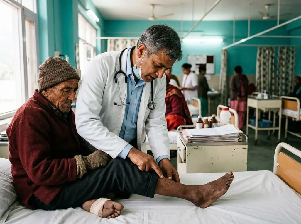
A recent cross-sectional study reveals that prolonged residence at high altitude significantly escalates foot frostbite risk among lowland migrants, with chronic mountain sickness acting as a significant physiological mediator through hyperviscosity and microcirculatory stasis.
Today
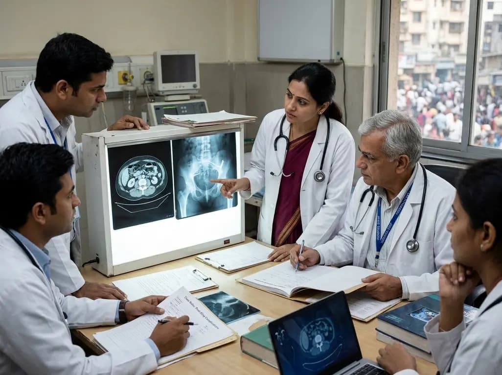
A litigation-derived series of 252 women highlights the risks of repeat revisions after midurethral sling surgery. Recurrent incontinence occurred in up to 60% of patients, and repeat revision rates reached 70%, emphasizing cautious patient selection and skilled multidisciplinary management.
Today
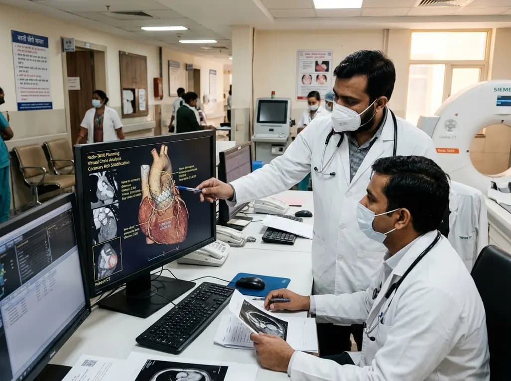
A multicenter study evaluating paired cardiac CT scans reveals that the virtual circle method reliably predicts index transcatheter valve frame expansion and changes in valve-to-aorta distance during redo-TAVR, offering accurate or conservative coronary risk stratification in 85% of cases.
Today
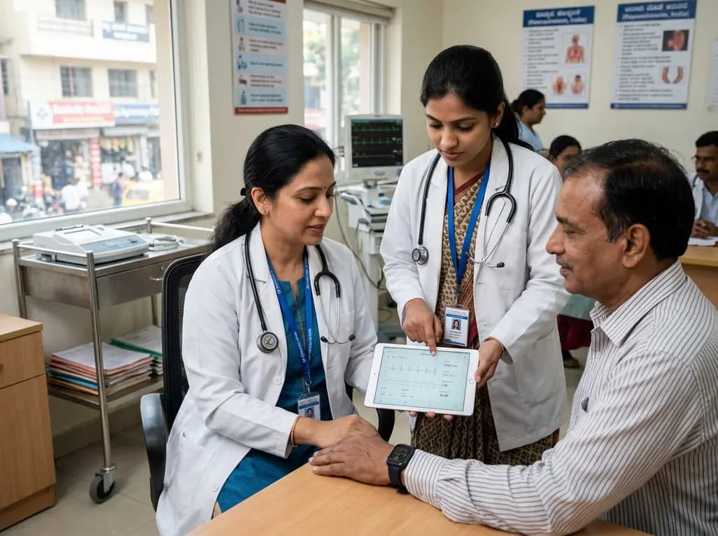
India is poised to transform chronic disease management through wearable health technology. Discover how continuous remote monitoring and predictive algorithms empower clinicians to detect early cardiovascular signals, personalize metabolic interventions, and deliver equitable preventive care across populations.
Today
