.webp&w=384&q=75)

Loading, please wait...
Loading, please wait...
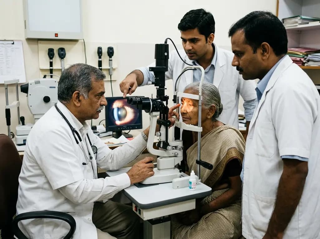
Chronic intraocular inflammation frequently causes severe structural ocular complications and permanent vision loss. Among its most challenging sequelae, uveitis-associated ocular hypotony represents a critical condition that requires prompt clinical intervention. While secondary ocular hypertension receives extensive clinical focus, persistent subnormal intraocular pressure induces devastating globe collapse. Consequently, affected eyes develop hypotony maculopathy, chorioretinal folds, optic disc edema, and premature cataracts. Pediatric patients, especially those with juvenile idiopathic arthritis, face a particularly high vulnerability. Therefore, this systematic review synthesizes current evidence on pathophysiology, clinical risk factors, multimodal diagnostic imaging, medical therapy, and advanced surgical management.
The pathophysiology of sustained ocular hypotony in uveitis involves secretory failure and structural distortion. Primarily, active intraocular inflammation releases a potent surge of inflammatory cytokines. These biochemical mediators disrupt the non-pigmented ciliary epithelium, causing marked hyposecretion and reduced aqueous humor production. Simultaneously, persistent inflammatory changes within the uveal tract accelerate uveoscleral outflow, thereby depleting intraocular volume rapidly. Over time, recurring cellular exudates form dense retrociliary cyclitic membranes. Consequently, these tractional membranes mechanically pull the ciliary body away from the scleral wall, inducing ciliary detachment. Furthermore, unmanaged long-standing inflammation causes irreversible ciliary tissue atrophy and progressive fibrosis. When intraocular pressure drops below critical thresholds, transscleral hydrostatic pressure gradients shift dramatically. As a result, patients develop choroidal expansion, chorioretinal folds, and progressive hypotony maculopathy. Thus, reversing both secretory shutdown and mechanical traction remains essential for restoring ocular structure.
Epidemiological data reveal that intraocular hypotony affects diverse uveitis populations across all age groups. In adult cohorts suffering from chronic non-infectious uveitis, the estimated prevalence typically hovers around ten percent. In contrast, pediatric cohorts demonstrate a substantially greater disease burden. Specifically, children with juvenile idiopathic arthritis-associated uveitis experience hypotony prevalence rates between ten and nineteen percent. Furthermore, clinical registries have identified several prominent risk factors that heighten patient vulnerability. Childhood disease onset strongly predisposes patients to severe ciliary body dysfunction and architectural collapse. Additionally, longer cumulative disease duration and persistent anterior chamber cell activity significantly increase hypotony risk. Severe vitreous haze, recurrent panuveitis, and band keratopathy also predict worse clinical outcomes. Notably, prior intraocular surgical procedures, particularly complex cataract extraction, represent major secondary risk triggers. Therefore, close surveillance of high-risk pediatric and adult patients is vital for early diagnosis.
Accurate clinical evaluation requires a meticulous diagnostic approach combining functional testing and structural imaging. Clinicians must first establish reliable intraocular pressure measurements using calibrated Goldmann applanation or dynamic contour tonometry. Subsequently, high-frequency ultrasound biomicroscopy serves as the gold standard for visualizing the anterior segment and retrociliary space. This imaging modality clearly identifies subtle cyclitic membranes, supraciliary fluid, and circumferential ciliary body detachment. In addition, anterior segment optical coherence tomography provides non-contact cross-sectional imaging of anterior chamber architecture. For posterior segment evaluation, spectral-domain optical coherence tomography reliably detects neurosensory retinal folds, macular thickening, and subretinal fluid. Fundus photography and fluorescein angiography further assist in assessing retinal vascular leakage and optic nerve congestion. Finally, posterior B-scan ultrasonography provides vital information regarding choroidal thickening and scleral infolding. Consequently, integrating these imaging modalities enables clinicians to differentiate reversible inflammatory hyposecretion from irreversible anatomical ciliary atrophy.
The primary medical goal in uveitic hypotony focuses on aggressive suppression of active inflammation to restore ciliary secretion. Clinicians generally initiate intensive topical, periocular, or systemic corticosteroids to rapidly halt inflammatory cascades. Furthermore, cycloplegic agents such as topical atropine help stabilize the blood-aqueous barrier and deepen the anterior chamber. When active inflammation persists despite corticosteroid administration, clinicians must promptly introduce steroid-sparing disease-modifying antirheumatic drugs. Specifically, antimetabolites such as methotrexate and mycophenolate mofetil offer reliable disease control while avoiding long-term steroid toxicity. Additionally, targeted biologic therapies, particularly tumor necrosis factor inhibitors like adalimumab and infliximab, demonstrate robust efficacy in refractory cases. If systemic inflammatory control alone fails to restore pressure, clinicians may explore secondary pharmacologic options. Topical ibopamine and high-dose prostaglandin analogs have occasionally stimulated pressure elevation in selected cases. Therefore, early multi-agent anti-inflammatory therapy remains the cornerstone of preserving ciliary architecture.
When conservative medical management fails to relieve structural traction, vitreoretinal surgery becomes necessary to prevent phthisis bulbi. Persistent cyclitic membranes exert severe mechanical traction that prevents ciliary body reattachment and functional recovery. Consequently, pars plana vitrectomy combined with meticulous cyclitic membrane dissection represents the definitive surgical approach. Vitreoretinal surgeons carefully sever fibrous traction bands to release the incarcerated ciliary processes. Furthermore, surgeons frequently utilize long-term intraocular tamponade with high-viscosity silicone oil to maintain internal globe volume. Clinicians also combine vitrectomy with sustained-release fluocinolone acetonide intravitreal implants. These implants deliver continuous local steroid therapy, suppressing persistent micro-inflammation while potentially promoting steroid-induced pressure elevation. However, visual recovery in chronic cases remains guarded due to permanent photoreceptor damage from long-standing hypotony maculopathy. Early surgical intervention before irreversible ciliary atrophy develops offers the best opportunity for anatomical and visual preservation.
Clinicians generally define chronic ocular hypotony as a persistent intraocular pressure below six to eight millimeters of mercury lasting for several consecutive weeks. This sustained pressure reduction triggers profound structural complications, including corneal decompensation, optic disc edema, and hypotony maculopathy. Furthermore, intraocular pressures falling below four millimeters of mercury carry the highest risk of irreversible visual impairment, requiring immediate multidisciplinary evaluation and aggressive clinical intervention.
Pediatric patients, particularly those diagnosed with juvenile idiopathic arthritis, exhibit higher hypotony rates due to aggressive, prolonged anterior segment inflammation. Moreover, younger ocular tissues demonstrate greater structural compliance, making the pediatric globe highly vulnerable to rapid architectural collapse. Additionally, children frequently experience delayed symptom reporting, which allows insidious cyclitic membrane proliferation and extensive ciliary body detachment to develop long before clinical detection occurs.
Vitreoretinal surgeons utilize silicone oil endotamponade as an indispensable mechanical and physiological stabilizer in eyes with refractory hypotony. Mechanistically, silicone oil provides essential internal structural support to prevent globe collapse and maintain ciliary body apposition against the sclera. Furthermore, silicone oil compartmentalizes the intraocular space, reducing inflammatory cytokine diffusion and facilitating a more stable, elevated intraocular pressure throughout extended post-surgical follow-up periods.
Disclaimer: This content is for informational and educational purposes only and does not constitute medical advice. Healthcare professionals should exercise their clinical judgment when interpreting and applying this information. Refer to the latest local and national guidelines for clinical practice.
References

Read summarized clinical updates, watch expert medical content, and earn CME certifications right from your smartphone.
Open your smartphone camera to scan and install.





Uveitis-associated ocular hypotony is a sight-threatening complication driven by ciliary hyposecretion, increased uveoscleral outflow, and cyclitic membranes. This systematic review synthesizes epidemiology, diagnostic imaging, stepwise medical immunosuppression, and modern vitreoretinal surgical techniques.
last month

Pancreatic cancer cachexia affects over 80% of patients, driving severe muscle wasting, systemic inflammation, and treatment intolerance. This review highlights key pathophysiological drivers, exocrine insufficiency, objective CT imaging, and emerging therapies like GDF-15 inhibitors to improve clinical outcomes.
Today

A scoping review reveals how pregnancy affects phonation through laryngeal edema and reduced phonation time rather than major acoustic pitch alterations. Learn how to identify, evaluate, and manage vocal symptoms in expectant mothers.
Today

According to WHO data, average lifespan reached 73.3 years in 2023, recovering from pandemic lows. However, healthy life expectancy lags at 62.8 years due to surging noncommunicable conditions like cardiovascular disease and dementia, demanding proactive primary screening and sustained chronic care.
Today

Researchers have engineered an artificial sensory-pain integrated receptor using a multi-threshold organic synaptic transistor. The system distinguishes benign touch from noxious stimuli, enabling safer, more responsive prosthetic limbs with built-in memory and simplified hardware architecture.
Today

Medical robotics is transitioning beyond operative suites into diagnostics, cardiology, and endocrinology. Explore how innovations in tele-robotics, AI integration, and nanomedicine are overcoming geographical barriers and transforming precision healthcare delivery across India's evolving disease landscape.
Today
