.webp&w=384&q=75)

Loading, please wait...
Loading, please wait...

Colorectal cancer screening relies heavily on the thorough mucosal inspection of the lower gastrointestinal tract. In recent years, healthcare providers have increasingly integrated artificial intelligence into endoscopy units to enhance diagnostic quality. Specifically, the clinical adoption of AI-CADe in colonoscopy represents a substantial technological evolution aimed at improving the adenoma detection rate. While clinical trials consistently highlight superior lesion discovery, real-world deployment depends heavily on endoscopist acceptance. Healthcare professionals must balance improved diagnostic sensitivity against daily operational friction. Therefore, understanding user perceptions among endoscopy teams provides critical guidance for hospital leaders and practicing gastroenterologists. This exploratory survey evaluates how clinical staff perceive automated detection tools before and after practical implementation.
The National Cancer Center conducted an exploratory cross-sectional study to examine user sentiment across two distinct implementation phases. Endoscopy staff members completed structured questionnaires before installation and one month after clinical rollout. Overall, twenty-nine healthcare professionals completed the pre-implementation phase, whereas twenty-five staff members finished the follow-up evaluation. The cohort comprised attending endoscopists, gastroenterology fellows, and specialized endoscopy nursing personnel. Consequently, the findings represent a multidisciplinary perspective on modern computer vision tools. Researchers designed the survey to capture baseline expectations alongside practical post-adoption reflections. Most participants expressed robust initial enthusiasm regarding automated lesion recognition. Furthermore, staff anticipated meaningful gains in adenoma identification and patient satisfaction. Importantly, these positive attitudes endured throughout the first month of hands-on procedural use. Clinicians noted that real-time visual assistance could enhance mucosal inspection quality. However, staff also recognized that successful deployment requires rigorous technical performance and smooth operational synergy.
The primary objective of automated detection software is increasing mucosal neoplasia discovery. In this study, respondents consistently affirmed that algorithmic assistance improves the adenoma detection rate. Endoscopists valued the software as a reliable second observer during challenging examinations. Specifically, the system helped clinicians identify subtle flat lesions and diminutive polyps that might escape standard visualization. As a result, endoscopists felt more confident in achieving comprehensive therapeutic polypectomy. Survey respondents highlighted that visual bounding boxes drew immediate attention to suspicious colonic folds. In addition, endoscopy nurses noted that digital assistance reduced operator fatigue during prolonged endoscopy lists. Consequently, procedural satisfaction scores remained high before and after technology installation. Many endoscopists believed that improved detection could directly lower interval cancer incidence. Nevertheless, clinicians emphasized that diagnostic sensitivity alone does not guarantee clinical excellence. Operators still need rigorous endoscopic withdrawal techniques and meticulous mucosal cleansing. Thus, technology functions as an adjunct rather than a surrogate for human skill.
Seamless operational integration represents a critical prerequisite for adopting novel medical devices. In routine endoscopy, rapid procedural turnover remains essential for maintaining institutional efficiency. Participants in this study shared notable feedback regarding procedural flow and equipment responsiveness. On one hand, staff reported general satisfaction with overall workflow impact during uncomplicated examinations. On the other hand, qualitative responses uncovered specific technical friction points. For example, several clinicians noted occasional delays in system detection during swift scope withdrawal. Moreover, repeated auditory and visual alerts occasionally distracted endoscopists during complex therapeutic maneuvers. When an algorithm repeatedly flags normal colonic folds, endoscopists must pause to re-examine the area. Consequently, these interruptions can prolong procedure times and disrupt the clinical rhythm. Nurses also expressed concerns regarding room setup complexity and monitor positioning. Therefore, manufacturers must prioritize responsive processing speeds and unobtrusive user interfaces. Enhancing system ergonomics ensures that automated detection assists clinicians without impeding routine clinical operations.
Despite evident clinical enthusiasm, respondents highlighted critical apprehensions regarding overdetection and user reliance. Survey participants frequently cited false-positive alarms as an ongoing operational frustration. In clinical practice, benign mucosal tags, retained fecal debris, and air bubbles can trigger automated notifications. Consequently, clinicians worry that false positives may prompt unnecessary tissue biopsies or redundant resections. These avoidable interventions increase pathology expenses, extend procedural duration, and elevate bleeding risks. Additionally, surveyed staff expressed valid concerns regarding psychological dependence on automated alerts. If trainees rely prematurely on automated guidance, they might fail to cultivate foundational visual inspection habits. Furthermore, experienced operators could develop complacency, reducing active attention in unflagged visual fields. Survey respondents identified diagnostic accuracy and false-positive rates as decisive factors governing sustained clinical utilization. Because unnecessary interventions burden healthcare systems, refining algorithmic specificity remains essential. Endoscopic societies must establish clear training standards to prevent cognitive deskilling among rising gastroenterologists.
Successful clinical integration of artificial intelligence demands deliberate governance and user-centered design. While software algorithms deliver impressive detection sensitivity, institutions must evaluate purchase expenses and ongoing maintenance costs. Indeed, financial feasibility and false-alarm frequency strongly determine long-term institutional adoption. Hospital administrators should involve nursing staff and junior clinicians early during equipment procurement. Moreover, endoscopy centers should implement structured onboarding protocols that teach endoscopists how to interpret real-time alerts critically. Rather than assuming the technology replaces clinical judgment, operators should treat it as an objective safety net. Future technological iterations must also integrate characterization modules to distinguish non-neoplastic hyperplastic polyps from true adenomas. Such optical biopsy features will minimize unnecessary polypectomies and curb tissue processing costs. Furthermore, longitudinal clinical registries must track real-world post-colonoscopy colorectal cancer rates over extended periods. In conclusion, algorithmic assistance holds tremendous promise for modern gastrointestinal endoscopy. By addressing usability feedback and refining alert precision, clinicians can optimize patient care while preserving diagnostic rigor.
Automated detection systems analyze video feeds in real time using deep convolutional neural networks. When the algorithm identifies a potential mucosal abnormality, it immediately highlights the suspicious area on the monitor. This visual prompt assists the endoscopist in identifying flat, diminutive, or poorly positioned polyps behind mucosal folds. Consequently, the technology acts as a vigilant secondary observer, significantly reducing adenoma miss rates and enhancing overall screening quality across diverse clinical settings.
False-positive alerts occur when algorithms misclassify normal anatomical structures, residual stool, or suction artifacts as neoplastic polyps. These persistent visual or auditory notifications can distract endoscopists during examination. Furthermore, repeated false alarms may prompt clinicians to perform unnecessary biopsies or resections on benign tissues. Such unneeded interventions inevitably prolong procedural duration, raise pathology processing expenses, and increase the risk of procedural complications such as post-polypectomy bleeding or colonic wall perforation.
Excessive reliance on automated detection systems creates potential risks of operator complacency and cognitive deskilling. When gastroenterology trainees rely heavily on automated visual prompts, they might fail to develop meticulous mucosal inspection discipline. Similarly, experienced clinicians may experience reduced vigilance in colonic segments where algorithms remain silent. To prevent technological dependence, academic institutions must establish structured endoscopy curricula that emphasize thorough manual inspection before integrating automated algorithmic assistance into everyday training.
Disclaimer: This content is for informational and educational purposes only. It does not constitute medical advice, diagnosis, or treatment recommendations. Healthcare professionals must exercise independent clinical judgment. Refer to the latest local and national guidelines for clinical practice.
References

Read summarized clinical updates, watch expert medical content, and earn CME certifications right from your smartphone.
Open your smartphone camera to scan and install.





A single-center exploratory survey evaluated endoscopy staff perceptions of AI-CADe in colonoscopy. Clinicians reported high satisfaction and enhanced adenoma detection but raised concerns about false-positive alerts, procedural workflow delays, and overreliance on automated systems.
Today
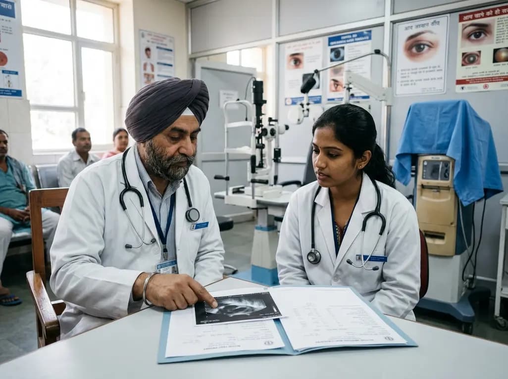
A multicenter study reveals that a significant proportion of HCV antibody-positive ophthalmic surgical patients remain untreated and eligible for direct-acting antivirals. Stratifying antibody titers and streamlining referral pathways can optimize hepatitis C micro-elimination efforts.
Today
Robotic single-port totally extraperitoneal (TEP) repair enables simultaneous excision of a canal of Nuck cyst and indirect hernia repair in women. Using the da Vinci SP platform and self-adhesive mesh, this approach offers minimal tissue trauma, low operative times, and rapid discharge without complications.
Today
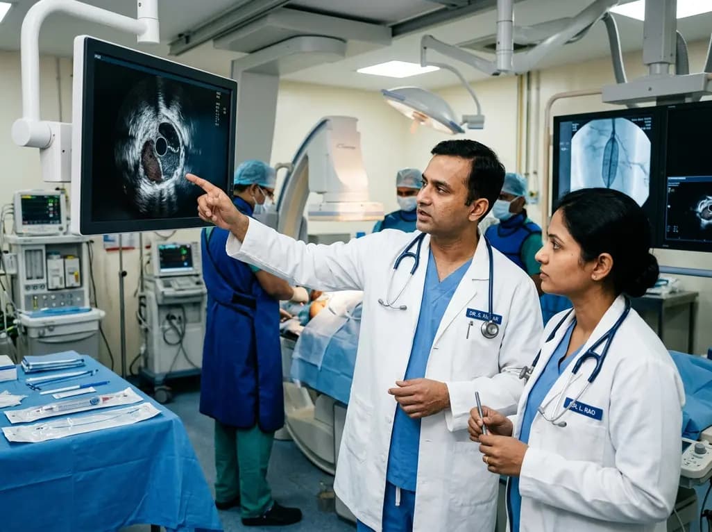
A multicenter REAL-LEAD registry sub-analysis evaluates how intravascular ultrasound-defined circumferential calcification and post-procedural minimum lumen area govern 1-year primary patency after drug-coated balloon angioplasty for femoropopliteal lesions.
Today

A systematic review and meta-analysis of 21 RCTs reveals that platelet-rich plasma provides small but statistically superior pain relief and functional improvements compared to hyaluronic acid at 12 months in knee osteoarthritis, influenced by platelet concentration and leukocyte enrichment.
Today

A multicenter real-world prospective study in Taiwan demonstrates that liraglutide effectively reduces body weight and BMI standard deviation score in adolescents with obesity. The therapy shows a favorable safety profile with mild gastrointestinal adverse events and zero treatment discontinuations due to intolerance.
Today
