.webp&w=384&q=75)

Loading, please wait...
Loading, please wait...

Groin masses in female patients often present a diagnostic dilemma for surgeons and primary care physicians. While indirect inguinal hernias occur frequently, an uncommon yet critical differential diagnosis is a canal of Nuck cyst. When these entities present concurrently, clinicians must address both the cystic lesion and the fascial defect simultaneously. Minimally invasive approaches continue to evolve, and recent advances have introduced robotic single-port totally extraperitoneal repair into modern surgical practice. This technique allows surgeons to achieve definitive excision and tension-free preperitoneal mesh placement through a single tiny incision. Consequently, patients benefit from reduced dissection trauma, less postoperative pain, and expedited clinical recovery.
The canal of Nuck represents a rudimentary embryological evagination of the parietal peritoneum in women. Typically, this peritoneal outpouching accompanies the round ligament of the uterus through the inguinal canal toward the labium majus. During normal fetal maturation, the structure obliterates completely before birth or within the first year of life. However, incomplete or failed obliteration results in a patent canal. If fluid accumulates due to an imbalance between secretion and absorption, a hydrocele or canal of Nuck cyst develops along the round ligament.
Anatomists generally categorize these anomalies into three distinct types. Type I describes an encysted hydrocele along the round ligament that lacks connection to the peritoneal cavity. Type II features free communication with the peritoneal space, functioning essentially like a congenital indirect hernia sac. Meanwhile, Type III presents as an hourglass or dumbbell configuration, where the deep inguinal ring constricts the middle of the fluid collection. Because the patent tract shares an intimate anatomical relationship with the internal ring, indirect inguinal hernias frequently coexist. Therefore, surgical teams must thoroughly inspect the entire myopectineal orifice during operative exploration.
Clinically, patients typically present with a unilateral, soft, fluctuant groin swelling. Many individuals report chronic or intermittent discomfort, which frequently worsens during prolonged standing, strenuous physical exertion, or increased intra-abdominal pressure. Because female groin swellings are comparatively rare, clinicians often misdiagnose this entity as an irreducible inguinal hernia, enlarged lymph node, femoral hernia, or soft-tissue lipoma. Furthermore, acute complications such as secondary infection, trauma-induced hemorrhage, or torsion can mimic a strangulated groin hernia, prompting emergency medical evaluation.
Consequently, high-resolution imaging plays an indispensable role in establishing the correct preoperative diagnosis. High-frequency ultrasonography serves as the initial diagnostic tool of choice. Sonographic examination typically reveals a well-circumscribed, anechoic or hypoechoic, thin-walled cystic mass within the inguinal canal without internal vascularity on Doppler evaluation. If the diagnosis remains indeterminate or if the lesion extends into the pelvis, magnetic resonance imaging provides exceptional soft-tissue differentiation. MRI clearly depicts the cyst extent, delineates its boundary against the round ligament, and excludes incarcerated intraperitoneal contents. Accurate preoperative imaging ensures meticulous operative planning and prevents unexpected findings in the theater.
Historically, surgeons managed canal of Nuck pathology via open anterior inguinal incisions. Although open surgery provides direct access, it requires significant tissue disruption, carries risks of chronic groin neuralgia, and often requires dividing the genital branch of the genitofemoral nerve. Subsequently, laparoscopic techniques such as transabdominal preperitoneal and totally extraperitoneal repairs gained substantial popularity. These laparoscopic modalities allow clear visualization of the deep inguinal ring while avoiding unnecessary peritoneal violation in totally extraperitoneal approaches.
Nevertheless, conventional laparoscopy faces mechanical limitations in narrow anatomical corridors. Rigid straight instruments can create awkward ergonomics and restricted angles during delicate cyst mobilization. The da Vinci Single-Port robotic system effectively overcomes these technical constraints. By consolidating three fully articulating, double-jointed instruments alongside a flexible high-definition stereoscopic camera within a single 25-millimeter cannula, the single-port platform delivers remarkable triangulation. Surgeons maneuver easily within confined retroperitoneal spaces without instrument clashing. Thus, robotic single-port totally extraperitoneal repair combines exceptional dexterity with the established benefits of preperitoneal mesh placement.
The operative procedure begins with an infraumbilical transverse incision to access the retrorectus and preperitoneal planes. After securing the specialized single-port access device, the surgeon establishes low-pressure carbon dioxide insufflation to develop the extraperitoneal working space. Next, the surgical team docks the da Vinci single-port system and introduces articulated robotic instruments under direct visualization. The robotic surgeon initiates dissection medial to the epigastric vessels, developing Retzius' space before extending lateral dissection toward Bogros' space.
Upon entering the inguinal canal, the surgeon carefully identifies the cystic mass closely adherent to the round ligament. Robotic wricked instruments allow gentle, precise dissection around the cyst wall, minimizing the risk of inadvertent fluid rupture. If an indirect hernia sac is present, the surgeon dissects it circumferentially and reduces it back into the extraperitoneal space. Once the team completely excises the cyst and ligates associated non-vital tissues, reconstruction begins. A self-adhesive 10 by 15-centimeter polypropylene mesh is introduced using the Hansol-roll technique. The surgeon deploys and positions the mesh widely across the myopectineal orifice without requiring traumatic tackers. Finally, the team evacuates the pneumo-preperitoneum under direct vision to ensure stable mesh orientation.
Published clinical outcomes show that robotic single-port totally extraperitoneal repair offers high operative efficiency and remarkable safety. Operative and console times average less than one hour in experienced hands. Because the procedure avoids entering the peritoneal cavity, it eliminates intra-abdominal adhesions and minimizes postoperative ileus. Furthermore, the absence of sharp penetrating fixators significantly lowers the incidence of chronic postoperative groin pain. Most patients achieve successful discharge on postoperative day one with minimal need for oral analgesics.
For general and robotic surgeons, recognizing the canal of Nuck cyst as a treatable entity remains crucial. Complete surgical excision is mandatory because incomplete aspiration or partial resection frequently leads to recurrent fluid collections and ongoing pain. Simultaneous tension-free mesh reinforcement prevents subsequent indirect or direct hernia recurrence. As robotic platforms become more widely accessible, single-port totally extraperitoneal repair represents an elegant, tissue-sparing approach for managing complex female groin pathologies safely and reproducibly.
A canal of Nuck cyst represents an abnormal fluid collection within a patent peritoneal evagination. In female embryos, this pouch accompanies the round ligament through the inguinal canal. When this peritoneal tract fails to close completely during development, peritoneal secretions accumulate. Consequently, adult women experience a cystic groin lump that may enlarge gradually or cause localized discomfort during exertion.
Conventional laparoscopic totally extraperitoneal repair requires multiple separate trocars and rigid straight instruments, which can clash within narrow surgical corridors. In contrast, the da Vinci single-port robotic platform introduces three fully articulating, wristed instruments and a flexible camera through a single small incision. This robotic system provides superior 3D visualization, improved dexterity, and streamlined preperitoneal dissection in tight spaces.
Simple needle aspiration or drainage provides only temporary relief because the secretory epithelial lining of the cyst remains intact. As a result, fluid inevitably reaccumulates, leading to symptom recurrence and potential secondary infection. Complete surgical excision of the cyst wall eradicates the underlying secretory tissue permanently. Additionally, concurrent mesh hernioplasty reinforces the abdominal wall against future inguinal hernia recurrence.
Disclaimer: This content is for informational and educational purposes only and does not substitute professional medical judgment, diagnosis, or treatment. Healthcare providers should verify clinical details and consult relevant medical literature. Refer to the latest local and national guidelines for clinical practice.
References
Lee CS et al. Robotic single-port totally extraperitoneal inguinal hernia repair with excision of a canal of Nuck cyst using the da Vinci Single-Port system: a video vignette. J Minim Invasive Surg. 2026 Sep 15. doi: 10.7602/jmis.2026.29.3.163. PMID: 42745705.
Long W, Han J, Milone L. Novel Use of Robotic Surgery in a Rare Case of Canal of Nuck Cyst: A Case Report and Literature Review. Am J Case Rep. 2025;26:e945112. doi: 10.12659/AJCR.945112.
Ural ÜM. Minimally invasive treatment of the cyst of the canal of Nuck with the laparoscopic total extraperitoneal approach. Northwest Med J. 2025;5(1):45-48. doi: 10.54307/NMJ.2025.109.

Read summarized clinical updates, watch expert medical content, and earn CME certifications right from your smartphone.
Open your smartphone camera to scan and install.





Robotic single-port totally extraperitoneal (TEP) repair enables simultaneous excision of a canal of Nuck cyst and indirect hernia repair in women. Using the da Vinci SP platform and self-adhesive mesh, this approach offers minimal tissue trauma, low operative times, and rapid discharge without complications.
Today

A multicenter study reveals that a significant proportion of HCV antibody-positive ophthalmic surgical patients remain untreated and eligible for direct-acting antivirals. Stratifying antibody titers and streamlining referral pathways can optimize hepatitis C micro-elimination efforts.
Today
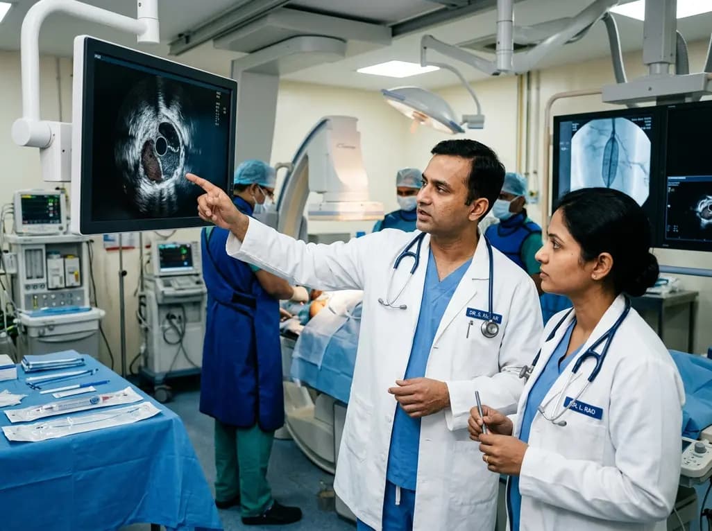
A multicenter REAL-LEAD registry sub-analysis evaluates how intravascular ultrasound-defined circumferential calcification and post-procedural minimum lumen area govern 1-year primary patency after drug-coated balloon angioplasty for femoropopliteal lesions.
Today

A systematic review and meta-analysis of 21 RCTs reveals that platelet-rich plasma provides small but statistically superior pain relief and functional improvements compared to hyaluronic acid at 12 months in knee osteoarthritis, influenced by platelet concentration and leukocyte enrichment.
Today

A multicenter real-world prospective study in Taiwan demonstrates that liraglutide effectively reduces body weight and BMI standard deviation score in adolescents with obesity. The therapy shows a favorable safety profile with mild gastrointestinal adverse events and zero treatment discontinuations due to intolerance.
Today
