

Loading, please wait...
Loading, please wait...

Acute sympathetic hyperactivity drives life-threatening clinical crises in both the cardiac and peripheral vascular domains. In critical care units, a refractory ventricular electrical storm and severe vascular vasospasm can quickly cause end-organ collapse. Consequently, clinicians urgently need rapid bedside interventions when conventional pharmacotherapy fails. The ultrasound-guided stellate ganglion block has rapidly emerged as a powerful rescue therapy. By interrupting the cervical sympathetic chain, clinicians can promptly modulate severe autonomic surges. Therefore, emergency physicians and intensivists now utilize this percutaneous approach to manage unstable ventricular arrhythmias and critical limb hypoperfusion effectively.
Ventricular electrical storm represents an acute arrhythmic state characterized by multiple sustained ventricular tachycardia episodes within twenty-four hours. Although standard advanced cardiac life support protocols prioritize antiarrhythmic medications and external cardioversion, heightened adrenergic drive frequently maintains recurrent arrhythmia loops. Furthermore, excessive endogenous catecholamines diminish ventricular fibrillation thresholds and exacerbate myocardial oxygen consumption. Similarly, acute upper limb ischemia involves severe arterial compromise where intense reflex vasospasm compounds mechanical luminal obstruction. In both clinical emergencies, peripheral vasoconstriction and arrhythmogenicity share a common pathophysiological driver: relentless sympathetic excitation.
Standard resuscitation often proves insufficient because circulating adrenaline continues to trigger cellular calcium overload. In addition, repeated electrical shocks amplify adrenergic surges, creating a self-perpetuating vicious cycle of electrical instability. Peripheral vascular beds respond to this adrenergic discharge with unrelenting microvascular spasm, thereby accelerating tissue necrosis. Consequently, interruption of sympathetic efferent output provides an indispensable physiological mechanism to break these refractory states before irreversible organ damage ensues.
Recent clinical evidence highlights the rapid efficacy of targeted autonomic modulation in the emergency department. Specifically, Patil and colleagues evaluated ultrasound-guided intervention at the C6 vertebral level in seven critical patients. The cohort included four individuals suffering from acute upper limb ischemia and three presenting with refractory ventricular tachycardia. Clinicians administered six to eight milliliters of 0.25% bupivacaine adjacent to the longus colli muscle under direct ultrasound visualization. Notably, every patient demonstrated tangible clinical improvement following local anesthetic delivery.
In patients with critical limb ischemia, the regional block produced dramatic reductions in ischemic rest pain. Furthermore, clinicians recorded visible restoration of limb warmth, accelerated capillary refill times, and recovered distal arterial waveforms. Meanwhile, patients presenting with recurrent ventricular tachycardia achieved substantial rhythm stabilization. The frequency of recurrent tachyarrhythmic events diminished precipitously, allowing hemodynamics to recover safely. Importantly, the operators documented no major procedural complications, confirming that precise sonographic needle guidance delivers exceptional safety during acute resuscitation.
Executing a safe sympathetic block requires rigorous anatomical understanding of the cervical fascial planes. The stellate ganglion typically resides anterior to the C7 and T1 transverse processes, resting medial to the vertebral artery. However, performing injection directly at the C7 level carries significant risk of accidental vertebral artery puncture or pneumothorax. Therefore, experts strongly recommend placing the local anesthetic deposit at the C6 vertebral level over the Chassaignac tubercle. At this level, the anterior tubercle forms a prominent bony landmark, shielding vital neurovascular structures effectively.
Clinicians position a high-frequency linear ultrasound probe transversely over the anterolateral neck to identify the internal jugular vein and carotid artery. Next, the practitioner visualizes the longus colli muscle resting directly on the prevertebral fascia. The needle enters in-plane from the lateral aspect, navigating cautiously between the carotid sheath and the cervical transverse process. Once the needle tip pierces the prevertebral fascia superficial to the longus colli muscle, negative aspiration excludes intravascular placement. Subsequently, real-time visualization of local anesthetic spread ensures robust drug distribution without entering the surrounding deep cervical vessels.
The therapeutic impact of cervical sympathetic blockade stems from profound neurocardiac and microvascular desensitization. Efferent sympathetic fibers from the stellate ganglion supply the sinoatrial node, conduction pathways, and ventricular myocardium. Left-sided sympathetic fibers directly control ventricular refractoriness and afterdepolarization thresholds. Consequently, local anesthetic blockade eliminates sympathetic hyperactivity, homogenizes ventricular action potential duration, and prevents trigger-induced re-entrant arrhythmias. In addition, this intervention reduces circulating norepinephrine levels, lowering myocardial wall tension and decreasing metabolic oxygen demand during unstable electrical storms.
Simultaneously, the blockade halts intense alpha-1 adrenergic vasoconstriction across the upper extremity vasculature. By disrupting sympathetic vasoconstrictor tone, the procedure prompts rapid smooth muscle relaxation within the brachial, radial, and ulnar vascular trees. As a result, collateral microvascular beds dilate immediately, delivering crucial oxygenated blood to ischemic tissue beds. This swift restoration of capillary perfusion prevents gangrenous tissue loss while surgical or interventional vascular teams mobilize. Thus, autonomic modulation simultaneously shields vulnerable cardiac tissue and saves threatened ischemic extremities through unified neurovascular pathways.
Clinicians must recognize that sympathetic blockade serves primarily as an invaluable temporizing bridge rather than definitive therapy. Although the local anesthetic dampens acute catecholamine toxicity, it does not correct the underlying mechanical or anatomical pathology. For instance, patients with acute coronary syndromes or structural heart disease still require urgent coronary revascularization or electrophysiological catheter ablation. Similarly, patients experiencing acute upper limb ischemia require formal vascular evaluation, such as surgical embolectomy, catheter-directed thrombolysis, or anticoagulation optimization. Nevertheless, the procedure buys essential time by breaking relentless physiological decay.
In resource-constrained settings or during off-peak hours, organizing definitive catheterization lab access or vascular surgery often involves logistical delays. During these precarious windows, patients frequently deteriorate into intractable circulatory collapse. By deploying ultrasound-guided regional anesthesia directly at the bedside, emergency physicians can halt recurrent defibrillator discharges and sustain tissue perfusion. Moreover, successful rhythm suppression stabilizes systemic blood pressure, facilitating safer patient transport to tertiary cardiac centers. Therefore, integrating this bedside skill into acute resuscitation algorithms dramatically optimizes outcomes during life-threatening cardiopulmonary emergencies.
Safety remains paramount whenever clinicians perform interventional procedures near the carotid sheath. Fortunately, ultrasound visualization significantly reduces the historic complications associated with blind landmark-based injections. Operators must continuously monitor the patient with pulse oximetry, continuous electrocardiography, and frequent non-invasive blood pressure assessments. Furthermore, clinicians must keep lipid emulsion therapy immediately available in the resuscitation bay to manage unintended local anesthetic systemic toxicity. Always aspirate before and intermittently during drug injection to prevent unintended intravascular administration into the vertebral or carotid vessels.
Physicians must also anticipate expected physiological manifestations of sympathetic blockade, such as ipsilateral Horner syndrome. Characterized by ptosis, miosis, and anhidrosis, Horner syndrome reliably confirms successful sympathetic disruption rather than an adverse complication. Conversely, transient hoarseness may occur if local anesthetic diffuses to the recurrent laryngeal nerve. Clinicians can minimize this diffusion by maintaining conservative injectate volumes between six and eight milliliters. In conclusion, adhering to precise ultrasound guidance and meticulous aspirating protocols guarantees optimal procedural safety and maximum clinical efficacy.
The procedure interrupts efferent cardiac sympathetic signals originating from the cervical sympathetic chain. High sympathetic tone shortens ventricular refractory periods and promotes triggered electrical activity during myocardial ischemia. By depositing local anesthetic around the sympathetic trunk, the block eliminates intense adrenergic stimulation. Consequently, this intervention homogenizes cardiac repolarization, elevates the ventricular fibrillation threshold, and stops recurrent re-entrant tachycardia circuits, restoring stable hemodynamics rapidly during life-threatening arrhythmic storms.
Clinicians predominantly perform the block at the C6 vertebral level rather than C7 to optimize procedural safety. At C6, the prominent anterior tubercle of Chassaignac protects underlying neurovascular structures. Furthermore, the vertebral artery usually traverses inside the transverse foramen at C6, significantly decreasing the risk of accidental arterial puncture. Depositing local anesthetic onto the prevertebral fascia covering the longus colli muscle allows caudal fascial spread down to the stellate ganglion reliably.
A successful block produces immediate clinical signs of sympathetic denervation, most notably ipsilateral Horner syndrome characterized by ptosis, miosis, and facial anhidrosis. In the ischemic limb, clinicians observe rapid peripheral vasodilatation, leading to noticeable cutaneous warming, accelerated capillary refill under two seconds, and visible reduction in resting ischemic pain. Furthermore, bedside Doppler ultrasonography confirms improved distal arterial flow signals, verifying restored microvascular perfusion before definitive surgical revascularization takes place.
Disclaimer: This content is for informational and educational purposes only... Refer to the latest local and national guidelines for clinical practice.
References

Read summarized clinical updates, watch expert medical content, and earn CME certifications right from your smartphone.
Open your smartphone camera to scan and install.





Ultrasound-guided stellate ganglion block emerges as a rapid bedside intervention for refractory ventricular electrical storms and acute upper limb ischemia. By blunting sympathetic overactivity, this targeted procedure restores perfusion and stabilizes lethal arrhythmias while bridging to definitive therapy.
Today

A health economic evaluation in Japan reveals that amenamevir is not cost-effective compared with valaciclovir for treating herpes zoster. Despite achieving similar clinical efficacy, amenamevir substantially increases drug expenditures, highlighting the critical role of valaciclovir in value-based viral care.
Today

The GESS-AEEM consensus establishes a clear clinical implementation framework for testosterone therapy in postmenopausal women with hypoactive sexual desire disorder, focusing on transdermal dosing, precise laboratory monitoring, and safety communication.
Today
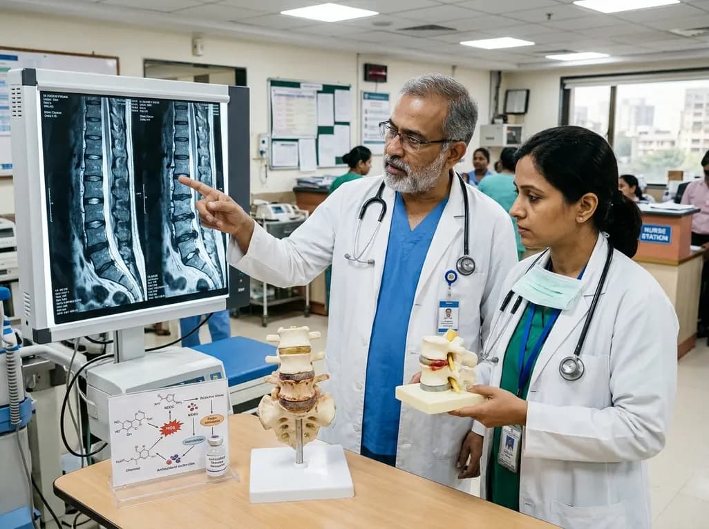
Intervertebral disc degeneration is a primary driver of chronic low back pain. Emerging evidence highlights reactive oxygen species and redox imbalance as central mechanisms. Targeted antioxidant strategies and biomaterial delivery systems offer promising disease-modifying therapies for spine health.
Today

A multicenter cohort study reveals that the 30-minute C-peptide response during OGTT independently predicts cystic fibrosis-related diabetes in patients with normal glucose tolerance, offering an early risk-stratification biomarker.
Today
