.webp&w=384&q=75)

Loading, please wait...
Loading, please wait...
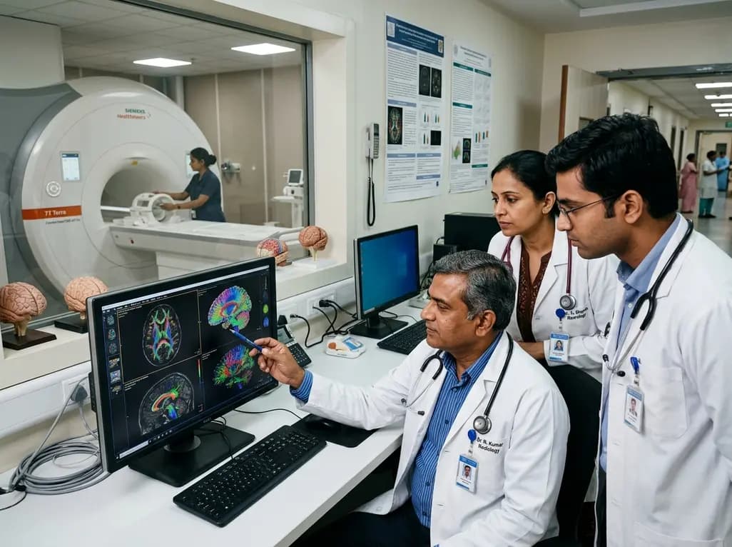
Advancements in neuroimaging continue to transform our understanding of white matter architecture in the central nervous system. Precise in vivo quantification of axonal caliber provides unprecedented insight into neural conduction velocity and structural connectivity. Recently, advanced noninvasive axon diameter mapping achieved a major milestone through the deployment of ultra-high-gradient diffusion magnetic resonance imaging. Researchers evaluating the next-generation Connectome 2.0 scanner demonstrated substantial improvements in sensitivity, spatial precision, and repeatability compared to earlier platforms. By probing restricted water diffusion within sub-micron cellular compartments, this innovative technology bridges the gap between conventional neuroimaging and microscopic histopathology. Consequently, it establishes a powerful foundation for detecting subtle neurodegenerative alterations long before macroscopic structural damage becomes evident.
Clinical MRI scanners typically operate with gradient strengths between 40 and 80 mT/m, which limits their sensitivity to restricted water diffusion inside smaller biological structures. Consequently, traditional diffusion tensor imaging primarily provides macroscopic tensor metrics rather than direct dimensions of axonal morphology. To overcome this fundamental physical boundary, neuroscientists developed the AxCaliber framework combined with spherical mean technique modeling. This biophysical model separates intra-axonal restricted diffusion from extra-axonal hindered compartments, enabling noninvasive caliber estimation across complex white matter tracts.
However, resolving smaller axons requires extremely strong gradient pulses applied over brief diffusion times to attenuate signal from larger extracellular spaces. On standard hardware, the theoretical resolution limit hovers well above the true mean diameter of human cerebral axons. Therefore, engineers engineered dedicated ultra-high-gradient systems capable of generating powerful diffusion encodings. In this benchmark comparative study, investigators evaluated 40 healthy adults to determine whether shifting from 300 mT/m to 500 mT/m gradient amplitudes could overcome historical resolution thresholds. Their results confirmed that higher gradient fields dramatically reduce the minimum detectable axon caliber from 3.6 μm down to 2.5 μm, opening new avenues for microstructural mapping.
The technological backbone of this breakthrough lies in the newly engineered Connectome 2.0 system. This cutting-edge human imaging platform features an unprecedented maximum gradient strength of 500 mT/m alongside a rapid slew rate of 600 T/m/s. In comparison, the original Connectome 1.0 platform delivered 300 mT/m with a slower slew rate of 200 T/m/s. Because gradient strength directly governs the diffusion-weighting b-value achievable within short timeframes, this massive increase provides decisive imaging advantages.
Specifically, the elevated gradient amplitudes permit a substantial reduction in echo time during pulse sequence execution. Shortened echo times directly mitigate T2 signal decay, leading to a marked boost in overall signal-to-noise ratio. Furthermore, the steep gradient slew rates reduce eddy-current artifacts and minimize geometric distortions that frequently degrade diffusion-weighted images. By combining short diffusion times with intense gradient pulses, the Connectome 2.0 scanner captures restricted water molecule displacements with extraordinary fidelity. As a result, biophysical models can isolate true intra-axonal diffusion signals without suffering from the noise contamination that typically confounds lower-gradient acquisitions.
To validate these hardware capabilities, researchers performed comparative evaluations across two cohorts comprising 20 matched participants imaged on Connectome 1.0 and 20 on Connectome 2.0. When analyzing the corticospinal tract, the Connectome 2.0 system yielded an estimated mean axon diameter of 2.66 ± 0.54 μm. In contrast, Connectome 1.0 produced an estimate of 3.35 ± 1.00 μm in the matched cohort. Statistical analysis demonstrated a significant reduction under Welch's t-test (p = 0.0110), reflecting superior sensitivity to smaller axonal calibers.
Histological studies have long established that the human corticospinal tract contains a diverse spectrum of fiber sizes, dominated by thin axons alongside fewer large-diameter fibers. Because lower gradient systems cannot resolve smaller calibers, their estimates become artificially skewed toward larger values. Thus, the lower measurements obtained via Connectome 2.0 reflect a more accurate biological representation rather than an imaging artifact. Although these group-level observations represent cross-sectional comparisons between matched cohorts, they provide robust evidence of enhanced diagnostic resolving power. Ultimately, this quantitative leap allows researchers to map white matter pathways with biological fidelity that approaches postmortem electron microscopy.
Beyond raw spatial resolution and sensitivity limits, clinical translation of any advanced neuroimaging metric hinges on measurement reproducibility. To rigorously assess this property, investigators conducted scan-rescan repeatability experiments involving seven healthy adult volunteers. Each individual underwent duplicate diffusion acquisitions under identical protocol parameters to quantify measurement stability across imaging sessions.
The results demonstrated a striking improvement in parameter reliability on the upgraded hardware. Specifically, the voxel-wise mean absolute difference in axon diameter estimates between scan and rescan dropped to 0.29 μm on Connectome 2.0, compared to 0.65 μm on Connectome 1.0. This more than twofold reduction in measurement variance underscores the robust stability afforded by higher signal-to-noise ratios and shorter echo times. Because biological fluctuations and hardware drift can obscure subtle disease progression, such high repeatability is vital. Clinicians and researchers can now track longitudinal microstructural changes with high statistical power, confident that observed variations represent true physiological shifts rather than scanner-induced noise.
The ability to quantify axonal microstructure noninvasively holds immense promise for clinical neurology and neurodegenerative disease research. Axonal loss and caliber changes represent primary pathological hallmarks across diverse conditions, including multiple sclerosis, amyotrophic lateral sclerosis, traumatic brain injury, and Alzheimer disease. In multiple sclerosis, for example, chronic demyelination frequently leads to axonal atrophy and metabolic collapse before overt lesion expansion occurs.
Similarly, in motor neuron diseases like amyotrophic lateral sclerosis, large myelinated motor axons degenerate preferentially during early stages. High-gradient diffusion MRI could enable early detection of this caliber-selective vulnerability, facilitating prompt therapeutic intervention. Moreover, in traumatic brain injury, diffuse axonal injury often escapes detection on standard structural scans despite persistent cognitive deficits. By quantifying axonal caliber distributions across projection, association, and commissural tracts, clinicians can better correlate structural disruption with neurological outcomes. Consequently, ultra-high-gradient diffusion imaging could serve as a noninvasive biomarker for disease staging, therapeutic monitoring, and clinical trial stratification.
While ultra-high-gradient systems like Connectome 2.0 currently remain specialized research platforms, their underlying principles are actively shaping the future of clinical neuroimaging. Gradient coil engineering and advanced radiofrequency hardware are steadily migrating into next-generation commercial hospital scanners. Furthermore, incorporating machine learning algorithms into reconstruction pipelines promises to shorten acquisition times, making microstructural protocols clinically feasible.
Additionally, ongoing research aims to integrate axon diameter mapping with complementary microstructural metrics, such as myelin volume fraction and g-ratio mapping. Combining these parameters will yield a comprehensive map of conduction velocity throughout the human connectome. As gradient technologies advance, clinicians will gain access to noninvasive virtual biopsies of cerebral tissue. This transformative capability will accelerate neuroscience discovery, enhance biomarker development, and refine targeted neurotherapies for complex brain disorders worldwide.
Ultra-high gradients generate intense diffusion weighting within very short time intervals. This physical capability allows imaging sequences to probe restricted water diffusion inside micron-scale axons, effectively overcoming the resolution limits of standard clinical scanners and enabling accurate measurement of small-caliber nerve fibers in vivo.
Connectome 2.0 delivers a maximum gradient strength of 500 mT/m and a slew rate of 600 T/m/s, whereas conventional clinical scanners achieve 40 to 80 mT/m. This dramatic hardware enhancement shortens echo times, boosts signal-to-noise ratios, and significantly enhances sensitivity to restricted microscopic water diffusion.
Disorders characterized by axonal pathology, including multiple sclerosis, amyotrophic lateral sclerosis, Alzheimer disease, and diffuse axonal traumatic brain injury, stand to benefit most. Measuring precise axon diameter changes enables earlier disease detection, improved progression monitoring, and quantitative evaluation of neuroprotective therapeutic responses in clinical trials.
Disclaimer: This content is for informational and educational purposes only and is not intended to serve as medical advice, diagnosis, or treatment. It does not replace clinical judgment or institutional protocols. Healthcare professionals should evaluate clinical findings within their specific practice environment and exercise independent judgment. Refer to the latest local and national guidelines for clinical practice.
References

Read summarized clinical updates, watch expert medical content, and earn CME certifications right from your smartphone.
Open your smartphone camera to scan and install.





A comparative human neuroimaging study reveals that ultra-high-gradient diffusion MRI at 500 mT/m improves axon diameter mapping sensitivity, resolving calibers down to 2.5 μm and significantly enhancing scan-rescan repeatability in the living human brain.
4 weeks back

Top-down CSF peptidomics uncovers novel endogenous peptide signatures and oxidative modifications differentiating multiple sclerosis from NMOSD, highlighting secretogranin-5 loss as a key biomarker of disease progression.
Today

Fetal foramen ovale restriction typically emerges in the third trimester with right heart enlargement. Evidence demonstrates it does not independently mandate cesarean delivery, and over 93% of postnatal cardiac findings resolve spontaneously within one year, supporting conservative obstetric management.
Today

The latest WHO Global Health Estimates highlight India's life expectancy reaching 71 years alongside a healthy lifespan of 60.6 years. While longevity rebounds post-pandemic, non-communicable conditions now account for three-quarters of top mortalities, underscoring urgent clinical shifts in routine primary care.
Today

New biomechanical research reveals that foot-strike angle significantly influences knee energy absorption during cutting tasks in female soccer players. Understanding these kinematic patterns offers clinicians valuable pathways for targeted ACL injury prevention and athletic retraining.
Today

A comprehensive review analyzes periconceptional GLP-1 receptor agonist exposure, revealing reassuring data on congenital anomalies and adverse perinatal outcomes while emphasizing the need for cautious patient counseling.
Today
