.webp&w=384&q=75)

Loading, please wait...
Loading, please wait...
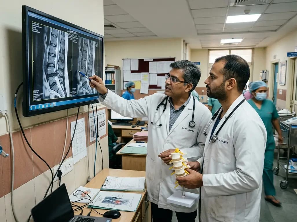
Degenerative spinal stenosis represents a major source of disability among aging populations globally. Historically, open surgical decompression yielded neural relief at the cost of paraspinal tissue trauma. However, modern endoscopic lumbar decompression provides comparable neural canal clearance through significantly smaller anatomical corridors. Spine surgeons now actively utilize both unilateral biportal endoscopy and uniportal single-port systems for bilateral canal enlargement. Consequently, rigorous comparative clinical trials are vital to determine which technical iteration offers superior operative efficiency and patient outcomes.
The prospective randomized controlled trial conducted by Liu and colleagues systematically investigated 110 patients afflicted with severe single-level lumbar spinal stenosis. Specifically, investigators selected individuals exhibiting Schizas grade C or grade D canal compromise. The research protocol randomized participants evenly into two cohorts of 55 patients each. One cohort underwent unilateral biportal endoscopic unilateral laminotomy for bilateral decompression, designated as UBE-ULBD. In contrast, the second group received single-port full-endoscopic unilateral laminectomy for bilateral decompression, termed Endo-ULBD. A single veteran spine surgeon executed all operative interventions to minimize technical operator bias. Furthermore, the researchers analyzed both intention-to-treat and per-protocol populations over a standardized 12-month postoperative duration. The primary endpoints captured pain severity via visual analogue scale scores, functional disability using the Oswestry Disability Index, and global patient satisfaction through modified Macnab criteria. Additionally, rigorous magnetic resonance imaging quantified dural sac cross-sectional area expansion and facet joint volume retention. As a result, this methodological framework established an exceptionally controlled baseline to assess subtle differences between biportal and uniportal spinal platforms.
Surgical execution differs fundamentally between biportal and uniportal visualization modalities. In biportal procedures, surgeons utilize independent working and viewing portals. Consequently, this configuration allows independent instrument triangulation and wider angulation across the central canal. Therefore, surgeons can maneuver conventional high-speed burrs and standard pituitary rongeurs with considerable mechanical freedom. In contrast, uniportal single-port endoscopy incorporates the endoscopic camera, continuous irrigation fluid, and surgical tools within a single coaxial sleeve. This unified arrangement minimizes skin incision size and diminishes overall surgical footprint. However, the coaxial working channel inherently restricts instrument trajectory and limits mechanical leverage during challenging bony resection. Operative timing data indicated that both techniques achieved comparable baseline procedural duration. Nevertheless, the biportal approach offered greater ergonomic ease when addressing severe contralateral lateral recess stenosis. Continuous saline irrigation maintained clear optical visualization throughout both operations. In addition, surgeons achieved effective homeostatic control without requiring heavy reliance on specialized radiofrequency wands. Ultimately, mastering the fluid outflow dynamics prevents dangerous intraoperative epidural pressure spikes in both endoscopic configurations.
Clinical recovery metrics revealed remarkable symptomatic improvements across both cohorts throughout the 12-month follow-up timeline. Patients experienced profound, rapid reductions in radicular leg discomfort and persistent axial low back pain. Statistically, visual analogue scale scores dropped precipitously within the first postoperative week and remained stable at one year. Similarly, functional disability scores on the Oswestry Disability Index showed drastic postoperative reductions in both cohorts. Modified Macnab ratings demonstrated excellent or good clinical outcomes in roughly ninety percent of all operated patients. Furthermore, intergroup statistical analyses detected no significant differences in long-term functional recovery between biportal and uniportal decompression groups. Both cohorts experienced early post-surgical ambulation within twenty-four hours due to minimal paraspinal tissue disruption. Consequently, patients avoided prolonged bed rest and required minimal postoperative opioid consumption. Moreover, patients reported prompt returns to routine personal activities and workplace responsibilities. These functional patterns affirm that both endoscopic approaches achieve adequate neural decompression. Thus, surgeon experience and institutional equipment availability should dictate procedural selection rather than anticipated differences in clinical symptom relief.
Postoperative magnetic resonance imaging verified outstanding morphological canal restoration in both treatment arms. Specifically, dural sac cross-sectional area expanded substantially from constricted preoperative dimensions to generous post-decompression volumes. Both modalities successfully cleared hypertrophied ligamentum flavum, encroaching osteophytes, and compressing lamina. However, detailed volumetric imaging revealed subtle anatomical distinctions regarding mechanical joint preservation. The biportal approach provided exceptional visibility of the contralateral lateral recess through over-the-top angled views. As a result, the operating surgeon effectively unroofed the contralateral traversing nerve root while avoiding excessive medial facetectomy. Meanwhile, uniportal endoscopy demonstrated superior facet joint preservation on the ipsilateral access side. Because the uniportal cannula maintains a strictly confined tubular trajectory, it avoids superfluous bone resection along the entry corridor. Preserving bilateral facet joint architecture remains essential to prevent secondary postoperative segmental instability and subsequent spondylolisthesis. In addition, dynamic flexion-extension radiographs confirmed stable spinal alignment without mechanical breakdown at the 12-month mark. Therefore, both techniques safely preserve spinal stability while providing wide neurological decompression.
Minimally invasive spine surgery prioritizes patient safety alongside effective neurological decompression. In this randomized cohort, overall complication rates remained remarkably low and statistically indistinguishable between groups. Notably, incidental durotomy occurred in a very small fraction of cases within both arms. When dural tears arose, surgeons managed them promptly using direct endoscopic repair, muscle patch application, or fibrin sealants without conversion to open surgery. Furthermore, neither cohort encountered catastrophic neurological deficits, deep surgical wound infections, or permanent motor weakness. Epidural hematoma formation represents another critical hazard during fluid-assisted spine surgery. Consequently, maintaining vigilant hemostasis and implementing meticulous postoperative drainage systems minimized symptomatic epidural fluid collections. In contrast to extensive open laminectomy, neither endoscopic group experienced significant paraspinal muscle denervation or severe postoperative muscular atrophy. Moreover, serum inflammatory markers demonstrated minimal surgical trauma in both cohorts. Ultimately, precise anatomical knowledge and disciplined irrigation management safeguard patients against rare complications such as increased intracranial pressure or fluid extravasation into the retroperitoneum.
The clinical findings carry immense practical significance for spine care across India and South Asia. Lumbar canal stenosis presents with high frequency among elderly Indian citizens, many of whom possess multiple medical comorbidities. Traditional open spine operations often entail extensive hospitalization, substantial blood loss, and prolonged postoperative convalescence. Conversely, minimally invasive endoscopic lumbar decompression allows early ambulation and short hospital stays, which drastically lowers nosocomial infection risks. Furthermore, biportal endoscopic systems offer an attractive financial entry point for Indian hospitals because surgeons can deploy standard arthroscopic video towers and conventional open spinal burrs. In contrast, uniportal endoscopic systems require proprietary optics and dedicated articulated instruments that carry higher capital expenditure costs. However, training facilities in India are expanding rapidly, enabling young orthopedic and neurosurgical trainees to master both techniques safely. Therefore, spine centers can tailor their endoscopic strategy to local institutional infrastructure and procedural familiarity. Ultimately, widespread adoption of these minimally invasive techniques will expand access to safe, durable canal decompression for aging patients across diverse socioeconomic demographics.
Biportal endoscopic lumbar decompression utilizes two separate independent portals: one for clear optical visualization and another for dedicated surgical instruments. This arrangement allows free instrument triangulation and familiar handling similar to open surgery. In contrast, uniportal endoscopy places the optics, irrigation inflow, and working instruments through a single integrated tubular channel. Consequently, uniportal surgery produces smaller cutaneous scars, whereas biportal surgery offers superior instrument freedom and ergonomic flexibility during complex bilateral canal clearance.
Both techniques successfully preserve facet joint integrity, but each possesses specific geometric advantages. Uniportal endoscopy excels at preserving ipsilateral facet joint volume because the single rigid tubular sleeve restricts extraneous bone resection along the entry corridor. Conversely, biportal endoscopy provides superior visualization and angulation across the midline. This panoramic view allows meticulous over-the-top contralateral undercut decompression, which effectively prevents inadvertent destabilization of the contralateral facet complex during severe lateral recess stenosis decompression.
Spine surgeons transitioning to biportal endoscopy often experience a smoother learning curve because two-portal mechanics closely mirror traditional open microsurgical triangulation. Surgeons can manipulate familiar high-speed drills and standard rongeurs freely. Conversely, single-port uniportal decompression requires adaptation to coaxial viewing and specialized linear instruments within a constrained channel. Regardless of platform, surgeons should initially undergo rigorous cadaveric training, master continuous fluid outflow management, and select straightforward single-level canal stenosis cases first.
Disclaimer: This content is for informational and educational purposes only... Refer to the latest local and national guidelines for clinical practice.
References

Read summarized clinical updates, watch expert medical content, and earn CME certifications right from your smartphone.
Open your smartphone camera to scan and install.





A prospective randomized controlled trial compared UBE-ULBD versus Endo-ULBD in 110 patients with lumbar spinal stenosis. Both endoscopic techniques achieved significant pain reduction, functional recovery, and dural expansion, demonstrating comparable clinical efficacy with distinct technical advantages.
last month

As monsoon seasons elevate arboviral disease threats, Tamil Nadu has initiated an ICMR-monitored evaluation of the tetravalent dengue vaccine in Tenkasi district. Clinicians emphasize clinical monitoring, public prevention, and high-quality early pediatric care to reduce disease morbidity across endemic regions.
Today

A comparative cohort study evaluates vNOTES vs TLH hysterectomy for benign conditions, demonstrating shorter operative duration, faster return to daily activities, and equivalent female sexual function scores without increased dyspareunia.
Today

Review how tailored exercise programs benefit frail older patients with heart failure. Explore pathophysiological mechanisms, individualized training prescriptions, and strategies to overcome adherence barriers in clinical practice.
Today

This comparative study evaluates posterior labral tears with and without glenohumeral instability, highlighting distinct demographic patterns, MRI underestimation in stable shoulders, and successful arthroscopic repair outcomes.
Today

A clinical study demonstrates that early intervention with tocilizumab, either as monotherapy or bridged with corticosteroids, achieves significant proptosis reduction and resolves active inflammation in treatment-naive thyroid eye disease.
Today
