.webp&w=384&q=75)

Loading, please wait...
Loading, please wait...
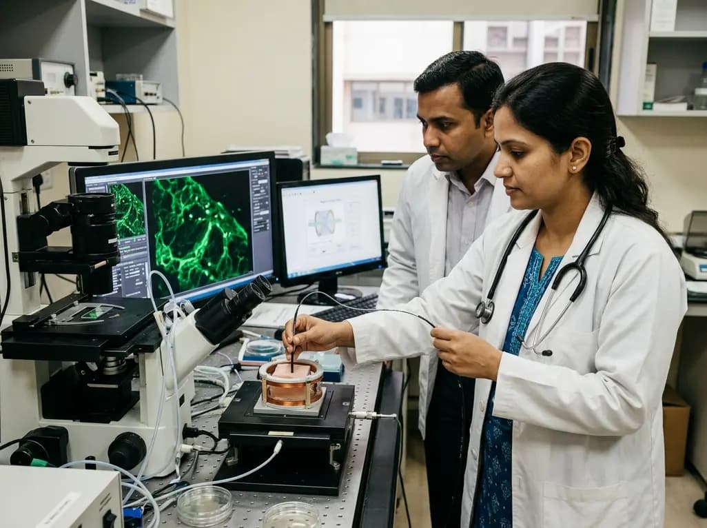
Magnetic microrobotics represents a transformative frontier in targeted medicine, offering unprecedented opportunities for minimally invasive interventions. Researchers aim to navigate untethered micro-agents through complex physiological fluid channels to deliver therapeutics directly to targeted diseased tissues. However, conventional micro-agent tracking predominantly relies on external imaging modalities such as magnetic resonance imaging and ultrasound, which frequently lack necessary optical resolution. To address these technical limitations, biomedical engineering investigators have integrated optical fluorescence techniques into flexible endoscopic tools. Specifically, two-photon endomicroscopy provides a breakthrough approach for acquiring high-resolution visual feedback during active microrobotic navigation. By combining ultrashort femtosecond laser pulses with fiber-optic flexible probes, this non-linear imaging modality captures fine cellular details deep within scattering biological tissues. Consequently, clinicians and researchers can monitor dynamic microrobotic maneuvers in real time with high fidelity. Furthermore, this technological convergence shifts microrobotics from passive observational tracking to active, feedback-driven surgical intervention. Therefore, integrating advanced optical endomicroscopy with magnetic propulsion creates promising new paradigms for targeted drug delivery and micro-scale surgery.
The primary hurdle in micro-agent navigation involves maintaining continuous visual tracking while operating within optically dense biological environments. Traditional single-photon fluorescence microscopy suffers from severe light scattering and out-of-focus background noise, which obscures tiny magnetic structures. In contrast, two-photon endomicroscopy utilizes localized near-infrared excitation, restricting fluorescence emission strictly to the optical focal volume. This localized excitation significantly enhances image contrast while minimizing photodamage to surrounding healthy biological tissue. Furthermore, flexible fiber bundles allow the miniaturized endomicroscope to access deep anatomical regions previously unreachable by standard microscope objectives. The integrated system captures dynamic micro-agent trajectories with high spatial and temporal fidelity during active propulsion. Consequently, operators obtain clear visualization of individual micro-agents as they travel through complex microchannels. Additionally, real-time optical feedback allows automated control algorithms to compute instantaneous position errors and adjust external magnetic driving fields. Thus, this imaging technique establishes a robust foundation for precision-guided therapies in minimally invasive surgical procedures.
In many biomedical scenarios, microrobotic operations involve heterogeneous populations containing active carrier agents alongside non-magnetic cellular payloads. Distinguishing these distinct components during active motion requires advanced multi-channel visualization techniques. Therefore, researchers incorporated dual-color fluorescence imaging capabilities into the endomicroscopic optical path. By labeling magnetic micro-agents and non-magnetic cellular entities with distinct fluorescent dyes, the system simultaneously tracks multiple biological components in real time. For instance, magnetic micro-carriers emit bright green fluorescence, whereas targeted cellular spheroids emit distinct red signals. Furthermore, separate spectral channels prevent cross-talk between signals, ensuring accurate identification during dynamic cellular interactions. Consequently, investigators can observe how magnetic forces influence neighboring unlabelled or distinctively tagged cells. Additionally, this multi-spectral approach allows real-time assessment of payload attachment, transport, and release mechanisms. Thus, dual-color endomicroscopy offers invaluable physiological insights into micro-agent behavior within complex, multi-component biological environments.
Optical tissue scattering remains a formidable obstacle for high-resolution micro-agent tracking within intact living mammalian organs. Standard optical tools lose focus within a few micrometers of tissue depth due to dense photon scattering. However, two-photon endomicroscopy effectively overcomes optical scattering by employing longer near-infrared excitation wavelengths. In recent experimental validations, researchers successfully imaged active magnetic micro-agents through a 140-micrometer-thick layer of rat mammary gland tissue. Notably, the magnetic micro-agents remained clearly detectable and sharp despite significant optical density in the surrounding tissue layer. Furthermore, the high signal-to-noise ratio allowed accurate tracking of micro-agent positions without losing signal intensity over time. Consequently, this depth capability demonstrates that optical guidance can function effectively beneath realistic anatomical barriers. Moreover, these findings confirm that two-photon fiber probes can navigate internal organ structures while maintaining high-resolution visualization. Therefore, deep tissue imaging bridges the critical gap between benchtop microfluidic testing and future clinical translational applications.
Precise movement control requires rigorous dynamic analysis of micro-agent kinematics across varying magnetic field actuation frequencies. To achieve controlled locomotion, external electromagnetic coils generate rotating magnetic fields that actuate helical or spherical micro-agents through fluidic media. Consequently, two-photon endomicroscopy captures rapid frame sequences, enabling detailed velocity and trajectory measurements. Investigators systematically analyzed micro-agent speed across multiple rotational actuation frequencies to identify optimal propulsion regimes. Furthermore, high-speed fluorescence acquisition revealed subtle hydrodynamic interactions between micro-agents and adjacent biological boundaries. Notably, when magnetic forces displaced HeLa cell spheroids from their initial alignment, real-time endomicroscopic feedback enabled rapid position re-centering. Operators adjusted the optical probe position to keep displaced cell spheroids strictly within the central field of view. Consequently, active image-guided compensation prevents loss of tracking during vigorous magnetic manipulation. Thus, dynamic motion monitoring ensures stable, accurate, and predictable microrobotic performance during complex tissue navigation tasks.
The successful combination of two-photon endomicroscopy and magnetic micro-agents represents a significant leap forward in biomedical engineering. By moving beyond passive observation, this technology establishes active, closed-loop image guidance for micro-scale interventions. Furthermore, future developments will focus on further miniaturizing probe dimensions and optimizing automated magnetic control algorithms. Clinicians anticipate that guided microrobots could eventually deliver localized chemotherapy directly to solid tumors, minimizing systemic side effects. Additionally, micro-agents could perform precise micro-suturing, localized bioteledelivery, or targeted intravascular debris clearance under direct optical control. Although clinical adoption requires extensive preclinical safety trials and regulatory evaluations, these preliminary results demonstrate incredible therapeutic potential. Ultimately, combining advanced non-linear optics with intelligent micro-scale robotics will transform minimally invasive surgical procedures, offering patients safer and highly targeted clinical solutions.
Two-photon endomicroscopy is an advanced optical imaging technique that uses localized near-infrared laser excitation to image micro-scale structures inside biological tissue. Unlike traditional modalities, it offers deep tissue penetration, high spatial resolution, and minimal photodamage. In microrobotics, this technique provides real-time fluorescence visual feedback, allowing operators to track, steer, and manipulate untethered magnetic micro-agents with extreme precision during complex minimally invasive procedures.
Dual-color fluorescence imaging utilizes two distinct fluorescent dyes to simultaneously label magnetic micro-agents and non-magnetic cellular payloads or tissue structures. By separating signals into different optical channels, researchers can clearly distinguish active robotic carriers from target tissue cells. This multi-spectral capability allows precise monitoring of carrier-payload interactions, verifying successful target attachment, guided transport, and localized drug release during dynamic magnetic manipulation within complex biological environments.
Yes, two-photon endomicroscopy excels at deep tissue imaging because near-infrared light scatters significantly less in biological tissue than visible light. Experimental study results demonstrated that magnetic micro-agents remain clearly detectable and trackable through a 140-micrometer-thick layer of rat mammary gland tissue. This key capability proves that optical guidance systems can operate successfully through dense anatomical barriers, supporting future in vivo diagnostic and therapeutic applications.
Disclaimer: This content is for informational and educational purposes only and does not constitute medical advice, diagnosis, or treatment. Healthcare professionals should rely on their clinical judgment and refer to the latest local and national guidelines for clinical practice.
References
Huaroto JJ et al. Two-Photon Endomicroscopy for Image-Guided Magnetic Micro-Agents. Small. 2026 Aug 11. doi: 10.1002/smll.75051. PMID: 42576800.
Huaroto JJ et al. Two-photon microscopy for microrobotics: Visualization of micro-agents below fixed tissue. PLoS ONE. 2023;18(8):e0289725.

Read summarized clinical updates, watch expert medical content, and earn CME certifications right from your smartphone.
Open your smartphone camera to scan and install.





Researchers demonstrate two-photon endomicroscopy for real-time fluorescence imaging and active guidance of magnetic micro-agents in deep biological tissue.
last month

A secondary analysis of an HIV cure-related trial in South Africa highlights the acceptability and safety of serial excisional lymph node and gut biopsies among young women. Clear communication, empathetic care, and altruistic motives helped mitigate initial apprehension and sustain longitudinal trial engagement.
Today

Explore the crucial link between dietary protein and immunocompetence. This review examines how low-protein nutrition shifts metabolic resources away from pathogen defense toward early reproduction, compromising host survival and immunity against severe bacterial infections.
Today
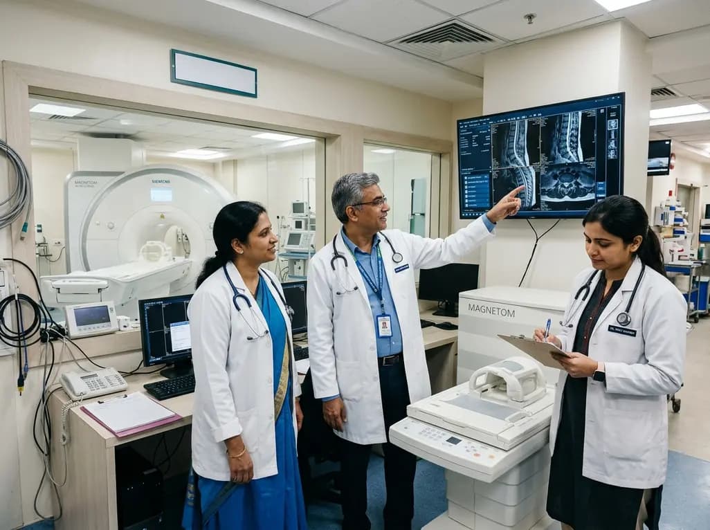
Under India's PLI initiative, domestic production of 57 critical medical devices has commenced, including MRI and CT scanners, cath labs, and heart valves. Global partnerships and local investments are expanding high-end technology, curbing import reliance, and strengthening clinical accessibility nationwide.
Today

A longitudinal study reveals that patients undergoing staged bilateral hip arthroscopy experience progressive contralateral labral deterioration during surgical intervals. However, structural degradation does not compromise 2-year postoperative clinical outcomes or patient-reported satisfaction scores.
Today

New immunological research reveals how PTPN22 forms a delayed inhibitory signaling complex to restrain T cell activation, explaining how the autoimmune-linked R620W variant disrupts feedback control and promotes persistent inflammation in conditions like rheumatoid arthritis and type 1 diabetes.
Today
