.webp&w=384&q=75)

Loading, please wait...
Loading, please wait...
Acute neurological deficits arising from vascular occlusion usually result from thromboembolic events related to atherosclerosis or cardiac arrhythmias. However, non-thrombotic etiologies present a critical clinical challenge. Tumor embolism stroke represents an exceptionally rare yet life-threatening cause of acute ischemic stroke. In this condition, neoplastic fragments detach from primary or metastatic lesions, enter the systemic arterial circulation, and occlude cerebral vasculature. While primary lung neoplasms and cardiac myxomas are the most frequent culprits, metastatic bone malignancies can occasionally seed the vasculature. This presentation often mimics standard atherothrombotic or cardioembolic ischemic stroke, creating significant diagnostic uncertainty during acute resuscitation. Prompt clinical evaluation and neurovascular imaging remain essential to minimize core infarct growth and preserve functional independence.
Furthermore, patients with an underlying oncologic history present unique management dilemmas. Intravenous thrombolysis may pose elevated risks of intracranial hemorrhage or systemic bleeding complications. Consequently, clinicians must maintain high suspicion when evaluating acute stroke symptoms in patients with a history of sarcoma or other aggressive malignancies. Rapid identification of atypical embolic sources is vital for guiding therapeutic strategies and preventing recurrent neurological events. Subsequent evaluation using advanced imaging and tissue sampling often provides essential clues. Understanding the unique mechanisms underlying tumor embolism stroke allows multidisciplinary care teams to deliver timely and targeted interventions.
A compelling clinical case involved a 46-year-old male who presented with sudden-onset left-sided hemiparesis and acute neurological impairment. Interestingly, the patient had undergone distal femoral amputation 11 years prior due to low-grade parosteal osteosarcoma. Emergency computed tomography angiography immediately demonstrated a focal occlusion in the right middle cerebral artery. The clinical presentation mirrored a classic acute large vessel occlusion stroke. Therefore, emergency teams prepared the patient for urgent endovascular intervention to restore cerebral blood flow.
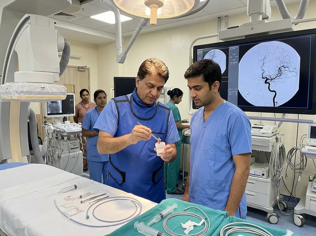
During endovascular procedure, neurointerventionalists successfully performed mechanical thrombectomy to recanalize the right middle cerebral artery. Rather than retrieving a standard red or white fibrin clot, the intervention retrieved a distinct white, gelatinous embolus. Postoperative magnetic resonance imaging confirmed acute cerebral infarctions in the right basal ganglia and temporal lobe. Simultaneously, systemic staging via chest computed tomography revealed extensive bilateral pulmonary metastases. Subsequent transthoracic echocardiography identified a prominent vegetative mass attached to the cardiac valve. This cardiac lesion provided the direct source for systemic arterial embolization into the intracranial circulation. Consequently, the clinical trajectory demonstrated how localized tumor growth within cardiopulmonary structures can trigger sudden cerebral arterial blockage.
Pathologic evaluation of retrieved embolic material remains indispensable when clinicians encounter unusual clot physical appearances. Histopathologic examination of the retrieved gelatinous mass revealed atypical pleomorphic tumor cells embedded within a complex matrix. Consequently, pathologists initiated detailed immunohistochemical analysis to establish the exact lineage of the metastatic cells. The tumor demonstrated strong nuclear positivity for SATB2 and SOX9, along with positive expression for CDK4. Additionally, nuclear staining revealed a Ki-67 proliferation index ranging between 10% and 20%, indicating active cellular proliferation.
To confirm the definitive diagnosis, molecular testing via fluorescence in situ hybridization was conducted on the specimen. The test confirmed MDM2 gene amplification, establishing the diagnosis of metastatic dedifferentiated parosteal osteosarcoma. Parosteal osteosarcoma typically behaves as a low-grade surface bone neoplasm with favorable long-term survival rates. However, high-grade dedifferentiation can develop decades after initial surgical resection. Recognizing these specific biomarkers allows clinical pathologists to differentiate osteosarcoma from soft tissue sarcomas or metastatic carcinomas. This transformation leads to aggressive metastatic dissemination, including pulmonary vascular invasion and secondary endocardial tumor seeding. Thus, thorough histological and genetic testing on embolectomy tissue provided essential insights into the underlying oncologic recurrence, directly altering the long-term oncologic management strategy for this patient.
The pathogenetic mechanism leading to cerebral occlusion in dedifferentiated parosteal osteosarcoma involves multiple step-wise cardiovascular events. Initially, dedifferentiated sarcoma cells metastasize to the pulmonary parenchyma through hematogenous spread. Centrally located pulmonary metastases can invade surrounding pulmonary veins, allowing malignant cells to access the left atrium and ventricle. Alternatively, tumor cells colonize cardiac structures, forming intra-cardiac masses or valvular tumor vegetations. As blood flows through the heart chambers under high mechanical pressure, tumor fragments break off into the systemic circulation.
Once detached, these tumor emboli travel through the aorta into the carotid arteries, eventually lodging in distal cerebral branch arteries such as the middle cerebral artery. Furthermore, tumor emboli induce local mechanical occlusion and secondary thrombosis. This dual mechanism exacerbates distal tissue ischemia and acute neurological dysfunction. Recognizing this pathophysiologic pathway emphasizes the importance of complete cardioembolic screening in cancer patients experiencing sudden stroke. Echocardiography, chest CT angiography, and systemic staging are crucial components of the post-stroke diagnostic workup. Identifying cardiac valvular tumor involvement helps clinicians anticipate potential recurrent embolic events and formulate appropriate systemic anticoagulation or oncologic therapies.
Mechanical thrombectomy has revolutionized the management of acute large vessel occlusion stroke. In cases involving tumor embolism stroke, endovascular intervention fulfills two vital clinical roles simultaneously. Therapeutically, mechanical thrombectomy physically retrieves the occluding mass, restores distal cerebral perfusion, and minimizes ischemic penumbral brain damage. Modern stent retrievers and aspiration catheters enable efficient recanalization even when dealing with dense, fibrous, or gelatinous tumor emboli. Re-establishing cerebral circulation remains the primary objective during the hyperacute phase of stroke management.
Diagnostically, endovascular thrombectomy provides intact tissue samples suitable for immediate microscopic and molecular examination. Obtaining tissue through mechanical thrombectomy often eliminates the need for invasive open surgical biopsies or complex systemic procedures. Consequently, neurointerventionalists directly contribute to definitive disease staging and oncologic diagnosis. However, clinicians must maintain caution because tumor emboli can exhibit different physical properties compared to standard thrombi. Gelatinous or calcified tumor fragments may resist standard aspiration or fragment during retrieval. Establishing standardized neurointerventional protocols for handling fragile tumor thrombi further enhances procedure safety and recanalization success rates. Therefore, neurointerventionalists should select appropriate thrombectomy techniques to minimize distal embolization. Integrating endovascular clot retrieval with rapid histopathological profiling ensures both prompt neurological rescue and accurate systemic disease management.
This rare clinical case provides valuable management insights for multidisciplinary medical teams caring for complex stroke patients. First, clinicians should consider tumor embolism stroke in any patient presenting with acute large vessel occlusion and a past history of malignancy, even after a prolonged disease-free interval. Parosteal osteosarcoma, although classically indolent, can undergo late dedifferentiation more than ten years after initial curative treatment. Therefore, clinicians must maintain lifelong vigilance and include systemic oncologic surveillance in long-term survivorship programs.
Second, when retrieved thrombectomy material exhibits atypical macroscopic features—such as a gelatinous, fleshy, or firm white consistency—routine pathological evaluation is imperative. Immediate immunophenotypic analysis using markers such as SATB2, SOX9, CDK4, and MDM2 FISH can rapidly confirm bone matrix sarcomas and guide targeted therapeutic decisions. Finally, comprehensive cardiac and pulmonary imaging must be performed promptly when unusual embolic etiologies are suspected. Transthoracic or transesophageal echocardiography combined with chest CT can uncover pulmonary venous invasion or intracardiac valvular lesions. By adopting a systematic, multidisciplinary approach combining neurointerventional therapy, pathology, cardiology, and oncology, healthcare teams can optimize patient outcomes in rare cardio-oncologic vascular emergencies.
Tumor embolism causes stroke when malignant tumor fragments detach from primary or metastatic lesions, enter the arterial circulation, and physically block cerebral arteries. These emboli often originate from lung tumors invading pulmonary veins or intracardiac tumor masses, interrupting distal blood flow and producing acute cerebral infarction.
Mechanical thrombectomy serves both therapeutic and diagnostic functions in tumor embolism stroke. While restoring cerebral arterial blood flow, the procedure physically retrieves the embolus. Pathologic and immunophenotypic evaluation of the retrieved tissue allows definitive diagnosis of the underlying tumor type without requiring additional invasive tissue biopsy procedures.
Low-grade osteosarcomas, such as parosteal osteosarcoma, can undergo high-grade dedifferentiation many years after initial surgical treatment. Long-term surveillance is necessary because dedifferentiated recurrences behave aggressively, metastasizing to the lungs and heart, and potentially causing severe vascular complications like tumor embolic stroke even decades later.
Disclaimer: This content is for informational and educational purposes only, and does not constitute medical advice, diagnosis, or treatment. Always seek the advice of a qualified healthcare provider with any questions you may have regarding a medical condition. Refer to the latest local and national guidelines for clinical practice.
References

Read summarized clinical updates, watch expert medical content, and earn CME certifications right from your smartphone.
Open your smartphone camera to scan and install.





A rare case of acute ischemic stroke caused by tumor embolism from dedifferentiated parosteal osteosarcoma highlights the crucial therapeutic and diagnostic role of mechanical thrombectomy.
2 months ago

Explore why official drug package inserts fail to reflect modern evidence for clozapine prescribing. Learn how legal standards of care, case law, slow titration, and vital adverse event monitoring empower clinicians to prioritize evidence-based medical practice over outdated manufacturer labeling.
Today

Occult maternal malignancy accounts for 3.82% of persistent unreportable NIPT results with genomic noise. Multiple chromosomal aberrations, notably on chromosome 18, warrant thorough oncological evaluation and whole-body MRI to ensure timely maternal diagnosis while preserving fetal care.
Today

This article reviews the forensic and clinical complexities of acute myocarditis arising on left ventricular hypertrophy in children, highlighting early diagnostic red flags and multidisciplinary postmortem autopsy protocols.
Today
A randomized controlled trial demonstrates that an eight-week dual-task training intervention significantly improves static and dynamic postural control over standard balance exercises in individuals with chronic ankle instability, addressing critical neurocognitive deficits during athletic and daily activities.
Today

A retrospective cohort study of 7,716 ICU patients from the MIMIC-IV database demonstrates that Gradient Boosting Decision Trees significantly outperform traditional SOFA scores in predicting in-hospital mortality among patients with sepsis and diabetes, with SHAP analysis providing clinical interpretability.
Today
