.webp&w=384&q=75)

Loading, please wait...
Loading, please wait...
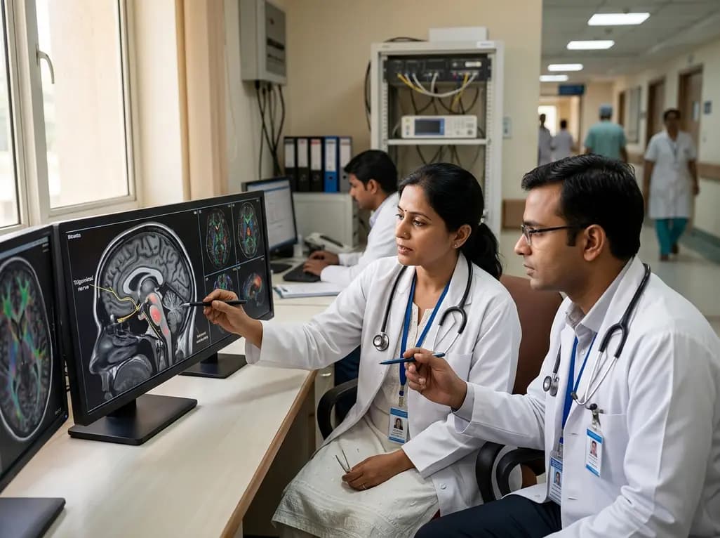
Migraine remains one of the most disabling neurological disorders globally, imposing significant physical, emotional, and socioeconomic burdens on affected individuals. Although clinicians recognize peripheral and central nervous system remodeling during migraine attacks, the exact biological underpinnings of cranial pain pathways remain incompletely understood. Groundbreaking research now provides direct in vivo evidence linking altered trigeminal nerve microstructure to localized neuroinflammation and central brainstem sensory modulation in human patients.
The trigeminovascular system serves as the primary anatomical conduit for transmitting craniofacial pain signals during migraine episodes. However, non-invasive assessment of subtle tissue alterations at the trigeminal root entry zone has historically challenged conventional clinical neuroimaging. To address this technological limitation, investigators evaluated 60 migraine patients alongside 20 age-matched healthy controls using ultra-high-field 7 Tesla diffusion tensor imaging.
Consequently, the researchers detected marked microstructural abnormalities at the trigeminal nerve root in patients suffering from migraine. Specifically, diffusion tensor metrics revealed significantly reduced fractional anisotropy within the affected nerve roots compared to healthy control participants. Because fractional anisotropy quantifies axonal alignment and myelin integrity, these findings confirm localized structural remodeling along the peripheral sensory pathway. Furthermore, the degree of microstructural disruption directly reflected the underlying biological chronicity of the disease.
Beyond structural changes, investigators sought to clarify whether localized cellular inflammation accompanies axonal disruption. To measure neuroinflammatory activity non-invasively, researchers performed positron emission tomography utilizing the radiotracer carbon-11-labeled PBR28. This advanced radioligand binds specifically to the translocator protein, which exhibits heightened expression in activated microglia, macrophages, and astrocytes during neuroinflammatory cascades.
Remarkably, the PET findings demonstrated elevated translocator protein binding at the trigeminal nerve root in migraineurs. Moreover, statistical analyses revealed a robust inverse correlation between fractional anisotropy and the PET signal. Patients with the lowest fractional anisotropy exhibited the most pronounced localized inflammatory activity. Therefore, these synchronized multimodal imaging results suggest that chronic or recurrent inflammatory cascades actively contribute to structural degradation of the cranial nerve pathway over time.
To examine functional central processing, investigators delivered innocuous electrical stimulation across the ophthalmic trigeminal nerve territory during ultra-high-field functional magnetic resonance imaging. Clinicians often observe generalized spinal trigeminal nucleus hyperexcitability when contrasting migraine cohorts against healthy controls. However, sub-analyses within the patient cohort revealed an intriguing neurobiological paradox.
Specifically, patients with more extensive structural disruption at the nerve root demonstrated diminished functional MRI activation within the ipsilateral spinal trigeminal nucleus. Thus, severe peripheral microstructural damage appears to impede normal afferent sensory transmission into the brainstem. Consequently, secondary central sensitisation may emerge as a compensatory adaptation to altered sensory input. This crucial insight underscores the dynamic interplay between peripheral nerve integrity and central trigeminal relay stations.
Interestingly, the neuroimaging abnormalities exhibited pronounced lateralization across the study cohort. Both microstructural alterations and localized neuroinflammatory signals manifested more robustly within the right trigeminal nerve root entry zone. Furthermore, corresponding functional brainstem responses showed parallel right-sided predominance during sensory testing.
Importantly, these imaging metrics aligned closely with clinical phenotype presentations. Patients with right headache dominance reported significantly greater overall migraine severity and headache burden compared to left headache-dominant participants. Therefore, these lateralized objective biomarkers substantiate patient-reported symptom distributions. As a result, assessing structural laterality provides clinicians with a valuable framework for understanding asymmetrical headache presentations in clinical neurology.
These multimodal discoveries provide essential translational perspectives for neurologists, radiologists, and headache specialists managing refractory migraine cases. Traditionally, medical practice classified migraine primarily as a functional, episodic neurochemical disorder lacking gross structural pathology. In contrast, modern high-resolution imaging demonstrates clear, quantifiable anatomical remodeling and localized neuroinflammation.
Additionally, these findings highlight why standard neuroimaging protocols often miss clinically meaningful changes in headache patients. Conventional 1.5 Tesla or 3 Tesla magnetic resonance protocols rarely capture subtle microstructural variations at cranial nerve root entry zones. Consequently, recognizing trigeminovascular neuroinflammation and structural degradation helps clinicians validate patient symptoms while laying the groundwork for targeted diagnostic strategies.
Understanding the interplay between neuroinflammation and trigeminal nerve integrity offers exciting therapeutic implications for future headache management. Currently, modern preventive therapeutics such as calcitonin gene-related peptide antagonists primarily target neurovascular signaling cascades. However, addressing chronic neuroinflammation and microstructural nerve degradation represents a vital, unexplored frontier.
Furthermore, identifying localized translocator protein elevation suggests that novel microglial-modulating agents could potentially halt or reverse nerve root remodeling. In addition, non-invasive neuromodulation targeting the trigeminal pathway may help restore balanced afferent signaling into brainstem nuclei. Ultimately, tracking fractional anisotropy and inflammatory biomarkers could guide individualized therapy selection and monitor treatment efficacy over longitudinal follow-up.
Altered microstructure reflects disrupted axonal organization and reduced myelin integrity along the nerve root. In diffusion tensor imaging, lower fractional anisotropy highlights localized tissue remodeling. This physical disruption correlates directly with prolonged neuroinflammation and altered sensory transmission into central brainstem nuclei.
Positron emission tomography demonstrates elevated translocator protein binding at the trigeminal nerve root, indicating microglial and macrophage activation. This inflammatory PET signal correlates inversely with fractional anisotropy, showing that higher neuroinflammation directly accompanies greater microstructural damage along the sensory nerve pathway.
Significant microstructural disruption at the nerve root impedes normal afferent electrical signaling into the spinal trigeminal nucleus. Consequently, severe axonal damage blunts immediate sensory responsiveness during innocuous cutaneous stimulation, leading to secondary compensatory sensory rewiring within central brainstem pathways.
Disclaimer: This content is for informational and educational purposes only and is not intended to serve as medical advice, diagnosis, or treatment. Healthcare professionals must exercise their independent clinical judgment when evaluating patient care. Refer to the latest local and national guidelines for clinical practice.
References

Read summarized clinical updates, watch expert medical content, and earn CME certifications right from your smartphone.
Open your smartphone camera to scan and install.





A landmark multimodal neuroimaging study reveals that trigeminal nerve microstructural alterations correlate directly with localized neuroinflammation and reduced brainstem sensory responses in patients with migraine.
last month

A comprehensive systematic review reveals that CT-based epicardial adipose tissue radiomics combined with machine learning holds significant promise for predicting atrial fibrillation occurrence and recurrence. However, technical heterogeneity and validation gaps currently limit immediate clinical adoption.
Today
A retrospective cohort study evaluated 1,000 low-risk singleton births to assess maternal and neonatal outcomes during office-hours versus off-hours. Off-hours deliveries were significantly associated with higher odds of intrapartum cesarean sections and episiotomies, highlighting critical obstetric staffing considerations.
Today
Procedural success in panvascular intervention does not guarantee vessel stability. The tri-ecological framework introduces vascular device suitcordance and the Suitcordance Score, matching mechanical, cellular, and physicochemical-immune dynamics to restore true vessel homeostasis and prevent late failures.
Today

Researchers have engineered an artificial sensory-pain integrated receptor using a multi-threshold organic synaptic transistor. The system distinguishes benign touch from noxious stimuli, enabling safer, more responsive prosthetic limbs with built-in memory and simplified hardware architecture.
Today

Medical robotics is transitioning beyond operative suites into diagnostics, cardiology, and endocrinology. Explore how innovations in tele-robotics, AI integration, and nanomedicine are overcoming geographical barriers and transforming precision healthcare delivery across India's evolving disease landscape.
Today
