.webp&w=384&q=75)

Loading, please wait...
Loading, please wait...

Diagnostic digital subtraction angiography remains the undisputed gold standard for evaluating complex cerebrovascular disorders. Historically, interventionalists relied on transfemoral catheterization for cerebral investigations. However, recent technological advancements have prompted widespread interest in transradial cerebral angiography. Neurointerventional specialists increasingly appreciate radial access because it allows immediate post-procedural ambulation and eliminates catastrophic groin hematomas. Nevertheless, many practitioners remain hesitant about potential increases in radiation exposure, procedural duration, and technical failure rates during complex arch navigation. A comprehensive retrospective cohort study recently analyzed 1,055 consecutive adult patients to address these critical clinical questions. The investigators directly compared right transradial access against conventional transfemoral access across standardized whole-brain angiography workflows. Consequently, the resulting clinical data offer crucial guidance regarding operator safety, diagnostic adequacy, and disease-specific technical efficiency in contemporary practice.
For decades, neurointerventional teams considered transfemoral access the universal standard for diagnostic cerebral catheterization. The wide caliber of the common femoral artery easily accommodates large introducer sheaths and provides straightforward access to the aortic arch. However, femoral puncture introduces substantial morbidity, including painful bed rest, delayed ambulation, and dangerous retroperitoneal hemorrhage. Interventional cardiologists shifted toward routine radial puncture years ago, documenting marked reductions in access-site complications, hospital stays, and overall mortality. Conversely, neuroendovascular operators adopted radial approaches more cautiously due to distinct anatomical constraints. Navigating the tortuous bovine arch, left common carotid artery, and distal vertebral pathways from the wrist presents notable technical hurdles. Additionally, anatomical variations such as subclavian loops can prolong procedure time and cause operator frustration during initial cases. Despite these historic reservations, modern radial-specific catheters and shaped guiding systems have transformed the clinical landscape. Experienced centers now achieve seamless supra-aortic vessel engagement across diverse patient anatomies. Furthermore, patient comfort and rapid recovery have driven intense institutional interest in transradial pathways across tertiary neurovascular centers. Therefore, the neurointerventional community now actively evaluates whether transradial catheterization can consistently match or exceed transfemoral performance without imposing extra radiation burdens on staff.
Radiation safety remains an essential priority for fluoroscopy operators, clinical technicians, and neurovascular patients. Earlier studies suggested that the transradial learning curve might unnecessarily elevate fluoroscopy time and cumulative radiation dose. However, recent evidence from a large cohort demonstrates that right transradial access actually minimizes radiation burdens during intracranial aneurysm evaluations. Specifically, operators performing transradial aneurysm assessments registered a mean total fluoroscopy time of only 235 seconds. In contrast, transfemoral procedures required an average of 278 seconds in the same diagnostic category. Moreover, total radiation exposure dose dropped significantly from 1074.0 milliGray in the femoral group to 877.8 milliGray in the radial cohort. Fluoroscopy-only radiation dose showed an identical favorable trend, recording 60.4 milliGray with radial access versus 73.7 milliGray with femoral access. Notably, these differences maintained rigorous statistical significance even after applying Holm-adjusted multi-comparison corrections. Therefore, structured transradial protocols do not inflate radiation exposure for routine aneurysm workups. In reality, favorable anatomical alignment between the right subclavian axis and anterior circulation vessels accelerates selective catheterization. Consequently, experienced teams capture pristine diagnostic series while substantially curbing overall ionizing radiation exposure to the patient and laboratory staff.
Diagnostic catheterization demands vary dramatically depending on the underlying neurovascular pathology. For instance, Moyamoya disease and complex brain arteriovenous malformations require complete six-vessel or bilateral external carotid evaluations. Selective catheterization of fine external carotid branches through radial routes historically raised concerns regarding catheter stability and torque control. In this multi-lesion study, investigators categorized procedures by anticipated procedural workflow rather than treating all cerebral angiograms uniformly. When assessing Moyamoya disease, procedural duration and radiation exposure between transradial and transfemoral cohorts showed no statistically significant differences. Similarly, patients presenting with brain arteriovenous malformations achieved equivalent radiation metrics across both procedural approaches. However, dural arteriovenous fistulas requiring extensive external carotid branch interrogation demonstrated distinctive technical considerations. The authors performed an exploratory evaluation in this specific subgroup to understand anatomical challenges. Although transfemoral access provided slightly more direct purchase for high-riding posterior branches, transradial navigation still attained adequate visualization in nearly all attempts. Interventionalists maintained reliable catheter control by employing dedicated microcatheters and specialized intermediate shapes. Thus, transradial approaches remain thoroughly viable across complex vascular architectures without compromising critical diagnostic information or patient safety.
A successful diagnostic angiogram must establish clear, high-resolution visualization of target vascular architecture without introducing serious procedural morbidity. Across the entire cohort of 1,055 patients, overall diagnostic adequacy achieved an extraordinary rate of 98.9 percent. Specifically, radial operators completed necessary diagnostic series in 635 consecutive patients while maintaining image resolution on par with conventional femoral runs. Furthermore, access-site safety strongly favored transradial entry. The transradial technique virtually eliminated large puncture hematomas, pseudoaneurysms, and retroperitoneal bleeding events that occasionally plague femoral approaches. Radial artery vasospasm occurred in a small minority of patients, but prompt intra-arterial vasodilators effectively counteracted constriction before causing technical failure. In addition, crossover from radial to femoral access occurred rarely, demonstrating the dependability of contemporary radial techniques. Patients receiving radial puncture were able to sit up, eat, and walk almost immediately after closure. Consequently, post-procedural nursing surveillance was markedly simplified, and recovery room congestion decreased significantly throughout the observation unit. These operational benefits highlight that transradial catheterization delivers excellent diagnostic confidence alongside unmatched procedural safety, lower post-procedure morbidity, and superior patient satisfaction.
These compelling clinical findings carry substantial practice implications for modern neurointerventional suites and hospital administrative teams. First, interventional services can confidently establish radial-first pathways as primary institutional policy for diagnostic neurovascular evaluations. Second, structured fellowship training and simulation models can rapidly accelerate the technical learning curve for developing operators. By mastering Simmons-2 catheter reconstitution and selective wire steering, trainees achieve rapid cannulation times comparable to experienced seniors. Moreover, centers in developing healthcare ecosystems like India can leverage transradial access to reduce overall hospitalization duration. Because transradial patients require minimal recovery time, departments can transition elective diagnostic angiograms into outpatient daycare procedures. This transition frees inpatient hospital beds, reduces medical staffing overhead, and lowers total healthcare expenditures for both families and hospitals. In addition, avoiding devastating vascular access complications prevents expensive emergency interventions and prolonged intensive care stays. Therefore, adopting transradial protocols aligns procedural safety with optimal health economics and institutional capacity. Interventional neuroradiology programs should systematically update their clinical pathways to embrace transradial catheterization as a first-line diagnostic standard for suitable patients.
Transradial access streamlines catheter advancement into anterior cerebral vessels by utilizing natural anatomical angles from the right arm. When experienced operators use dedicated radial curves, vessel cannulation proceeds rapidly without excessive catheter manipulation. Consequently, operators achieve shorter fluoroscopy times and lower cumulative radiation doses, particularly during intracranial aneurysm evaluations.
No, transradial access preserves excellent image quality and overall diagnostic confidence. In large clinical cohorts, diagnostic adequacy reaches approximately 99 percent across varied cerebrovascular pathologies. Catheter stability remains robust, allowing precise contrast delivery and pristine digital subtraction series that match the diagnostic accuracy of conventional transfemoral angiography.
Operators effectively prevent radial artery spasm by administering a dedicated intra-arterial cocktail immediately after placing the vascular sheath. This formulation typically contains verapamil, nitroglycerin, and unfractionated heparin. Furthermore, using hydrophilic coated sheaths, minimizing wire exchanges, and maintaining light conscious sedation help relax the radial muscular wall throughout the procedure.
Disclaimer: This content is for informational and educational purposes only and should not be considered professional medical advice. Healthcare professionals must rely on independent clinical judgment and institutional protocols. Refer to the latest local and national guidelines for clinical practice.
References

Read summarized clinical updates, watch expert medical content, and earn CME certifications right from your smartphone.
Open your smartphone camera to scan and install.





A landmark cohort study of 1,055 patients reveals that transradial cerebral angiography significantly reduces radiation dose and fluoroscopy time in intracranial aneurysm evaluations while maintaining 98.9% diagnostic adequacy compared to transfemoral access across varied neurovascular pathologies.
Today

A North Texas pilot study reveals that combining virtual physician consultations with in-person house calls safely manages adult pneumonia. By utilizing CRB-65 severity scores, clinicians effectively triaged patients, providing home-based recovery while directing higher-risk individuals to emergency care.
Today

A quasi-experimental study demonstrates that culturally tailored educational interventions significantly boost HPV knowledge and vaccination intentions. These findings provide actionable strategies for healthcare providers in India to overcome vaccine hesitancy and advance cervical cancer prevention.
Today

A comprehensive meta-analysis evaluates the association between Body Mass Index and outcomes following abdominal aortic aneurysm repair. Discover how overweight and obesity correlate with lower mortality while underweight status confers heightened risk, challenging conventional preoperative risk stratification.
Today

The SOLARIO trial shows that a short course (≤7 days) of systemic antibiotic therapy is noninferior to long courses (≥4 weeks) in orthopedic infections treated with local antibiotic carriers, with significantly fewer adverse events.
Today
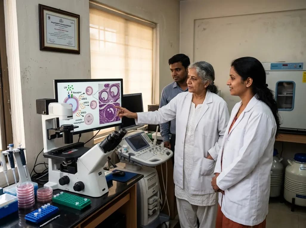
New research reveals that preovulatory follicles communicate via LH-induced paracrine signaling. Diffusible EGF ligands epiregulin and amphiregulin coordinate oocyte meiotic resumption across neighboring follicles, challenging traditional concepts of follicular autonomy.
Today
