.webp&w=384&q=75)

Loading, please wait...
Loading, please wait...
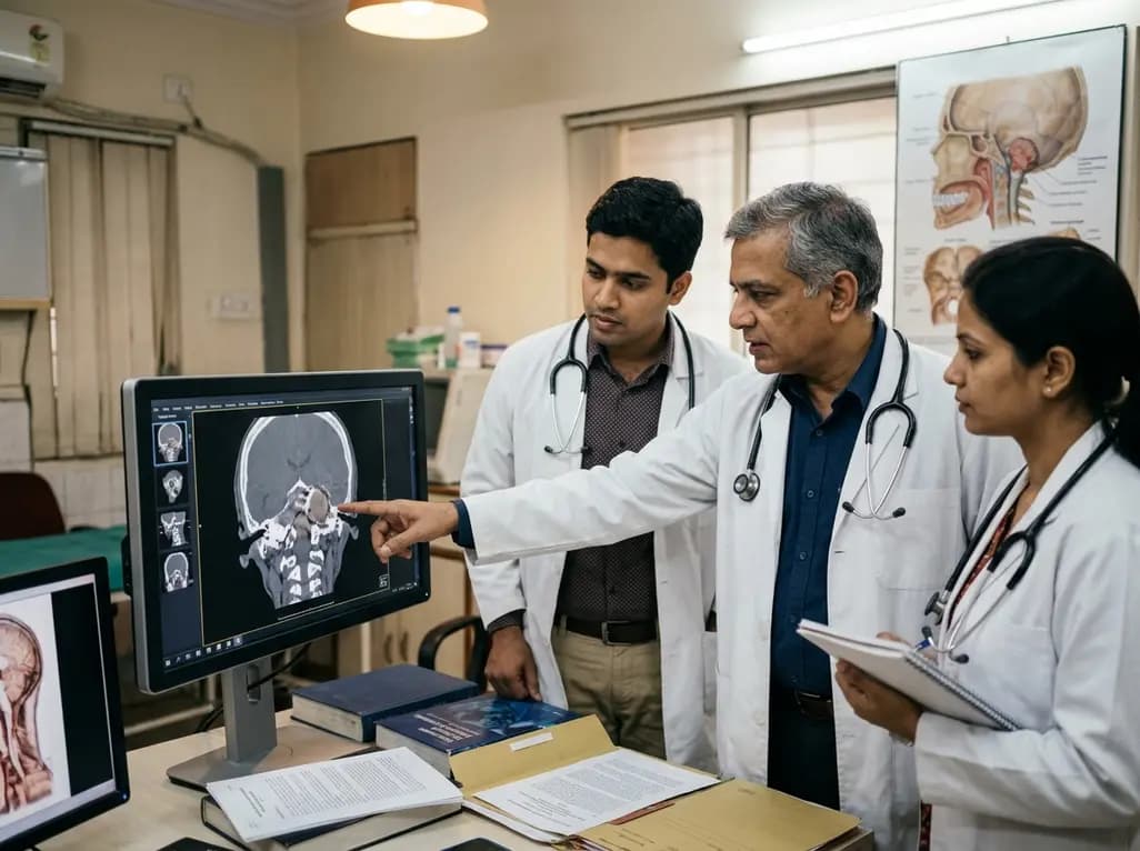
Surgical management of ventral skull base lesions presents formidable anatomical and technical hurdles for modern surgical teams. Practitioners frequently encounter critical neurovascular structures tightly clustered within deeply seated cranial compartments. Historically, open transfacial or transmandibular osteotomies provided necessary surgical exposure for these complex lesions. However, these extensive traditional approaches caused substantial patient morbidity, noticeable facial scarring, and prolonged functional recovery. Over the past two decades, minimally invasive skull base surgery has completely reshaped this clinical landscape. The endoscopic endonasal corridor rapidly became the primary route for many midline ventral skull base lesions. Nevertheless, significant anatomical limitations persist when pathologies extend inferior to the nasopalatine line or lateral to the pterygoid plates. Consequently, surgical teams increasingly utilize the transoral endoscopic approach to safely address these complex anatomical regions. Through natural mucosal corridors, endoscopic techniques provide direct visualization and uncompromised illumination of deep structures. Furthermore, this approach avoids external facial incisions, thereby preserving cosmetic integrity and aerodigestive neuromuscular function. Multidisciplinary collaboration between otorhinolaryngologists and neurosurgeons plays an essential role in successfully navigating these intricate corridors. By combining high-definition visualization with angled endoscopes, surgeons can resect lesions previously deemed inaccessible without destructive osteotomies. Therefore, understanding specific mucosal incisions and corridors remains critical for optimizing operative efficacy.
The transoral endoscopic approach encompasses several distinct surgical corridors tailored to targeted skull base compartments. Specifically, surgeons exploit three main anatomical pathways based on tumor epicenter, size, and vector of extension. First, the transoral transclival and retropharyngeal corridor provides a direct midline trajectory to the craniovertebral junction. Consequently, this midline corridor exposes the lower clivus, anterior arch of the atlas, and the odontoid process. Therefore, surgeons successfully manage ventral compressive pathologies and notochordal remnants without requiring brain retraction. Additionally, the transoral sublabial transmaxillary corridor provides wide lateral and superior access across the middle skull base. By opening the anterior maxillary sinus wall through a gingivobuccal incision, surgeons achieve direct entry into the infratemporal fossa. Furthermore, this transmaxillary corridor preserves nasal airflow dynamics and avoids external facial split procedures entirely. In addition, surgeons frequently utilize transoral palatal and lateral pharyngeal corridors to target the lower parapharyngeal space and pterygoid region. These precise lateral pathways offer exceptional working angles for extensive lateral pathology. Thus, selecting the optimal corridor depends on accurate preoperative cross-sectional imaging and meticulous anatomical triangulation.
Executing safe transoral interventions requires precise mucosal incisions matched to underlying bony and muscular anatomy. In midline craniovertebral junction lesions, surgeons utilize a linear posterior pharyngeal wall incision. Subsequently, they dissect the prevertebral musculature laterally, creating an optimal operative corridor while protecting the carotid arteries. For infratemporal fossa lesions, surgeons prefer a horizontal transoral sublabial incision. This specific incision facilitates an anterior maxillotomy, unlocking direct visualization of the maxillary antrum and pterygomaxillary fissure. Meanwhile, addressing pathologies in the lower parapharyngeal space and greater wing of the sphenoid requires tailored regional incisions. Specifically, surgeons employ palatal, pterygomandibular trigone, or anterior tonsillar pillar incisions. These lateral incisions grant direct access to deep muscular and fascial compartments while sparing major neurovascular trunks. Furthermore, precise soft tissue elevation preserves vascularized mucosal flaps for primary layered closure. Layered mucosal closure significantly reduces postoperative pharyngeal dehiscence and dead space formation. Consequently, careful incision planning guarantees adequate target exposure while minimizing structural disruption and long-term functional impairment.
A diverse spectrum of benign and malignant pathologies arises within ventral and lateral skull base compartments. Among midline extradural lesions, clival chordomas and basilar invagination represent common indications for anterior decompression. In these specific cases, the transoral corridor provides a direct decompression path without traversing contaminated paranasal cavities. Furthermore, surgeons effectively resect benign lesions such as schwannomas, juvenile angiofibromas, and pleomorphic adenomas situated in the infratemporal fossa. Conversely, malignant neoplasms like carcinomas and sarcomas present distinct surgical challenges due to local tissue infiltration. Malignant tumors frequently infiltrate deep fascial planes and encase critical vessels, demanding aggressive oncologic clearance. Consequently, surgeons must achieve clear microscopic margins while protecting the internal carotid artery and lower cranial nerves. However, aggressive tumor biology dictates higher recurrence risks in malignant cohorts compared to benign counterparts. Therefore, comprehensive multidisciplinary skull base teams must integrate adjuvant radiotherapy or systemic therapy into postoperative treatment regimens. This collaborative therapeutic model ensures optimal disease control and promotes long-term survival.
Complex skull base neoplasms often expand across multiple anatomical compartments simultaneously. In such scenarios, single-corridor access may prove insufficient for complete and safe tumor extirpation. Consequently, surgeons frequently adopt multiportal strategies combining endoscopic endonasal and transoral routes. This combined modular approach provides complementary optical angles and multi-instrument triangulation during difficult resections. For example, an endonasal telescope can illuminate superior clival extensions while transoral instruments resect the inferior tumor bulk. Furthermore, multiportal approaches overcome instrument crowding, which frequently limits narrow single-port procedures. Surgeons can introduce navigation probes, suctions, and dissection instruments through separate anatomical entry points simultaneously. Additionally, real-time intraoperative image guidance and Doppler ultrasonography improve surgical safety near vital vascular structures. Meticulous dynamic coordination between two operating surgeons ensures fluid instrument exchange and continuous operative field visualization. Thus, modular multiportal interventions significantly expand the surgical envelope for highly challenging and extensive ventral skull base lesions.
Long-term tertiary center experiences demonstrate that transoral endoscopic techniques achieve reliable decompression and favorable clinical outcomes. Benign lesions consistently exhibit excellent local control rates following complete surgical excision. However, malignant lesions display substantial recurrence rates, requiring vigilant and systematic postoperative surveillance. Magnetic resonance imaging at scheduled intervals remains essential to detect early tumor regrowth and guide timely secondary interventions. Additionally, surgical teams must remain vigilant regarding potential perioperative complications. Cerebrospinal fluid leak represents a critical concern, particularly when extensive dural resection is necessary. To mitigate this risk, surgeons execute robust multilayered reconstructions using autologous fascia, fat grafts, and vascularized flaps. Furthermore, velopharyngeal insufficiency and pharyngeal wound dehiscence require strict postoperative swallowing protocols and tailored nutritional support. Overall, rigorous patient selection, refined microsurgical technique, and proactive complication management make transoral endoscopic approaches highly effective for select ventral skull base disorders.
Surgeons primarily indicate this approach for ventral skull base lesions, including clival chordomas, basilar invagination, and infratemporal fossa tumors. Additionally, it offers exceptional access to neoplasms involving the lower parapharyngeal space and pterygoid region when traditional endonasal access cannot reach beneath the nasopalatine line.
Transoral endoscopic access utilizes natural anatomical corridors, completely eliminating visible facial scars and major transfacial osteotomies. Furthermore, modern high-definition endoscopes provide superior illumination and magnification of critical neurovascular structures. Consequently, patients experience reduced surgical trauma, less postoperative pain, and faster overall recovery times.
Teams prevent and manage cerebrospinal fluid leaks by performing meticulous multilayered closures using autologous fascia lata, fat grafts, and vascularized mucosal flaps. Additionally, intraoperative tissue sealants and temporary postoperative lumbar drainage are frequently utilized to promote watertight healing and protect underlying neural structures.
Disclaimer: This content is for informational and educational purposes only and should not be considered medical advice. Always consult a qualified healthcare professional regarding any medical condition or treatment. Refer to the latest local and national guidelines for clinical practice.
References

Read summarized clinical updates, watch expert medical content, and earn CME certifications right from your smartphone.
Open your smartphone camera to scan and install.





A 10-year study evaluates the transoral endoscopic approach for skull base lesions across 71 patients, detailing tailored mucosal corridors for the craniovertebral junction, infratemporal fossa, and parapharyngeal space.
last month
A laboratory technician's death in Siberia from an unverified respiratory pathogen has renewed global vigilance for pneumonic plague. Clinicians must recognize early presentations of Yersinia pestis, enforce immediate respiratory isolation, and start early bactericidal therapy to prevent severe secondary spread.
Today

Tripura has launched a comprehensive statewide oncology campaign to address late-stage breast cancer presentations. By strengthening community screening for women aged 30 and older and expanding District Day Care Cancer Centres, the initiative improves early clinical detection and decentralized chemotherapy access.
Today
The US FDA has granted approval to the SELUTION SLR drug-eluting balloon for treating coronary in-stent restenosis. This milestone introduces a sustained-release sirolimus platform that eliminates the need for additional permanent metallic stents while maintaining robust vascular healing and luminal patency.
Today

The Thumbs-Out Classification (TOC) is a reliable screening tool for detecting thumb-in-palm deformity in children with cerebral palsy. Utilizing traffic-light tiers for radial abduction, TOC enables clinicians to monitor upper limb function and initiate timely therapeutic or surgical interventions.
Today

A comprehensive review analyzes periconceptional GLP-1 receptor agonist exposure, revealing reassuring data on congenital anomalies and adverse perinatal outcomes while emphasizing the need for cautious patient counseling.
Today
