

Loading, please wait...
Loading, please wait...

Transcatheter device closure represents an established therapy for secundum atrial septal defects in modern cardiology. However, structural heart teams recognize that mechanical complications can occasionally arise years after deployment. Specifically, septal occluder erosion stands out as a serious, life-threatening complication following percutaneous intervention. While clinicians typically expect tissue erosion within twelve months, extraordinary cases can present decades later. Therefore, physicians must maintain vigilance when adult patients with prior closure devices present with unexplained systemic findings.
Interventional cardiologists select nitinol occluders because these devices facilitate rapid endothelialization across the atrial septum. Nevertheless, ongoing cardiac motion creates persistent mechanical friction between rigid metallic frames and compliant atrial walls. Specifically, dynamic pulsation against adjacent structures can gradually cause micro-abrasions in surrounding myocardial tissue. Consequently, deficient aortic or superior rims substantially increase the risk of friction against the aortic root. In particular, absent retro-aortic tissue allows the device flange to impinge directly upon the aortic wall. Furthermore, device oversizing generates excessive radial tension, which progressively weakens contiguous margins. Most acute erosions trigger rapid hemopericardium and pericardial tamponade shortly after implantation. However, slow mechanical abrasion can proceed indolently without causing sudden pericardial rupture. Over extended periods, dense fibrous capsules often contain the localized tissue breakdown. As a result, chronic erosion can form abnormal fistulous channels rather than catastrophic cardiac collapse. Thus, clinicians must recognize that mechanical device wear remains possible throughout a patient's lifespan.
A landmark clinical report recently documented cardiac device erosion occurring more than two decades after implantation. Remarkably, this interval represents the longest documented timeline between initial device placement and subsequent mechanical erosion. Rather than presenting with severe chest pain or syncope, the patient developed progressive fatigue and malaise. Subsequent diagnostic testing revealed pronounced intravascular hemolysis resulting from abnormal intracardiac blood flow. When the device eroded into the adjacent right atrium, it formed an abnormal fistulous pathway. Consequently, red blood cells encountered severe mechanical shear stress while passing over exposed metallic wire mesh. Therefore, laboratory results confirmed severe anemia, elevated lactate dehydrogenase, depleted haptoglobin, and circulating schistocytes. Because these clinical features mimic hematologic disorders, clinicians can easily misdiagnose the underlying etiology. In addition, subtle shunts can evade detection if physicians do not perform targeted echocardiography. Hence, physicians must always consider structural device disruption whenever an occluder recipient develops unexplained hemolytic anemia.
Comprehensive multimodality cardiovascular imaging plays an essential role in confirming late device-related complications. Although standard transthoracic echocardiography provides an initial view, it often misses subtle localized tissue erosions. Therefore, transesophageal echocardiography remains the primary diagnostic modality to delineate structural integrity. Multiplane transesophageal views clearly demonstrate high-velocity flow jets, disc positioning, and tissue defects around the device. Additionally, three-dimensional echocardiography provides detailed anatomical perspectives of adjacent cardiac valves and aortic margins. Furthermore, cardiac computed tomography angiography delivers exceptional spatial resolution to examine metallic framework geometry. Electrocardiogram-gated computed tomography accurately excludes wire fracture, detects pseudoaneurysms, and maps fistulous tracts. Similarly, cardiac magnetic resonance imaging can quantify regurgitant volumes without administering iodinated contrast media. Consequently, combining these imaging techniques enables heart teams to choose the optimal corrective strategy. Ultimately, precise anatomical mapping determines whether a transcatheter approach remains safe and viable.
Historically, cardiothoracic surgeons treated cardiac erosion through open median sternotomy with cardiopulmonary bypass support. Surgical intervention permits direct device explantation, tissue debridement, and autologous pericardial patch reconstruction. However, redo cardiac surgery carries notable morbidity, increased transfusion requirements, and extended recovery times. Therefore, interventional cardiologists increasingly evaluate percutaneous catheter-based approaches for suitable structural fistulas. In this published case, operators successfully repaired the right atrial fistula using a transcatheter occlusion device. Specifically, the intervention team deployed an occluder directly across the fistulous communication via femoral venous access. They carefully confirmed stable positioning under continuous echocardiographic guidance. This targeted intervention immediately sealed the defect and eliminated high-velocity turbulent blood flow. Consequently, erythrocyte destruction stopped completely, which resolved the patient's severe intravascular hemolysis. Furthermore, post-procedure evaluations demonstrated stable device placement without compromise to adjacent intracardiac structures. Thus, transcatheter closure provides an effective, minimally invasive therapeutic solution for highly selected patients.
Transcatheter defect closure remains widespread across cardiac catheterization laboratories throughout India. While interventional cardiologists achieve outstanding procedural success, long-term follow-up remains a significant clinical challenge. Many Indian patients default on outpatient appointments once their acute symptoms resolve after initial intervention. Consequently, subtle late mechanical complications can progress silently for years before detection. Therefore, healthcare institutions must establish structured lifelong registries for adult congenital heart disease patients. In addition, primary care physicians must learn to identify subtle red flag signs promptly. New systolic murmurs, exertional breathlessness, dark urine, or unprovoked anemia necessitate urgent echocardiographic assessment. Furthermore, interventionalists must strictly adhere to sizing recommendations and avoid aggressive device oversizing during initial closure. Thorough pre-procedural rim assessment remains crucial to minimize long-term frictional forces against aortic margins. Ultimately, combining cautious device selection with dedicated long-term clinical monitoring protects patients across decades.
Septal occluder erosion develops late when continuous cardiac contractility generates repetitive mechanical friction between rigid metallic struts and adjacent thin cardiac tissues. Over multiple decades, constant cyclic motion can gradually wear through contiguous atrial walls or aortic margins. Deficient anatomical rims, mild device oversizing, and localized fibrous remodeling significantly heighten this mechanical wear. Consequently, delayed microscopic abrasions progressively coalesce into macroscopic tissue perforations or fistulas years after initial placement.
Mechanical hemolysis occurs when structural device erosion creates an abnormal fistulous channel with high-velocity turbulent blood flow. As red blood cells accelerate across exposed nitinol wires, severe mechanical shear stress ruptures the erythrocyte membranes. Patients typically present with progressive fatigue, jaundice, dark cola-colored urine, and unprovoked anemia. Diagnostic laboratory evaluations reliably show elevated serum lactate dehydrogenase, marked reticulocytosis, severely depressed haptoglobin levels, and prominent fragmented red cells, or schistocytes, on peripheral blood smears.
Transcatheter closure cannot resolve every late erosion defect because procedural feasibility depends entirely on individual patient anatomy. Interventional cardiologists carefully reserve catheter-based techniques for stable patients with discrete, well-defined fistulous tracts that feature adequate landing margins. Conversely, extensive aortic root lacerations, free pericardial perforations, severe device instability, or active infective endocarditis mandate urgent surgical intervention. Multidisciplinary heart teams must always evaluate comprehensive imaging before selecting transcatheter intervention over open sternotomy and device explantation.
Disclaimer: This content is for informational and educational purposes only... Refer to the latest local and national guidelines for clinical practice.
References

Read summarized clinical updates, watch expert medical content, and earn CME certifications right from your smartphone.
Open your smartphone camera to scan and install.





A landmark case reveals transcatheter closure of an atrial septal occluder erosion presenting with hemolysis two decades after implantation. This report highlights the longest documented latency for device erosion, emphasizing multimodality imaging, minimally invasive fistula repair, and lifelong patient surveillance.
Today
A health economic evaluation in Japan reveals that amenamevir is not cost-effective compared with valaciclovir for treating herpes zoster. Despite achieving similar clinical efficacy, amenamevir substantially increases drug expenditures, highlighting the critical role of valaciclovir in value-based viral care.
Today

The GESS-AEEM consensus establishes a clear clinical implementation framework for testosterone therapy in postmenopausal women with hypoactive sexual desire disorder, focusing on transdermal dosing, precise laboratory monitoring, and safety communication.
Today
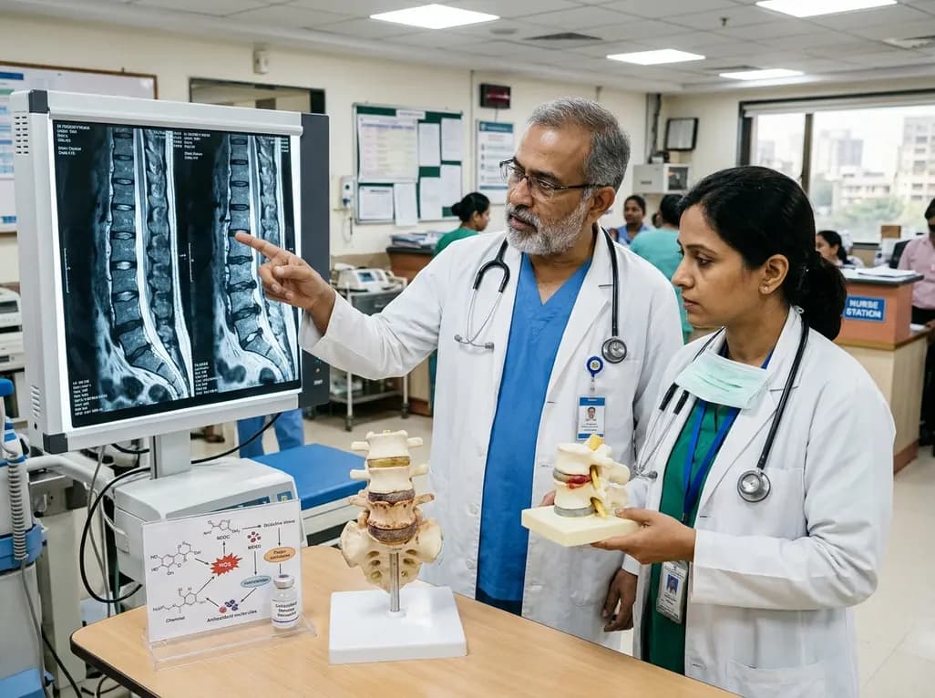
Intervertebral disc degeneration is a primary driver of chronic low back pain. Emerging evidence highlights reactive oxygen species and redox imbalance as central mechanisms. Targeted antioxidant strategies and biomaterial delivery systems offer promising disease-modifying therapies for spine health.
Today

A multicenter cohort study reveals that the 30-minute C-peptide response during OGTT independently predicts cystic fibrosis-related diabetes in patients with normal glucose tolerance, offering an early risk-stratification biomarker.
Today
