

Loading, please wait...
Loading, please wait...

Clinicians increasingly recognize Parkinson's disease as a multifaceted neurodegenerative disorder that encompasses both debilitating motor deficits and profound non-motor impairments. However, conventional diagnostic tools and scalp electroencephalograms often fail to capture real-time cortical network dynamics outside primary motor pathways. The clinical application of concurrent TMS-EEG in Parkinson's disease offers an unprecedented window into cortical excitability and reciprocal connectivity. By delivering localized magnetic pulses while simultaneously recording millisecond-level electrophysiological responses, researchers can directly assess both motor and cognitive neural circuits.
Investigating the pathophysiological boundaries of Parkinson's disease requires evaluating diverse functional networks. Researchers recently conducted a comprehensive neurophysiological evaluation involving forty-six individuals with Parkinson's disease and twenty-seven matched healthy control participants. Specifically, the investigators applied single-pulse transcranial magnetic stimulation over two distinct anatomical nodes: the primary motor cortex and the dorsolateral prefrontal cortex. Meanwhile, high-density electroencephalography recorded transcranial magnetic stimulation-evoked potentials across the entire scalp. This dual-site stimulation protocol allowed the research team to compare local reactivity against distributed network responses. Consequently, the study captured precise corticothalamic and corticocortical transmission characteristics in both cohorts. In addition, the investigators quantified global mean field amplitude alongside time-frequency oscillatory powers. These multimodal metrics illuminated distinct neurophysiological fingerprints distinguishing patients from healthy individuals. Therefore, concurrent neurostimulation provides deep mechanistic clarity that standard neuroimaging cannot supply alone. Clinicians can thus track circuit-specific dysfunction across multiple cerebral domains with remarkable temporal precision. Furthermore, these findings emphasize that Parkinsonian pathology extends well beyond isolated nigrostriatal degeneration. Cortical circuitry exhibits distinct compensatory and pathological alterations that directly govern daily functional capability.
When investigators delivered pulses to the primary motor cortex, patients with Parkinson's disease exhibited a significantly elevated P30 amplitude compared to healthy individuals. Furthermore, this magnified early evoked component demonstrated a strong positive correlation with motor disability scores. Because early evoked potentials reflect local cortical excitability and rapid intracortical interneuron signaling, elevated P30 responses suggest loss of physiological inhibition. In healthy brains, balanced gamma-aminobutyric acid neurotransmission dampens initial pyramidal cell discharges. However, striatal dopamine depletion disrupts basal ganglia outflow, causing profound downstream disinhibition within the primary motor strip. Consequently, aberrant corticospinal facilitation produces exaggerated cortical responses to exogenous magnetic pulses. Notably, the linear relationship between P30 amplitude and motor impairment suggests that this electrophysiological metric tracks disease burden reliably. Clinicians often struggle to measure fluctuating motor symptoms objectively during pharmacological management. Therefore, quantifying early motor potentials could establish a robust functional biomarker for monitoring progression. In addition, these objective electrophysiological markers may guide individualized adjustments of dopaminergic pharmacotherapy and rehabilitative interventions. Ultimately, tracking cortical hyper-excitability provides vital mechanistic feedback that complements subjective clinical rating scales.
Beyond classic motor pathology, non-motor symptoms such as depression and cognitive slowing heavily impair patient quality of life. The study evaluated prefrontal circuitry by stimulating the dorsolateral prefrontal cortex directly. Following prefrontal stimulation, patients demonstrated an increased area under the curve for global mean field amplitude at the P30 and P60 time points. Moreover, these heightened prefrontal evoked responses correlated significantly with clinical depression severity scores within the patient cohort. Consequently, this observation indicates that affective disturbances in Parkinson's disease arise from measurable neurophysiological alterations in frontostriatal networks. Previously, clinicians often viewed depressive symptoms as secondary psychological reactions to chronic motor disability. However, these electrophysiological data confirm an organic, circuit-based etiology rooted in prefrontal cortical hyperexcitability. In addition, impaired monoaminergic regulation in the dorsolateral prefrontal cortex alters cortical-subcortical communication loops. Therefore, prefrontal evoked potentials provide an objective metric for evaluating neuropsychiatric comorbidities in movement disorders. Understanding these neurobiological mechanisms helps clinicians tailor targeted psychiatric interventions, including targeted repetitive transcranial stimulation protocols. Consequently, physicians can differentiate neurobiologically driven mood disorders from situational distress with greater confidence.
In addition to evoked potential waveforms, oscillatory dynamics provide crucial information regarding functional brain states. The investigation revealed significant power reductions across both alpha and beta frequency bands during magnetic stimulation in patients. Furthermore, distinct topographical patterns emerged depending strictly upon whether stimulation engaged the motor or prefrontal cortex. In healthy individuals, robust beta oscillations facilitate resting postural maintenance, while alpha rhythms coordinate sensory gating. However, pathologically altered oscillatory activity reflects disrupted functional connectivity across large-scale cerebral loops. When dopaminergic denervation destabilizes basal ganglia-thalamocortical loops, normal cortical synchrony deteriorates rapidly. Consequently, widespread spectral power reduction signifies an inability to sustain coherent communication between distant cortical hubs. These electrophysiological deficits align with executive dysfunctions and bradykinetic motor features commonly encountered in clinical practice. Therefore, spectral power analysis offers complementary diagnostic value alongside classic time-domain evoked components. Moreover, analyzing frequency-specific perturbations enables investigators to assess how disease progression damages dynamic oscillatory coordination throughout the entire brain. As a result, tracking time-frequency spectral alterations could serve as a non-invasive index of cortical network disintegration.
These electrophysiological discoveries carry immediate implications for modern neuromodulatory strategies in neurological practice. Repetitive transcranial magnetic stimulation and deep brain stimulation represent cornerstone advanced therapies for refractory Parkinson's disease. However, selecting optimal anatomical stimulation targets and adjusting stimulation frequencies has historically relied upon empirical trial-and-error. By characterizing patient-specific abnormalities in both motor and prefrontal cortices, clinicians can design personalized therapeutic protocols. For instance, discovering concurrent primary motor and prefrontal hyperactivity supports dual-target neuromodulation approaches. Similarly, monitoring evoked potentials before and after therapeutic interventions allows clinicians to quantify cortical normalization objectively. Furthermore, these electrophysiological biomarkers could dramatically improve patient stratification in neuroprotective clinical trials. Patients exhibiting elevated P30 amplitudes or prefrontal hyper-reactivity might receive targeted therapies earlier in their clinical trajectory. Accordingly, integrating neurostimulation with real-time electroencephalography bridges the gap between fundamental laboratory neuroscience and everyday movement disorder management. Ultimately, this translational paradigm accelerates the development of precision medicine tailored to individual neurophysiological profiles. Consequently, patients stand to gain more durable symptom control and superior preservation of cognitive health.
Although these findings mark significant progress, transitioning advanced neurophysiological techniques into routine clinical practice requires standardized protocols. Future longitudinal studies must examine whether evoked potential elevations precede detectable motor manifestations in prodromal cohorts. In addition, researchers should evaluate how fluctuating dopaminergic medication states modulate these electrophysiological signatures over time. Expanding clinical registries across diverse demographic populations, including Indian cohorts, will establish essential normative reference values. Moreover, integrating automated machine learning algorithms with multichannel electrophysiological data could streamline real-time clinical analysis. Consequently, busy tertiary neurology centers could rapidly screen patients for subclinical affective and motor circuit dysfunction. Clinicians could thereby initiate prophylactic neurorehabilitation before debilitating disability becomes irreversible. As hardware systems become more portable and cost-effective, bedside cortical excitability testing will become increasingly feasible. Therefore, combining neurostimulation with high-density electroencephalography heralds a transformative era in neurodegenerative disease diagnostics. Movement disorder specialists will increasingly rely on functional cortical measurements to deliver comprehensive, compassionate, and personalized neurological care. Thus, continuous technological refinement will firmly establish electrophysiological mapping as an essential clinical tool.
Concurrent TMS-EEG directly probes cortical excitability and functional network connectivity beyond simple motor pathways. By delivering magnetic pulses to specific regions like the motor or prefrontal cortex, clinicians can measure immediate millisecond-level electrophysiological responses, identifying network disruptions that underpin both motor disability and non-motor complications.
The elevated P30 amplitude following primary motor cortex stimulation indicates cortical disinhibition driven by aberrant basal ganglia-thalamocortical signaling. Because this amplitude correlates positively with motor symptom severity, it serves as an objective, quantifiable electrophysiological biomarker for tracking motor disability and monitoring response to therapeutic interventions.
Prefrontal stimulation over the dorsolateral prefrontal cortex reveals increased global mean field amplitude at P30 and P60, which significantly correlates with depression scores. This relationship demonstrates that affective symptoms in Parkinson's disease stem from measurable frontostriatal network hyperexcitability rather than purely reactive psychological distress.
Disclaimer: This content is for informational and educational purposes only. It is not intended to be a substitute for professional medical advice, diagnosis, or treatment. Always seek the advice of your physician or other qualified health provider with any questions you may have regarding a medical condition. Refer to the latest local and national guidelines for clinical practice.
References

Read summarized clinical updates, watch expert medical content, and earn CME certifications right from your smartphone.
Open your smartphone camera to scan and install.





A landmark study evaluates concurrent TMS-EEG in Parkinson's disease across motor and prefrontal cortices, revealing elevated P30 amplitudes linked to motor deficits, prefrontal hyper-reactivity associated with depression scores, and widespread spectral power desynchronization.
Today
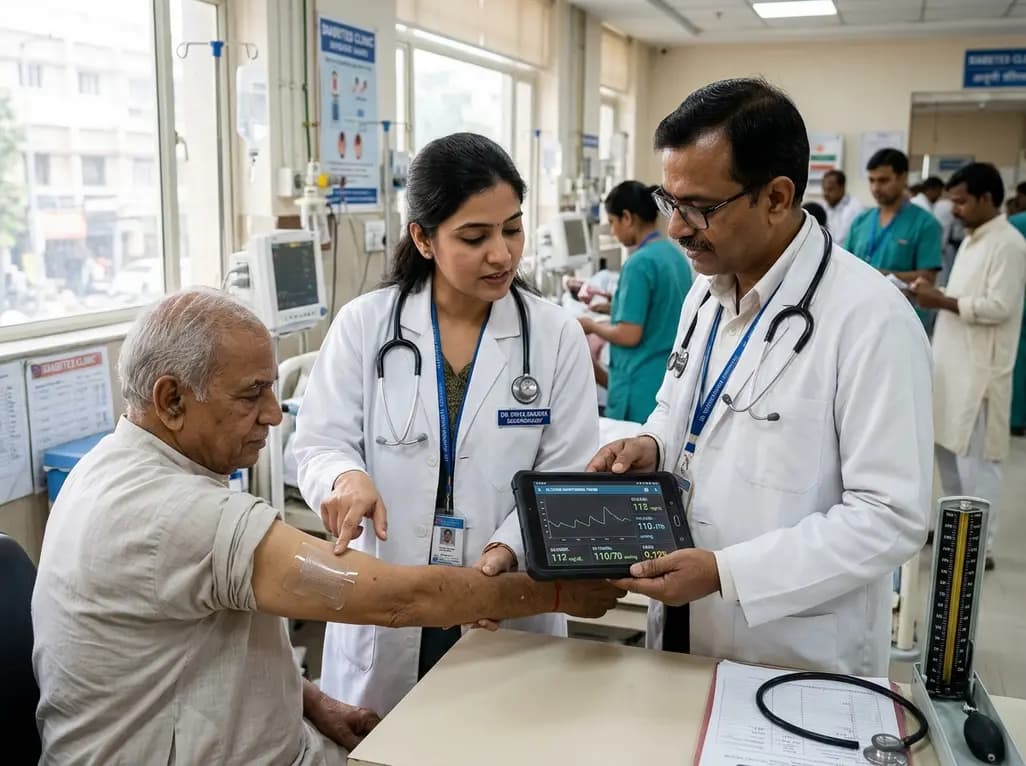
A breakthrough ultrathin multimodal sensor decouples physical hemodynamic monitoring from disposable electrochemical calibration, achieving long-term wearable glucose prediction with 81.36% accuracy and a 9.12% MARD. This offers a robust, drift-free solution for outpatient metabolic surveillance.
Today

A clinical review of menopause and neurology highlights the neurobiological impacts of midlife hormonal shifts on cognition, epilepsy, migraine, multiple sclerosis, and stroke, emphasizing safe hormone therapy prescribing.
Today

Co-administering antiseizure medications and DOACs frequently causes pharmacokinetic interactions, subtherapeutic drug levels, and thromboembolic failures. This review highlights enzyme induction risks, unpredictable outcomes with levetiracetam, and the vital role of therapeutic drug monitoring.
Today

A comparative review of tension band wiring and intramedullary screw fixation in thumb metacarpophalangeal arthrodesis, evaluating union rates, hardware complications, and clinical outcomes.
Today

Following regulatory scrutiny from FSSAI regarding promotional representations and lean muscle preservation claims, Dr. Reddy's Laboratories and Nestlé Health Science are currently reviewing the branding and marketing strategy for their joint nutritional supplement Celevida GLP+.
Today
