

Loading, please wait...
Loading, please wait...

Spontaneous intracerebral hemorrhage represents one of the most catastrophic forms of acute cerebrovascular disease worldwide. Although surgical evacuation offers lifesaving decompression for selected cohorts, clinical trajectories remain heterogeneous and difficult to anticipate. Clinicians continuously search for reliable prognostic indicators that reflect underlying physiological resilience before taking patients to the operating theatre. Recent clinical evidence demonstrates that preoperative temporal muscle thickness serves as an objective, non-invasive surrogate of whole-body skeletal muscle mass and physiological reserve. By assessing cranial computed tomography scans already acquired during admission, surgical teams can rapidly gauge baseline frailty without requiring supplemental radiation or costly metabolic assessments. Consequently, integrating neuromuscular markers into acute decision-making models offers a promising avenue to refine surgical patient selection and individualize aggressive post-operative neurocritical support.
Sarcopenia reflects an age-related or disease-induced depletion of skeletal muscle volume and strength. In acute neurological emergencies, clinicians rarely possess the luxury of performing whole-body dual-energy x-ray absorptiometry or bioimpedance tests. Fortunately, standard cranial computed tomography routinely visualizes cranial musculature with high anatomical resolution. Measuring temporal muscle thickness and calculating the corresponding temporal muscle index offer a pragmatic window into systemic muscle wasting. The masticatory muscles receive cranial motor innervation and experience ongoing nutritional, hormonal, and metabolic demands comparable to somatic musculature. Therefore, reduced temporal muscle cross-sectional bulk serves as an immediate visual marker of latent sarcopenic vulnerability. Furthermore, neuroradiologists and emergency neurosurgeons can standardize these axial measurements at predefined cranial landmarks within seconds. This rapid radiological assessment bridges the gap between bedside geriatric assessment and urgent surgical neurotrauma intervention.
A recent cohort investigation evaluated patients undergoing emergent surgical evacuation of spontaneous supratentorial intracerebral hemorrhage between 2014 and 2022. Investigators measured baseline temporal muscle parameters from admission computed tomography scans to evaluate post-operative overall survival across a median follow-up of 36 months. Patients categorized into the low muscle status group experienced a striking 67 percent mortality rate compared to roughly 30 percent in patients exhibiting preserved muscle bulk. Univariate Cox regression analysis revealed hazard ratios greater than 2.3 for both metrics among sarcopenic individuals. Crucially, this robust association persisted even after investigators adjusted the multivariate model for classical neurosurgical predictors, including the admission Glasgow Coma Scale, pupillary status, and systemic blood pressure. These findings prove that systemic physiological exhaustion independently dictates post-surgical survival beyond initial hematoma volume and neurological damage alone.
Why does reduced craniofacial muscle bulk correlate so strongly with long-term mortality after neurosurgical decompression? Skeletal muscle tissue operates as an active endocrine and metabolic organ rather than a passive mechanical engine. Specifically, healthy muscle reserves preserve metabolic homeostasis during prolonged hypercatabolic states triggered by acute brain injury. Patients with established sarcopenia lack amino acid reservoirs necessary for wound repair, cellular immunity, and systemic immune defenses. As a result, these frail individuals face heightened susceptibility to secondary hospital-acquired infections, aspiration pneumonia, ventilator dependency, and septic shock during intensive care. Furthermore, systemic muscle loss frequently coexists with subclinical microvascular damage, chronic systemic inflammation, and latent cardiopulmonary vulnerability. Therefore, whenever an elderly or malnourished patient experiences catastrophic cerebral bleeding, low muscular baseline capacity drastically accelerates multi-organ failure and delays neurocognitive recovery.
Neurosurgical teams can immediately translate these imaging metrics into everyday triage protocols. The admission cranial computed tomography scan remains universally available, eliminating extra diagnostic delays or additional costs. When clinicians measure temporal muscle thickness at the level of the orbital roof or Sylvian fissure, they acquire actionable risk stratification data before entering the operating room. However, surgical teams must not interpret low muscle parameters as an absolute contraindication to hematoma evacuation. Instead, surgical teams should view these measurements as objective frailty warnings. For instance, identifying low muscle reserves alerts intensivists to potential extubation failures, prolonged rehabilitation needs, and heightened post-operative mortality. By combining structural stroke indices with objective sarcopenic measures, neurointensivists can communicate realistic prognostic expectations to families and establish proactive recovery milestones early.
Identifying sarcopenic vulnerability early enables targeted post-operative interventions that significantly enhance rehabilitation outcomes. Neurocritical care teams should initiate customized nutritional supplementation promptly following surgical decompression to mitigate excessive catabolic tissue destruction. Enteral formulas enriched with high biological value proteins, essential branched-chain amino acids, and micronutrients may attenuate secondary muscle wasting in vulnerable patients. Furthermore, physical therapists and rehabilitation physicians should introduce bedside mobilization protocols as soon as intracranial pressures stabilize. Simultaneously, speech therapists must perform aggressive swallowing evaluations, because masticatory and pharyngeal muscle weakness often increases aspiration risks. Thus, incorporating craniofacial muscular metrics transforms standard neuroimaging into a comprehensive multidisciplinary roadmap, guiding aggressive physical therapy, nutritional support, and tailored neurocritical rehabilitation.
Clinicians measure temporal muscle thickness on standard axial computed tomography slices, typically perpendicular to the muscle axis at the level of the orbital roof or Sylvian fissure. Bilateral measurements are averaged or indexed to intracranial area to minimize anatomical variation and achieve reliable systemic muscle estimates.
Baseline sarcopenia diminishes essential physiological reserves needed to survive acute systemic stress and prolonged bed rest. Sarcopenic patients experience impaired immune function, severe metabolic depletion, and delayed tissue healing, substantially increasing their risk of post-operative pneumonia, sepsis, prolonged mechanical ventilation, and subsequent long-term mortality.
Yes, clinicians can readily combine the temporal muscle index with established scoring tools like the intracerebral hemorrhage score. Multivariate clinical data demonstrate that temporal muscle parameters provide independent, supplemental prognostic information, thereby enhancing overall outcome prediction without requiring supplemental radiological examinations or specialized laboratory equipment.
Disclaimer: This content is for informational and educational purposes only and does not constitute medical advice, diagnosis, or treatment. Healthcare professionals should rely on their independent clinical judgment and verify all medical decisions against current protocols. Refer to the latest local and national guidelines for clinical practice.
References
Pesonen EK et al. Temporal muscle thickness and index predict overall survival after surgical evacuation of spontaneous intracerebral hemorrhage. Clin Neurol Neurosurg. 2025 Jul. doi: 10.1016/j.clineuro.2025.108962. PMID: 40367879.
Mohajerani E et al. Temporal muscle thickness as a feasible sarcopenia marker and outcome predictor after aneurysmal subarachnoid hemorrhage. Acta Neurochir (Wien). 2025. doi: 10.1007/s00701-025-06562-z. PMID: 40439824.
Steindl A et al. Sarcopenia in Neurological Patients: Standard Values for Temporal Muscle Thickness and Muscle Strength Evaluation. J Clin Med. 2020;9(5):1272. doi: 10.3390/jcm9051272.
Dubinski D et al. Frailty in Traumatic Brain Injury—The Significance of Temporal Muscle Thickness. J Clin Med. 2023;12(24):7550. doi: 10.3390/jcm12247550.

Read summarized clinical updates, watch expert medical content, and earn CME certifications right from your smartphone.
Open your smartphone camera to scan and install.





A landmark study demonstrates that preoperative temporal muscle thickness (TMT) and index (TMI) independently predict long-term overall survival in patients undergoing surgical evacuation of spontaneous intracerebral hemorrhage, providing an accessible CT-based biomarker for risk stratification.
Today

The 2025 Australian meningococcal surveillance report shows a 13% decline in invasive meningococcal disease notifications to 118 cases. Genogroup B accounted for 82.2% of typed infections, predominantly impacting infants and young adults, highlighting critical vaccination and rapid diagnostic imperatives.
Today
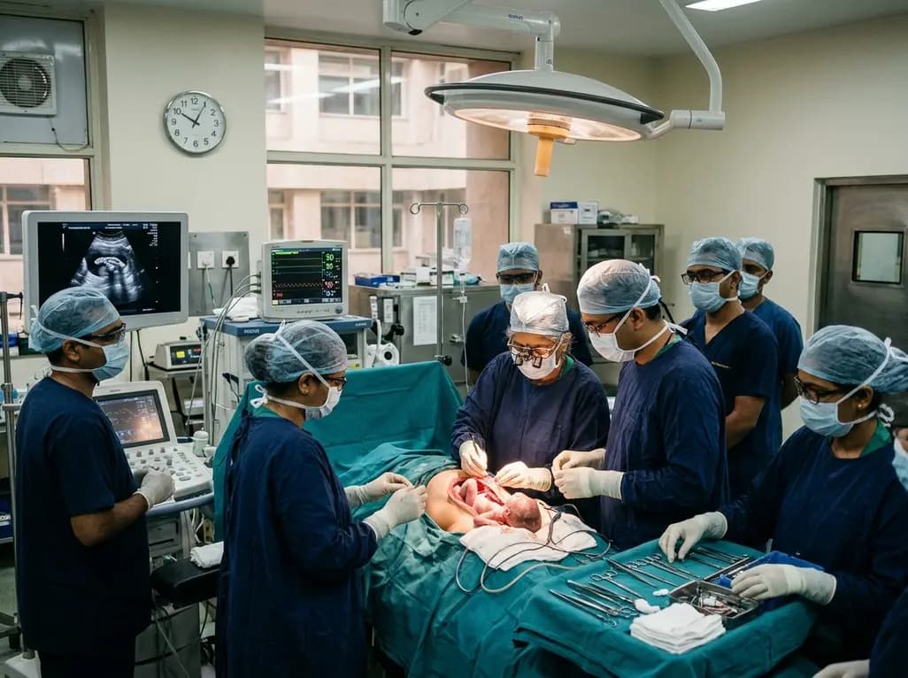
Doctors at AIIMS New Delhi have achieved a historic milestone by performing India's first open prenatal myelomeningocele repair on a 25-week unborn fetus. This landmark multidisciplinary procedure provides vital neuroprotective benefits, lowers hydrocephalus risks, and transforms maternal-fetal medicine nationwide.
Today
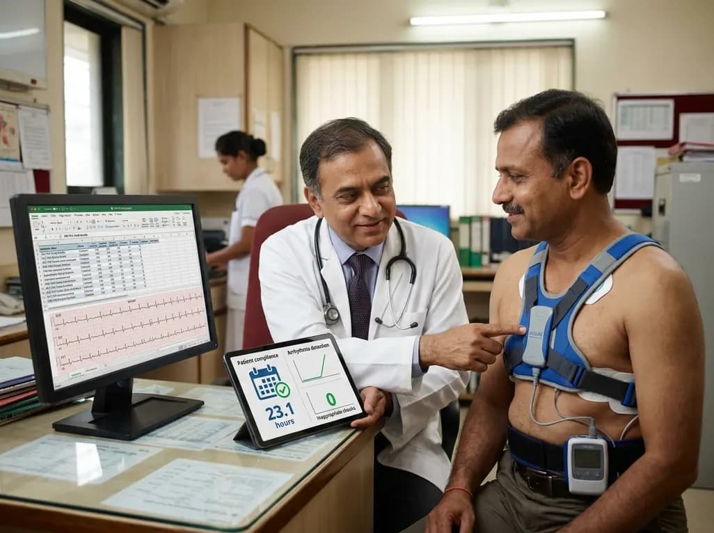
The ACE-PAS registry evaluated 21,612 patients fitted with the ASSURE wearable cardioverter defibrillator. The study demonstrated a 100% ventricular arrhythmia conversion rate, an inappropriate shock incidence of 0.0065 per patient-month, and high daily compliance of 23.1 hours, confirming real-world safety.
Today

A clinical overview of resveratrol supplementation in mitigating exercise-induced muscle damage. This narrative review explores physiological mechanisms, reductions in soreness markers like creatine kinase, dosage strategies, and bioavailability enhancements for sports medicine and nutrition practitioners.
Today

Childhood insulinoma is a rare cause of recurrent hyperinsulinemic hypoglycemia that often eludes early diagnosis. Advanced GLP-1 receptor-targeted PET/CT imaging using 68Ga-exendin-4 complements anatomic MRI to localize occult pancreatic lesions, facilitating focused, curative surgical enucleation.
Today
