

Loading, please wait...
Loading, please wait...
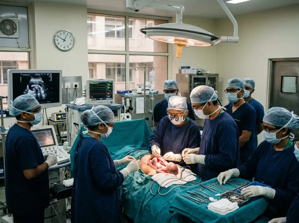
Doctors at AIIMS New Delhi have performed India's first fetal surgery for spina bifida. Specifically, the surgical team operated on an unborn fetus at twenty-five weeks of gestation. Consequently, this pioneering milestone establishes advanced in utero neurosurgical capabilities across the country. Myelomeningocele represents the most devastating form of open spinal dysraphism. In this condition, an abnormal vertebral gap exposes neural tissue to progressive mechanical trauma. Furthermore, direct exposure to amniotic fluid damages delicate spinal roots throughout pregnancy. Therefore, timely maternal-fetal intervention aims to protect neural integrity before irreversible injury occurs.
Myelomeningocele develops during primary neurulation when the embryonic neural tube fails to close completely. Consequently, the spinal cord and surrounding meninges herniate through a posterior skeletal defect. Historically, researchers explained congenital neural damage through the two-hit hypothesis. The first hit represents abnormal embryological neurulation during the initial four weeks of gestation. However, the second hit involves progressive mechanical and chemical destruction of exposed neural structures. Continuous exposure to amniotic fluid triggers inflammatory degradation of fragile unmyelinated axons. Additionally, dynamic hydrostatic trauma against the uterine wall exacerbates progressive neurologic loss. Persistent cerebrospinal fluid leakage through the spinal defect also leads to hindbrain herniation. Consequently, downward displacement of cerebellar tonsils produces the classic Chiari II malformation. This anatomical displacement obstructs normal ventricular drainage, precipitating severe progressive hydrocephalus. Thus, affected infants frequently develop lifelong lower extremity paralysis, neurogenic bladder, and orthopaedic deformities. Epidemiological studies indicate that neural tube defects remain a substantial public health concern across India. Recent systematic reviews demonstrate a pooled prevalence approaching 9.46 cases per 1,000 live births. Therefore, early identification and specialized surgical intervention represent crucial clinical priorities.
On November 12, an interdisciplinary surgical contingent at AIIMS achieved this breakthrough. Specifically, the six-hour procedure took place at the prestigious AIIMS Neurosciences Centre. The patient underwent more than five weeks of meticulous preoperative workup and clinical simulation. Maternal-fetal medicine specialists completed extensive radiological evaluations using high-resolution ultrasound and magnetic resonance imaging. Furthermore, non-directive psychological counselling ensured that the prospective parents completely understood all potential outcomes. Neurosurgical lead Professor Deepak Gupta coordinated seamlessly with maternal-fetal medicine lead Professor Neena Malhotra. Additionally, Professor K Aparna Sharma spearheaded fetal surveillance and initial patient screening. The surgical team carefully exposed the gravid uterus through a specialized lower midline laparotomy. Surgeons then created an exact uterine hysterotomy to directly access the fetal lumbosacral region. Operating under high-power microdissection, neurosurgeons separated the placode from surrounding dysplastic skin. Subsequently, they achieved watertight neural tube closure and mobilized paraspinal muscles. Plastic surgery teams simultaneously assisted in complex multilayered soft-tissue closure. Consequently, both the mother and fetus remained hemodynamically stable throughout postoperative recovery. Crucially, the central government funded the entire procedure under the Janani Shishu Suraksha Karyakram initiative.
The rationale for intrauterine repair rests firmly on the landmark Management of Myelomeningocele Study. Published as a randomized clinical trial, the MOMS trial transformed global pediatric neurosurgery. Prior to this study, surgeons universally deferred myelomeningocele repair until after birth. However, postnatal repair failed to prevent irreversible intrauterine neurological degeneration. The trial compared open prenatal surgical repair before 26 weeks against standard postnatal intervention. Strikingly, the results revealed profound neuroprotective benefits favoring the prenatal intervention group. For example, the need for cerebrospinal fluid shunting decreased dramatically from 82% to 40%. Furthermore, fetal closure prompted significant anatomic reversal of hindbrain herniation in over two-thirds of patients. In addition, children receiving prenatal repair exhibited substantially superior lower-limb motor function. Indeed, the probability of independent ambulation at thirty months nearly doubled compared to controls. Long-term follow-up studies confirm preserved executive functioning and reduced hindbrain-related mortality. Nevertheless, pediatric surgeons emphasize that fetal repair improves functional outcomes rather than providing a complete cure. Accordingly, families require honest, transparent discussions regarding persistent baseline disabilities.
Executing open maternal-fetal interventions requires extraordinary coordination across diverse hospital services. At AIIMS, more than thirty specialized clinicians and nurses participated in this monumental effort. Specifically, obstetric anaesthetists and neuroanaesthetists managed complex dual-patient physiological dynamics simultaneously. They maintained profound uterine relaxation to prevent maternal uterine contractions during open operative manipulation. At the same time, clinicians carefully maintained normal maternal blood pressure to preserve uteroplacental perfusion. Continuous intraoperative ultrasonography guided hysterotomy site selection away from the anterior placental edge. Moreover, ultrasound surveillance monitored fetal heart rate stability and umbilical arterial flow continuously. Surgeons continually replenished warmed lactated Ringer solution inside the amniotic cavity. This fluid replacement effectively prevented fetal hypothermia and umbilical cord compression. Neonatologists stood fully prepared with emergency resuscitative capabilities throughout the surgical timeline. Plastic surgeons facilitated primary myofascial flap advancement to ensure durable cutaneous coverage over exposed dura. Consequently, seamless interdisciplinary communication ensured zero maternal or fetal intraoperative mortality. Ultimately, institutional synergy serves as the foundational cornerstone for high-risk fetal surgical centers.
Despite significant neurofunctional benefits, open fetal interventions introduce considerable obstetric and maternal risks. For instance, hysterotomy substantially increases the likelihood of spontaneous preterm labor and early membrane rupture. In the MOMS cohort, the average gestational age at delivery was thirty-four weeks. Additionally, mothers face genuine risks of uterine dehiscence, chorioamnionitis, and intraoperative hemorrhage. Because of the uterine hysterotomy scar, all subsequent deliveries necessitate planned caesarean sections. Therefore, clinicians must enforce rigorous inclusion criteria before scheduling in utero surgery. Ideal candidates must present with an isolated open defect located between T1 and S1 vertebrae. Furthermore, anatomical confirmation of Chiari II malformation remains a prerequisite for eligibility. Clinicians strictly exclude cases with severe spinal kyphosis, chromosomal aneuploidies, or associated non-neural malformations. Maternal factors like severe obesity, prior preeclampsia, and uncontrolled systemic illness also preclude surgical candidacy. Thus, multidisciplinary teams must weigh maternal safety equally against potential neurological gains for the child. Close post-discharge surveillance remains imperative to manage cervical shortening and detect premature uterine irritability.
The successful milestone at AIIMS marks a monumental shift for maternal-fetal medicine in India. Previously, families diagnosed with fetal myelomeningocele faced only termination or postnatal palliative surgical options. Postnatal closure frequently left children with severe paraplegia and permanent shunt dependency. However, this landmark surgery demonstrates that public healthcare institutions can deliver world-class fetal interventions. Providing this sophisticated surgery under Janani Shishu Suraksha Karyakram ensures exceptional health equity. Consequently, underprivileged families can access cutting-edge in utero therapies without crippling financial strain. AIIMS now aims to establish a structured nationwide fetal care and referral network. This structured program will train obstetricians in early second-trimester anomaly ultrasound screening. In addition, prompt identification within the twenty-six-week therapeutic window will optimize patient referral pathways. Nevertheless, preventive public health initiatives such as preconception folic acid supplementation remain foundational. By merging primary neural defect prevention with advanced intrauterine surgery, healthcare systems can transform infant outcomes. Ultimately, Indian fetal medicine has entered a new era of proactive in utero healing.
Q1: Why is fetal surgery for myelomeningocele preferred over traditional repair after birth?
Performing surgery before birth arrests the progressive chemical and mechanical destruction of exposed spinal nerves caused by continuous contact with amniotic fluid. Clinical evidence from the landmark MOMS trial demonstrates that in utero closure significantly reduces the need for ventriculoperitoneal shunt placement by half and markedly reverses hindbrain herniation. Furthermore, prenatal repair doubles the likelihood of independent ambulation at thirty months, whereas waiting for postnatal surgery often permits irreversible neurological degeneration.
Q2: What gestational age window and clinical criteria determine eligibility for prenatal spina bifida repair?
Eligible candidates typically undergo surgical intervention between nineteen and twenty-five weeks and six days of gestation. Clinicians require an isolated open lumbosacral defect between T1 and S1, accompanied by documented hindbrain herniation. Additionally, fetuses must demonstrate a normal karyotype without other major anatomical anomalies. Mothers must have a healthy uterus without prior classical hysterotomies, placental issues, or serious systemic comorbidities, ensuring minimal risk during deep anaesthesia and open hysterotomy.
Q3: What maternal and obstetric risks are associated with undergoing open intrauterine fetal surgery?
Open fetal surgery introduces tangible obstetric hazards, predominantly premature rupture of membranes and early preterm labor, with an average delivery occurring around thirty-four weeks. Additionally, mothers encounter risks of uterine dehiscence, placental abruption, and intraoperative hemorrhage requiring blood transfusion. Because the procedure entails a surgical incision into the uterine wall, all subsequent deliveries mandate scheduled caesarean delivery to avert catastrophic uterine rupture during labor, requiring lifelong obstetric vigilance.
Disclaimer: This content is for informational and educational purposes only. It does not constitute medical advice or replace professional judgment. Refer to the latest local and national guidelines for clinical practice.
References

Read summarized clinical updates, watch expert medical content, and earn CME certifications right from your smartphone.
Open your smartphone camera to scan and install.





Doctors at AIIMS New Delhi have achieved a historic milestone by performing India's first open prenatal myelomeningocele repair on a 25-week unborn fetus. This landmark multidisciplinary procedure provides vital neuroprotective benefits, lowers hydrocephalus risks, and transforms maternal-fetal medicine nationwide.
Today

Acute uncompensable heat stress impairs the gut barrier, increasing small intestinal permeability. A landmark physiological trial reveals that elevated baseline oestradiol correlates with augmented intestinal barrier disruption in females, offering vital insights into heat illness and systemic endotoxemia.
Today
A randomized crossover trial reveals that daily fresh watermelon consumption significantly reduces baseline TMAO levels and favorably modulates lipid profiles, lowering triglycerides and LDL cholesterol in adults with overweight and obesity.
Today

Arthroscopic physeal-sparing suture-anchor fixation provides a safe, tissue-preserving treatment for pediatric femoral-side PCL avulsions. By anchoring strictly within the epiphysis, this technique restores native knee stability while eliminating the risk of growth arrest associated with transphyseal reconstruction.
Today
A large-scale retrospective cohort study demonstrates that GLP-1 receptor agonists significantly reduce all-cause hospitalizations, alcohol-related admissions, major cardiovascular events, and mortality in patients with overweight or obesity and alcohol use disorder, providing profound clinical insights.
Today
