

Loading, please wait...
Loading, please wait...

Managing temperature extremes in aSAH presents a persistent clinical challenge during neurocritical resuscitation. Aneurysmal subarachnoid hemorrhage triggers acute physiological derangements that destabilize endogenous temperature control. Consequently, hospitalized patients frequently develop extreme thermal fluctuations during the initial week of intensive care. These temperature deviations arise from systemic infectious complications, marked systemic inflammation, or direct damage to hypothalamic regulatory pathways. Erythrocyte breakdown products within the subarachnoid space stimulate intense neuroinflammatory responses and microglial activation. Furthermore, circulating pro-inflammatory cytokines reset central hypothalamic set points, driving recurrent febrile episodes despite conventional antipyretic administration. Conversely, certain severely compromised patients display spontaneous hypothermia without therapeutic cooling interventions. This unintended hypothermia often reflects extensive brainstem injury, profound dysautonomia, or progressive cardiovascular shock. In both scenarios, uncorrected temperature extremes exacerbate ongoing ischemic cellular distress. Neurointensivists increasingly recognize that systemic temperature is an active modifier of neurological recovery rather than an inert clinical biomarker. Therefore, identifying precise thermal thresholds and maintaining continuous physiological stability remain essential clinical goals. Diligent thermal regulation during early critical care helps prevent secondary brain insult and preserves functional autonomy in vulnerable post-hemorrhagic patients across neurocritical care settings.
Compelling quantitative findings from a post-hoc analysis of the multicenter Earlydrain trial illuminate how body temperature governs neurological prognosis. Investigators evaluated longitudinal physiological profiles across 287 aneurysmal subarachnoid hemorrhage patients throughout their first eight intensive care days. The researchers employed Generalized Estimating Equations to examine multiple clinically relevant temperature thresholds. Crucially, statistical models adjusted for hemorrhage severity, patient age, intracerebral hemorrhage, intraventricular extension, systemic infection, and lumbar drainage utilization. In the univariate analysis, every investigated temperature boundary predicted unfavorable functional outcomes on the modified Rankin Scale at 180 days. Specifically, fever thresholds above 38.0 °C, 38.2 °C, and 39.0 °C consistently correlated with poor recovery. Similarly, spontaneous hypothermia below 36.0 °C signaled adverse clinical outcomes. In adjusted multivariable models, extreme deviations demonstrated profound independent prognostic relevance. Temperatures exceeding 39.0 °C increased the adjusted odds of unfavorable neurological outcomes by 2.60-fold. Even more strikingly, spontaneous hypothermia below 36.0 °C produced an adjusted odds ratio of 3.82 for poor functional recovery. Interestingly, the occurrence of secondary cerebral infarctions showed no direct statistical relationship with temperature extremes. This crucial distinction suggests that extreme temperatures worsen outcome by intensifying microvascular cellular vulnerability rather than precipitating large-vessel occlusive infarctions.
Severe hyperthermia imposes severe metabolic burdens on injured cerebral tissue. When brain temperature exceeds physiological norms, cerebral metabolic oxygen consumption escalates sharply. Consequently, injured neurons burn through cellular energy reserves, accelerating intracellular ATP depletion. Because damaged cerebral vasculature exhibits impaired autoregulation, cerebral blood flow cannot sufficiently compensate for this hypermetabolic state. Therefore, critical metabolic mismatches develop within vulnerable perilesional zones. In addition, severe fever triggers massive presynaptic releases of glutamate into extracellular spaces. This toxic excitotoxic accumulation promotes excessive intracellular calcium influx, activating destructive intracellular proteases, endonucleases, and lipases. Hyperthermia also disrupts blood-brain barrier integrity, allowing systemic fluid and inflammatory molecules into cerebral tissues. As a result, vasogenic brain edema worsens, precipitating elevated intracranial pressure and compromising capillary perfusion. Pro-inflammatory cytokines and reactive oxygen species multiply exponentially under sustained hyperthermic conditions. Furthermore, hyperthermia exacerbates cortical spreading depolarizations, propagating waves of metabolic exhaustion across peri-infarct tissue. Although infections frequently cause fevers in intensive care, sterile neurogenic fever causes identical physiological harm. Thus, neurocritical teams must aggressively treat elevated core temperatures to prevent preventable neuronal necrosis and optimize long-term cognitive outcomes in recovering intensive care patients.
While hyperthermia draws immediate clinician focus, spontaneous hypothermia presents an equally lethal clinical condition. In the Earlydrain trial cohort, temperatures falling below 36.0 °C conferred a striking 3.82-fold increase in poor six-month recovery. However, clinicians must clearly distinguish therapeutic hypothermia from spontaneous systemic hypothermia. Therapeutic cooling protocols deliberately reduce core temperatures while controlling hemodynamics, shivering, and systemic electrolytes. In contrast, spontaneous hypothermia in non-cooled subarachnoid hemorrhage patients reflects catastrophic endogenous failure. Extensive blood extravasation damaging hypothalamic nuclei or severe brainstem compression impairs central thermoregulatory homeostasis. Furthermore, spontaneous cooling frequently accompanies refractory septic shock, pituitary-adrenal crisis, or metabolic collapse. Hypothermia also induces serious systemic morbidities that jeopardize patient survival. Specifically, subnormal temperatures impair platelet aggregation, inhibit coagulation factor cascades, and increase secondary hemorrhagic risk. Systemic cooling also precipitates cardiac dysrhythmias, suppresses immune cell phagocytosis, and blunts typical febrile warning signs of hospital-acquired infection. Consequently, unexpected spontaneous hypothermia signals profound physiological exhaustion rather than protective metabolic slowing. When an intensive care patient drifts below 36.0 °C, clinicians must investigate underlying shock, sepsis, or neuro-endocrine failure. Therefore, intensive care protocols should mandate active, controlled rewarming alongside rigorous cardiovascular resuscitation.
Implementing effective temperature control in Indian intensive care units demands tailored multidisciplinary strategies. Many critical care facilities encounter tropical climates, high baseline nosocomial infection rates, and antimicrobial resistance challenges. Consequently, clinicians must reliably differentiate central neurogenic fever from hospital-acquired pneumonia, line sepsis, or ventriculitis. Routine laboratory surveillance, including procalcitonin kinetics and serial cerebrospinal fluid analysis, helps guide targeted antimicrobial stewardship. Furthermore, neurointensivists must establish strict normothermia protocols targeting core temperatures between 36.0 °C and 37.5 °C. First-line interventions typically include scheduled intravenous paracetamol and simple physical surface cooling measures. However, pharmacological antipyretics often fail in treatment-resistant central pyrexia. In such scenarios, advanced surface cooling wraps or intravascular heat-exchange catheters achieve precise closed-loop thermal stability. In addition, critical care teams must aggressively prevent and treat shivering. Shivering dramatically increases systemic metabolic demands, triggers lactic acidosis, and spikes intracranial pressure. Clinicians can manage shivering using validated shivering assessment scales, surface counter-warming, intravenous magnesium infusions, and judicious low-dose sedatives. Continuous core temperature monitoring through bladder or esophageal sensors remains essential to avoid dangerous thermal overshoots. Ultimately, institutionalizing standardized targeted temperature management pathways empowers Indian critical care teams to minimize secondary brain injury and improve functional recovery.
Extreme temperatures disrupt metabolic equilibrium and worsen secondary neuronal injury in aneurysmal subarachnoid hemorrhage. Hyperthermia accelerates cerebral oxygen consumption, amplifies neuroinflammation, and compromises blood-brain barrier integrity. Conversely, spontaneous hypothermia often reflects catastrophic hypothalamic dysfunction or severe systemic decompensation. Consequently, both extremes directly exacerbate cellular distress and substantially reduce the likelihood of favorable functional recovery at six months.
Recent clinical data show that temperature extremes do not significantly alter the rates of secondary macroscopic infarctions. Instead, hyperthermia amplifies neuronal vulnerability and cellular distress within borderline ischemic zones without directly precipitating structural arterial occlusions. Therefore, clinicians must focus on mitigating microvascular stress and metabolic mismatch rather than expecting antipyretic interventions alone to eliminate secondary infarction risks.
Intensive care teams should implement strict normothermia protocols targeting core temperatures between 36.0 °C and 37.5 °C. Clinicians typically initiate baseline antipyretic pharmacotherapy and prompt diagnostic workups for systemic infections. In refractory central fevers, advanced surface or intravascular targeted temperature management devices provide precise thermal control, preventing secondary metabolic crises while minimizing shivering through appropriate counter-warming and sedatives.
Disclaimer: This content is for informational and educational purposes only... Refer to the latest local and national guidelines for clinical practice.
References

Read summarized clinical updates, watch expert medical content, and earn CME certifications right from your smartphone.
Open your smartphone camera to scan and install.





A post-hoc analysis of the Earlydrain trial reveals that temperature extremes in aneurysmal subarachnoid hemorrhage—both fever above 39°C and hypothermia below 36°C—significantly worsen 180-day functional recovery, underscoring the critical necessity of maintaining targeted normothermia in the ICU.
Today
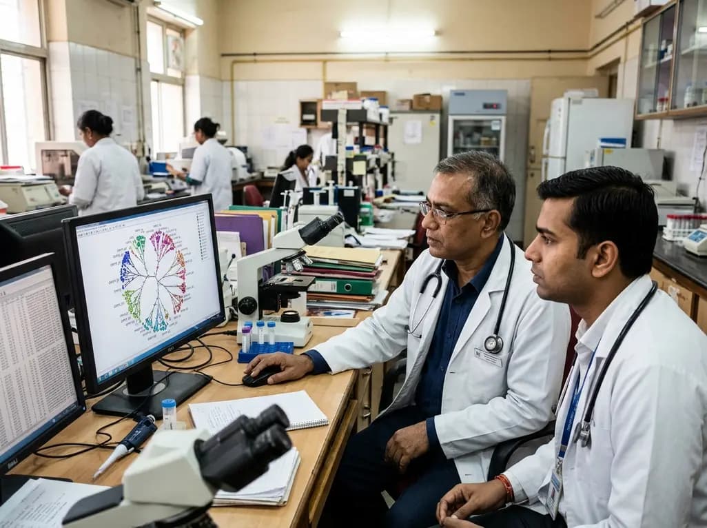
A cross-sectional molecular epidemiological study evaluates HIV-1 transmission clusters and pretreatment drug resistance among treatment-naïve men who have sex with men. Findings highlight prominent CRF07_BC transmission and significant assortative mixing, informing targeted public health and antiretroviral strategies.
Today

A new study evaluates a multilingual digital health platform combining rule-based cervical cancer risk assessment, retrieval-augmented generation AI, and geospatial navigation. The tool dramatically reduces clinical hallucinations while providing actionable health guidance for underserved populations.
Today
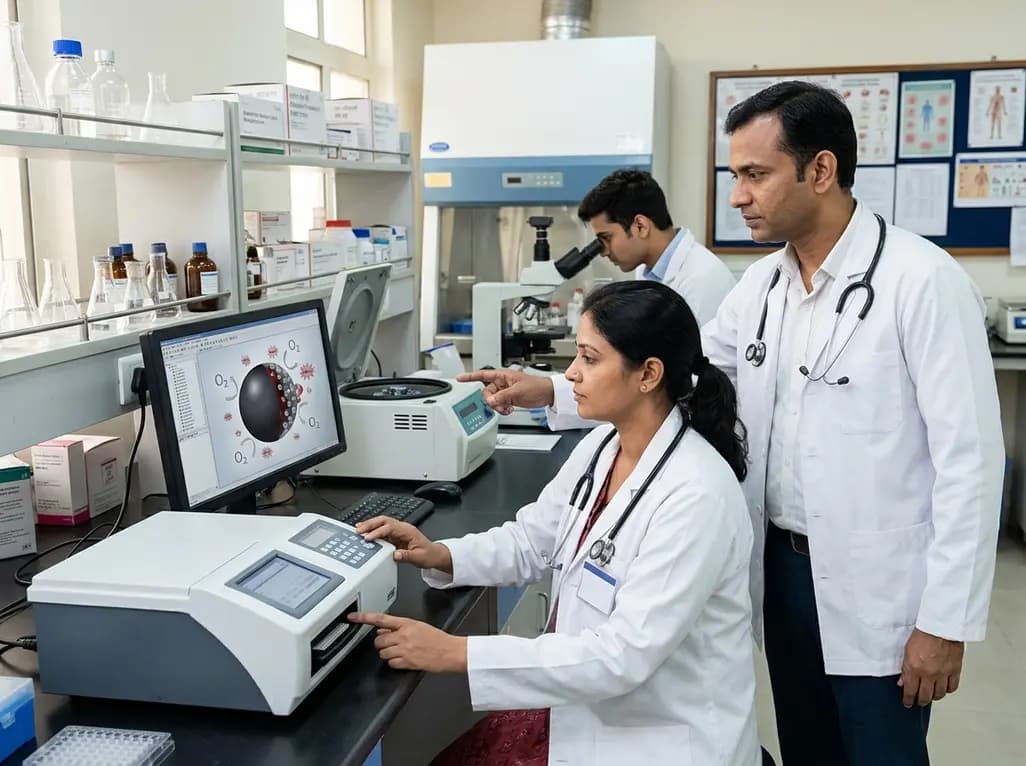
A novel MOF-based platform encapsulates hemoglobin with catalytic platinum and ceria nanozymes to deliver oxygen while scavenging toxic reactive oxygen species. This study highlights key design principles for managing hypoxia and reperfusion injury while resolving crucial stability challenges.
Today
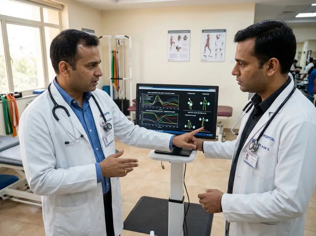
A vector coding study compares elite and sub-elite weightlifters during maximal snatch attempts, revealing that performance differences stem from phase-dependent knee-ankle coordination during transition and turnover phases rather than overall trajectory patterns.
Today

A pilot feasibility study demonstrates that a newly developed mobile app for bolus insulin dose adjustment significantly improves dosing confidence, treatment adherence, and hypoglycemia prevention for patients with type 1 diabetes while reducing provider workload in resource-limited clinical settings.
Today
