.webp&w=384&q=75)

Loading, please wait...
Loading, please wait...

Evaluating drug-resistant focal epilepsy requires an intricate understanding of how electrophysiological signals manifest as observable clinical behaviors. Particularly, prefrontal seizures present unique diagnostic dilemmas for neurologists and epileptologists worldwide. The human prefrontal cortex possesses extensive reciprocal connections with subcortical structures and adjacent cortical areas. Consequently, epileptic activity arising from these regions propagates with extreme rapidity. This rapid spread often obscures the true epileptogenic zone during routine video-EEG monitoring. Clinicians frequently encounter bizarre hypermotor behaviors, autonomic changes, or abrupt loss of contact without distinct lateralizing cues. Traditional linear models often fail to capture these multidimensional electroclinical correlations. Fortunately, modern computational neuroscience provides sophisticated frameworks to decipher these intricate patterns. Applying graph theory to stereoelectroencephalographic data allows clinicians to parse complex semiological phenomena quantitatively and trace their underlying neuroanatomical origins with enhanced clarity.
Historically, frontal lobe epilepsy has remained among the most difficult presentations to localize accurately prior to surgical intervention. Prefrontal seizures exhibit broad behavioral variability that can easily mimic non-epileptic events or temporal lobe episodes. Moreover, rapid interhemispheric spread through the corpus callosum and anterior commissure frequently clouds scalp electroencephalography. Therefore, patients often undergo stereoelectroencephalography, commonly termed SEEG, to directly map the epileptogenic network. Even with intracerebral electrodes, determining which electrophysiological discharge produces a specific semiological feature remains challenging. Traditional visual inspections rely heavily on subjective expert interpretations. In contrast, advanced network modeling provides an objective mathematical framework to examine these interactions. By treating clinical signs as observable nodes, investigators can quantify the statistical interdependence of discrete behaviors. Thus, network science bridges the longstanding gap between bedside semiology and invasive intracranial neurophysiology.
Graph theory conceptualizes complex biological systems as sets of nodes connected by functional edges. In a pivotal proof-of-concept study, researchers analyzed stereoelectroencephalographic data from forty-two patients suffering from pharmacoresistant focal epilepsy involving the prefrontal cortex. Expert neurophysiologists meticulously scored semiological events and simultaneous intracerebral electrical activities. Subsequently, the investigators constructed two distinct computational graphs: a semiological network and a hybrid electroclinical network. Centrality metrics, including strength and betweenness centrality, identified the most influential components within these interconnected systems. Highly central nodes exert disproportionate control over information flow throughout the neural network. Consequently, identifying these hub features clarifies which clinical signs reliably indicate core seizure dynamics. This quantitative approach fundamentally transforms descriptive semiology into an objective, data-driven diagnostic asset for comprehensive presurgical evaluations.
The semiological network analysis revealed that impairment of consciousness serves as the single most central clinical feature. During prefrontal seizures, patients frequently display sudden alterations in awareness and responsiveness. Although clinicians historically focused on striking motor stereotypies, this mathematical modeling highlights awareness disruption as the primary structural hub tying other signs together. Furthermore, impaired contact consistently correlated with the propagation of discharges across widespread frontoparietal networks. When seizure activity disrupts these extensive frontoparietal association cortices, conscious processing rapidly degrades. Therefore, recognizing loss of consciousness as a primary network hub assists clinicians in distinguishing prefrontal onset from localized motor strip discharges. Additionally, tracking the precise latency from electrical onset to awareness loss provides indispensable information regarding network recruitment speed.
In the hybrid network analysis, intracranial electrophysiological activities were coupled directly with clinical semiology to evaluate structural-functional hubs. Notably, the anterior cingulate area emerged as the most central brain activity feature within this integrated model. This anatomically defined hub incorporated Brodmann area 32 and the rostral division of Brodmann area 24. The anterior cingulate cortex maintains extensive structural links to the limbic system, prefrontal cognitive circuits, and motor execution pathways. As a result, epileptogenic discharges within this region rapidly synchronize motor and autonomic manifestations while altering conscious awareness. Furthermore, its elevated centrality suggests that anterior cingulate involvement represents an essential gateway for seizure spread across frontal subdomains. Identifying this structure as an electroclinical linchpin enables clinicians to refine invasive electrode implantation strategies and target curative resections more accurately.
These network-derived findings offer immediate practical value for tertiary epilepsy care centers managing surgical candidates. Surgical success in frontal lobe epilepsy historically lags behind temporal resections, largely due to incomplete network delineation. By identifying primary electroclinical hubs, surgical teams can tailor resection margins or laser interstitial thermal therapy to disrupt core propagation pathways. Moreover, symptom network models provide a foundational architecture for integrating artificial intelligence into automated video-EEG analysis. Machine learning algorithms can automatically detect, quantify, and map semiological sequences into validated network graphs in real time. In addition, this framework can readily expand to larger patient cohorts and other challenging focal epilepsies, such as insular or parietal variants. Ultimately, quantitative network science elevates clinical neurophysiology, providing clinicians with rigorous, objective tools to optimize long-term seizure freedom.
Symptom network analysis provides a mathematical framework that links distinct clinical manifestations directly with intracerebral electrophysiological activity. Consequently, clinicians can identify central driver nodes rather than evaluating symptoms in isolation. This approach refines presurgical mapping, enhances diagnostic precision, and clarifies rapid propagation pathways in complex focal drug-resistant epilepsies.
Impairment of consciousness frequently dominates because prefrontal ictal discharges rapidly recruit bilateral corticocortical networks and frontoparietal awareness circuits. Furthermore, the anterior cingulate cortex actively synchronizes wide neural territories, disrupting conscious processing early. Therefore, even focal prefrontal discharges manifest prominently as acute awareness impairment, masking more subtle localized semiological indicators.
Stereoelectroencephalography allows precise, three-dimensional recording of deep sulcal and mesial frontal structures that conventional surface EEG misses. Consequently, it captures high-frequency ictal discharges from critical hubs like the anterior cingulate cortex. This direct intracranial recording clarifies electroclinical concordance, guiding targeted surgical resections or neuromodulatory interventions with superior clinical outcomes.
Disclaimer: This content is for informational and educational purposes only. It is not intended to substitute for professional medical advice, diagnosis, or treatment. Always seek the advice of your physician or another qualified healthcare provider with any questions you may have regarding a medical condition or treatment options. Never disregard professional medical advice or delay in seeking it because of something you have read. While we strive to ensure the information provided is accurate and up to date, medical knowledge evolves rapidly, and clinical guidelines may change. Any reliance on the information provided is solely at your own risk. Refer to the latest local and national guidelines for clinical practice.
References
Gauld C et al. Symptom network analysis of prefrontal seizures. Epilepsia. 2025 Jul. doi: 10.1111/epi.18372. PMID: 40105434.
Bonini F et al. Frontal lobe seizures: from clinical semiology to localization. Epilepsia. 2014 Feb;55(2):264-77. doi: 10.1111/epi.12500.
Bartolomei F et al. Defining epileptogenic networks: Contribution of SEEG and mathematical modeling. Epilepsia. 2017 Jul;58(7):1134-1147. doi: 10.1111/epi.13791.

Read summarized clinical updates, watch expert medical content, and earn CME certifications right from your smartphone.
Open your smartphone camera to scan and install.





Prefrontal seizures present complex diagnostic challenges due to rapid propagation. A groundbreaking study in Epilepsia utilizes symptom and hybrid network analysis with stereoelectroencephalography to identify key semiological and anatomical hubs, highlighting critical clinical targets for epilepsy surgery.
Today
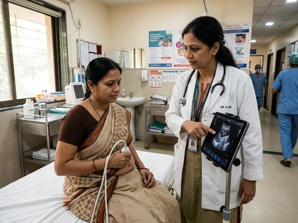
Self-performed ultrasound empowers patients to capture diagnostic-quality scans for remote clinical review. This review evaluates its accuracy, technological challenges, and integration hurdles.
Today
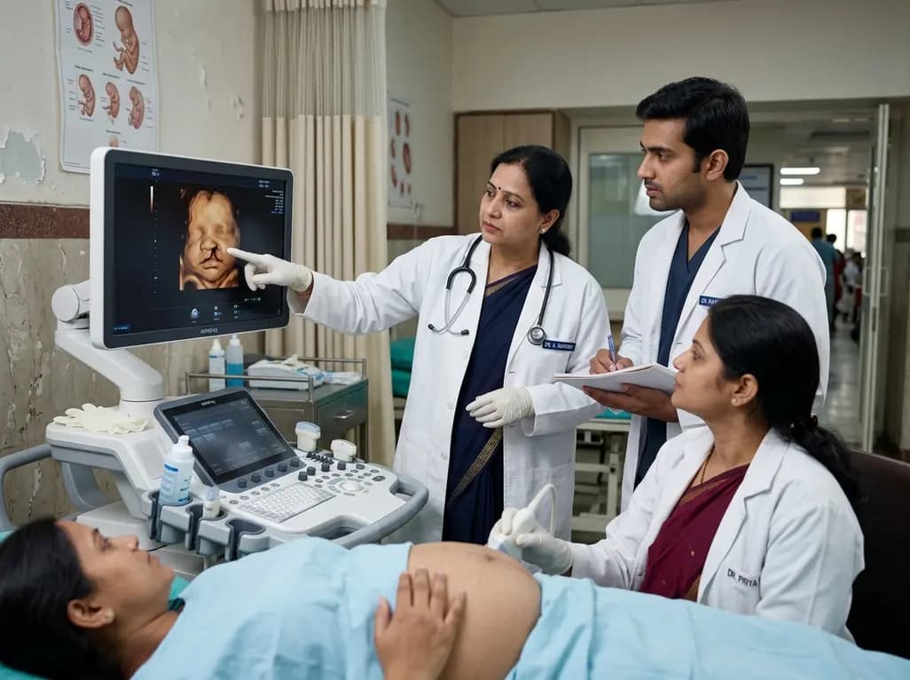
A 5-year retrospective study evaluates prenatal ultrasound detection of fetal cleft lip and palate. While overall detection reached 87%, accuracy varied from 99.3% in combined clefts to 50% in isolated cases. Combined defects showed elevated risks of concurrent structural anomalies and chromosomal abnormalities.
Today

An updated systematic review and meta-analysis of 7,875 patients reveals that routine microaxial left ventricular support during nonemergent high-risk PCI does not reduce mortality or ischemic complications, while significantly increasing major bleeding events.
Yesterday
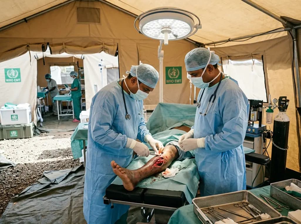
Following Cyclone Chido, a WHO EMT-2 field hospital performed 268 surgeries in Mayotte. While acute trauma is expected initially, 98% of cases were septic wounds of the extremities requiring secondary intention healing, revealing crucial operational insights for disaster surgical preparedness.
Today

The SED-SEEN-SEEP consensus outlines an Integrated Care Pathway for preclinical type 1 diabetes, structuring target screening, autoantibody confirmation, metabolic staging, and proactive follow-up to prevent diabetic ketoacidosis and facilitate early disease-modifying intervention.
Yesterday
