

Loading, please wait...
Loading, please wait...

Managing sylvian fissure arteriovenous malformations represents one of the most demanding tasks in vascular neurosurgery. These complex lesions lie adjacent to essential speech, motor, and sensory cortices within the lateral sulcus of the brain. Because sylvian fissure arteriovenous malformations exhibit distinct angioarchitectural features and fragile hemodynamic balances, clinical teams often struggle to determine the ideal interventional strategy. Recently, an extensive PRISMA-compliant systematic review evaluated the contemporary evidence for microsurgery, stereotactic radiosurgery, and endovascular embolization across global centers. Consequently, clinicians now have clearer comparative insights into therapeutic efficacy, angiographic obliteration benchmarks, and procedural safety profiles.
The sylvian fissure separates the frontal and parietal lobes from the temporal lobe, creating intricate anatomical corridors. Historically, surgical literature categorized sylvian fissure arteriovenous malformations under inconsistent and vague anatomical labels, greatly complicating direct evidence synthesis. Furthermore, these malformations derive their primary arterial supply directly from branches of the middle cerebral artery. Therefore, surgical dissection requires exceptional microsurgical skill to preserve en passage vessels supplying adjacent eloquent brain parenchyma. Additionally, venous drainage configurations vary significantly between superficial middle cerebral veins and deep basal cerebral pathways. When deep venous outflow predominates, surgical complexity and ischemic hazards increase markedly. For instance, deep sylvian lesions frequently demonstrate higher Spetzler-Martin grades and elevated rupture propensities during follow-up. Recent evidence highlights that earlier studies frequently aggregated disparate vascular cohorts, obscuring true anatomical behavior. Consequently, neurosurgeons emphasize strict anatomical categorization and detailed angiographic appraisal before selecting any invasive intervention.
Microsurgical resection represents the established curative standard for accessible vascular malformations located within the sylvian cisterns. Systematic evidence reveals that dedicated surgical cohorts achieve exceptional angiographic obliteration success. Specifically, reviewed cohorts reported complete angiographic resection in 8 out of 8, 40 out of 41, and 94 out of 94 patients across specialized operative series. Consequently, microsurgery provides immediate curative protection against devastating future intracranial rupture. However, extensive dissection within eloquent perisylvian territories carries measurable neurological risks. Transient postoperative deficits occurred in 4 of 8, 4 of 28, 9 of 94, and 18 of 41 surgical patients. Additionally, postoperative hematoma formation developed in two individuals in the largest evaluated series. Most neurological deficits resolved during follow-up; nevertheless, careful subarachnoid dissection remains paramount to avoid permanent disability. Neurosurgeons frequently utilize intraoperative neuromonitoring and awake mapping to safeguard essential language pathways. Thus, microsurgery delivers reliable cure when multidisciplinary teams carefully select accessible lesions.
Stereotactic radiosurgery provides an effective alternative for compact, unruptured lesions situated in surgically prohibitive eloquent brain parenchyma. However, radiosurgery does not achieve immediate lesion closure, introducing a distinct latency phase spanning several years. In a dedicated radiosurgical cohort of 87 patients, actuarial obliteration reached 35 percent at three years. Furthermore, obliteration rates increased substantially to 60 percent at five years and 76 percent at ten years. Meanwhile, patients remain continuously exposed to intracranial hemorrhage risks while the abnormal vessel walls gradually thrombose. The systematic review identified a cumulative post-radiosurgery bleeding rate of 5 percent across ten years of follow-up. Therefore, the annual hemorrhage risk averages approximately 1 percent during the latency interval. In contrast to open surgery, stereotactic radiosurgery avoids acute mechanical brain retraction and tissue trauma. Consequently, clinicians must counsel patients thoroughly regarding the prolonged radiological surveillance required after radiation delivery.
Endovascular techniques have advanced significantly through refined flow-directed microcatheters and modern liquid embolic agents. Nevertheless, the systematic review identified no dedicated clinical series demonstrating curative endovascular therapy for true sylvian lesions. Instead, existing reports described broader middle cerebral artery cohorts without isolated sylvian-specific angiographic cure. Therefore, endovascular embolization does not represent a stand-alone curative modality for these complex malformations. In clinical practice, neurointerventionists employ embolization primarily as a targeted preoperative or preradiosurgical adjunct. Specifically, targeted embolization can seal high-flow fistulas, reduce overall nidus volume, and occlude associated intranidal aneurysms. Consequently, partial embolization facilitates safer subsequent microsurgical resection and lowers intraoperative hemorrhage. However, incomplete embolization introduces potential risks, including catheterization complications and altered hemodynamic stress. As a result, endovascular interventions require careful multidisciplinary planning within comprehensive neurovascular boards to prevent avoidable procedural complications.
Formulating an optimal treatment plan for sylvian malformations demands a personalized, multidisciplinary approach. Clinicians cannot infer comparative superiority among modalities because published cohorts involve fundamentally different baseline patient risks and anatomical locations. For example, surgical series generally select accessible superficial nidi, whereas radiosurgical cohorts encompass deeper, higher-risk malformations. Moreover, healthcare centers in diverse regional settings face specific resource variations, including differing access to dedicated radiosurgery facilities. In specialized neurosurgical centers, hybrid operating suites provide excellent platforms for integrated intraoperative angiography and resection. Additionally, clinicians must carefully weigh patient age, hemorrhagic presentation, and functional nidus architecture before recommending intervention. For ruptured, surgically accessible malformations, open microsurgery offers prompt hematoma evacuation, decompression, and reliable cure. Conversely, unruptured deep lesions often favor cautious radiosurgical management or active clinical observation. Ultimately, collaborative case assessment ensures that patients receive the safest evidence-based intervention suited to their specific clinical circumstances.
The systematic analysis emphasizes urgent needs for standardized reporting and classification within future neurovascular research. Crucially, past studies frequently combined sylvian, insular, and opercular malformations under vague anatomical categories. Consequently, clinicians encounter significant difficulty isolating lesion-specific outcomes from published literature. To address these deficiencies, future investigations must establish uniform anatomical definitions based on high-resolution digital subtraction angiography. Furthermore, prospective studies should report standardized denominators, uniform follow-up intervals, and validated functional scales. In addition, prospective multicenter registries will clarify whether partial embolization before radiosurgery truly enhances long-term vascular occlusion. As modern hemodynamic imaging methods evolve, quantitative blood-flow profiling will refine patient stratification and risk assessment. Therefore, international neurovascular societies must champion collaborative prospective registries across clinical academic centers. Through rigorous methodology and standardized data collection, clinicians can optimize management strategies for these challenging cerebrovascular anomalies.
Sylvian fissure arteriovenous malformations are vascular anomalies nestled within the lateral sulcus, directly engaging branches of the middle cerebral artery. They lie adjacent to critical motor, sensory, and language cortices. Their complex configuration requires precise anatomical differentiation from deep insular or superficial lobar lesions to guide safe intervention.
Stereotactic radiosurgery achieves gradual lesion obliteration, reaching approximately 35 percent at three years, 60 percent at five years, and 76 percent at ten years. However, patients carry an annual post-treatment hemorrhage risk of roughly 1 percent during this latency period before complete angiographic occlusion occurs.
Endovascular embolization currently acts primarily as an adjunctive treatment rather than a curative monotherapy for true sylvian malformations. Neurointerventionalists utilize embolization preoperatively to diminish high-flow feeders, eliminate associated aneurysms, and reduce nidus volume, thereby enhancing the safety and efficacy of subsequent microsurgical excision or radiosurgery.
Disclaimer: This content is for informational and educational purposes only and does not constitute medical advice. Healthcare professionals should integrate these insights with clinical judgment and institutional protocols. Patients must consult qualified specialists before making medical decisions. Neither authors nor publishers assume liability for actions taken based on this content. Refer to the latest local and national guidelines for clinical practice.
References
Konovalov AN et al. Microsurgery, stereotactic radiosurgery and endovascular treatment of sylvian fissure arteriovenous malformations: a systematic review. Neurosurg Rev. 2026 Nov 14. doi: 10.1007/s10143-026-04579-1. PMID: 43026378.
Tarokhian A, Sabahi M, Dmytriw AA, Arjipour M. Sylvian fissure arteriovenous malformations: case series and systematic review of the literature. Neuroradiol J. 2021;34(6):578-587.
Bowden G, Kano H, Tonetti D, Niranjan A, Flickinger J, Lunsford LD. Stereotactic radiosurgery for sylvian fissure arteriovenous malformations with emphasis on hemorrhage risks and seizure outcomes. J Neurosurg. 2014;121(5):1070-1077.

Read summarized clinical updates, watch expert medical content, and earn CME certifications right from your smartphone.
Open your smartphone camera to scan and install.





A 2026 systematic review evaluates microsurgery, stereotactic radiosurgery, and endovascular therapy for sylvian fissure arteriovenous malformations, highlighting surgical resection efficacy, radiosurgical obliteration latency, and the adjunctive role of embolization in preserving neurological function.
Today

The 2025 Australian meningococcal surveillance report shows a 13% decline in invasive meningococcal disease notifications to 118 cases. Genogroup B accounted for 82.2% of typed infections, predominantly impacting infants and young adults, highlighting critical vaccination and rapid diagnostic imperatives.
Today
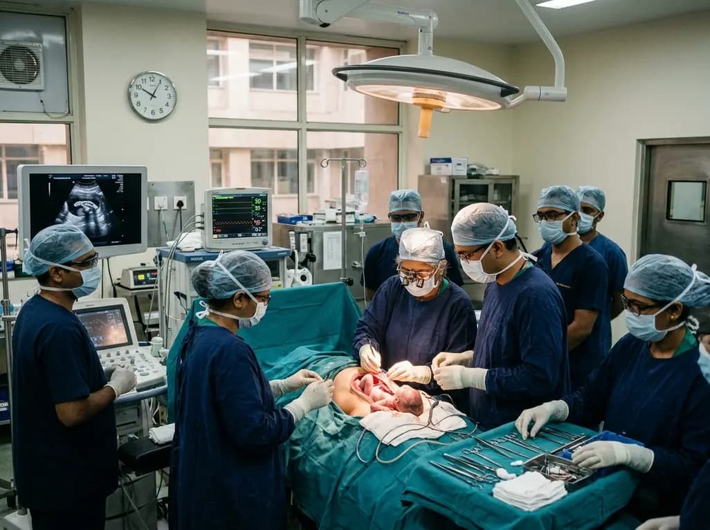
Doctors at AIIMS New Delhi have achieved a historic milestone by performing India's first open prenatal myelomeningocele repair on a 25-week unborn fetus. This landmark multidisciplinary procedure provides vital neuroprotective benefits, lowers hydrocephalus risks, and transforms maternal-fetal medicine nationwide.
Today

The ACE-PAS registry evaluated 21,612 patients fitted with the ASSURE wearable cardioverter defibrillator. The study demonstrated a 100% ventricular arrhythmia conversion rate, an inappropriate shock incidence of 0.0065 per patient-month, and high daily compliance of 23.1 hours, confirming real-world safety.
Today

A clinical overview of resveratrol supplementation in mitigating exercise-induced muscle damage. This narrative review explores physiological mechanisms, reductions in soreness markers like creatine kinase, dosage strategies, and bioavailability enhancements for sports medicine and nutrition practitioners.
Today

Childhood insulinoma is a rare cause of recurrent hyperinsulinemic hypoglycemia that often eludes early diagnosis. Advanced GLP-1 receptor-targeted PET/CT imaging using 68Ga-exendin-4 complements anatomic MRI to localize occult pancreatic lesions, facilitating focused, curative surgical enucleation.
Today
