.webp&w=384&q=75)

Loading, please wait...
Loading, please wait...
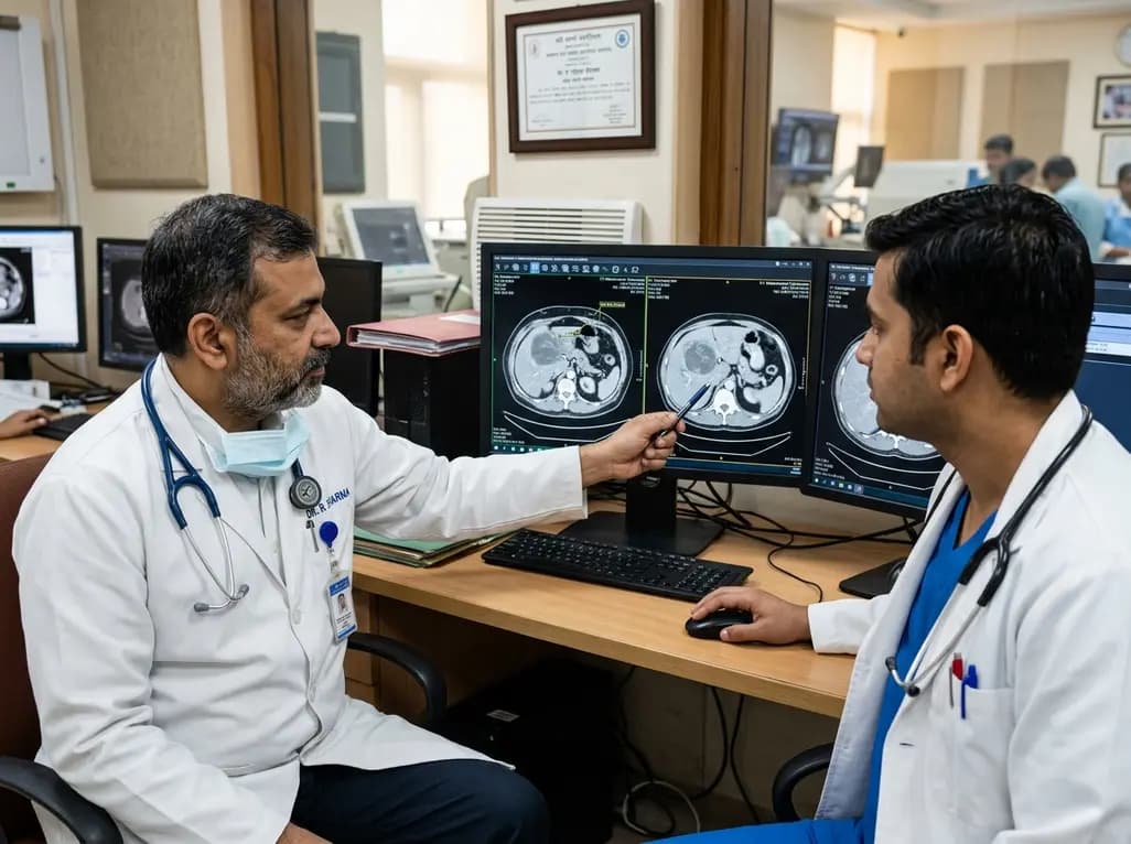
Evaluating reliable prognostic markers remains vital when managing advanced hepatocellular carcinoma during modern immunotherapy regimens. Clinicians historically relied on serum alpha-fetoprotein trends to monitor treatment responses and forecast patient survival. However, emerging radiological research highlights that measuring subcutaneous fat attenuation on follow-up computed tomography offers critical clinical intelligence. Longitudinal body composition changes reflect systemic metabolic responses, host systemic inflammation, and underlying tumor biology during immune checkpoint inhibitor treatment. Consequently, combining tissue attenuation dynamics with biochemical markers yields superior prognostic precision compared to traditional standalone biomarkers. In clinical oncology, identifying early physiological signals during immune checkpoint blockade is essential because standard tumor response endpoints often manifest months after therapy initiation. Hepatocellular carcinoma presents complex clinical challenges due to coexisting chronic liver disease, underlying cirrhosis, and variable systemic inflammation. While alpha-fetoprotein dynamics remain standard in clinical monitoring, serum biomarkers alone do not fully capture host systemic responses. Body composition metrics offer non-invasive insight into host metabolic homeostasis. Evaluating these radiological parameters provides clinicians with holistic predictive models, ensuring timely risk stratification and personalized management strategies for patients suffering from advanced liver disease.
Subcutaneous adipose tissue attenuation serves as an accessible radiomic biomarker derived from routine follow-up computed tomography imaging. Radiologists evaluate these quantitative density alterations on portal venous phase scans at the L3 vertebral level. Standard Hounsfield unit measurements reflect alterations in adipocyte lipid content, vascularity, and extracellular matrix remodeling. Consequently, subtle changes in subcutaneous tissue radiodensity capture early host metabolic shifts long before gross anatomical size changes become clinically visible. Furthermore, because contrast-enhanced CT scans represent standard oncological monitoring tools, evaluating subcutaneous fat attenuation requires no additional imaging procedures or expenses. Therefore, integrating routine body composition assessment into existing imaging protocols provides clinicians with effortless, objective metabolic data. Abdominal adipose depots are sensitive indicators of systemic biological stress, cachexia, and metabolic flux. When patients receive systemic immunotherapy, immune activation and cytokine release influence adipose tissue turnover and lipid distribution. Standard anatomical evaluation based solely on tumor diameters may fail to reflect these early metabolic adaptations. By utilizing computerized density assessments of subcutaneous fat, radiologists quantify microstructural modifications within peripheral tissue depots, offering valuable quantitative imaging biomarkers that enhance clinical decision-making.
While serum alpha-fetoprotein dynamics provide useful early indications of tumor burden changes, they occasionally lack sensitivity or specificity in non-secreting tumors. Recent clinical trial data and real-world cohort validation demonstrate that adding subcutaneous fat attenuation changes significantly enhances overall survival prediction. Patients exhibiting specific longitudinal shifts in subcutaneous tissue radiodensity achieved superior prognostic stratification beyond alpha-fetoprotein measurements alone. Specifically, multi-variable statistical models incorporating both radiological fat attenuation dynamics and serum biochemical responses demonstrated improved C-index values for overall survival. Consequently, evaluating early adipose tissue changes helps oncologists distinguish high-risk patients who might require treatment adjustments from those demonstrating favorable long-term survival trajectories. Combining biochemical and radiological dynamics creates a comprehensive prognostic framework that mitigates individual biomarker limitations. In clinical cohorts, patients who demonstrate persistent tumor marker elevation alongside unfavorable fat attenuation shifts represent a particularly vulnerable subgroup. Conversely, favorable subcutaneous tissue changes may reassure clinicians when alpha-fetoprotein trends yield ambiguous results. Incorporating both modalities into predictive models enhances prognostic accuracy for overall survival, providing clinicians with greater confidence when formulating long-term therapeutic strategies.
The biological link between subcutaneous fat attenuation and patient outcomes highlights the complex interaction between host systemic metabolism and cancer immunotherapy. Immune checkpoint inhibitors rely on robust host immune responses, which remain intimately linked with systemic nutritional status and systemic inflammatory signaling. Progressive changes in subcutaneous fat density frequently correlate with systemic lipid mobilization, systemic inflammation, and cancer-induced metabolic reprogramming. Therefore, monitoring adipose tissue quality provides vital insights into the host metabolic environment during immune activation. Understanding these dynamic physiological changes allows clinicians to better appreciate why specific patients derive prolonged survival benefits while others experience early disease progression. Adipose tissue functions as an active endocrine organ involved in immune modulation. Systemic inflammation triggered by malignant tumors or immune checkpoint inhibition alters lipid metabolism, influencing subcutaneous fat composition and radiodensity. Consequently, tracking longitudinal changes in fat attenuation allows oncologists to monitor host metabolic resilience throughout the therapeutic journey. Identifying early metabolic decompensation enables timely supportive care interventions, such as nutritional optimization or anti-inflammatory support, which may ultimately improve overall treatment tolerance and clinical outcomes.
Translating body composition metrics into routine clinical practice represents a major advancement in personalized liver cancer care. Modern automated radiomic software enables fast, highly reproducible segmentation of abdominal fat depots from standard clinical CT scans. By embedding automated adipose tissue attenuation analysis into routine radiology reporting templates, multidisciplinary care teams gain valuable prognostic insights without increasing clinical burden. Furthermore, future prospective clinical trials will help establish standardized attenuation cutoffs to further refine risk prediction algorithms. As liver cancer management continues evolving toward precision medicine, combining radiomic fat assessments with conventional tumor markers will optimize therapeutic decision-making for patients worldwide. Standardizing quantitative image analysis across radiology departments will facilitate seamless integration into routine clinical workflows. Automated software tools can perform L3 slice selection and tissue segmentation within seconds, eliminating observer variability and saving precious clinical time. Furthermore, combining body composition analysis with circulating biomarkers could refine candidate selection for clinical trials testing second-line therapies. Ultimately, utilizing non-invasive CT-derived markers bridges radiological reporting and oncological care, supporting multidisciplinary teams in delivering tailored, patient-centered therapy.
Subcutaneous fat attenuation reflects subtle underlying alterations in host tissue metabolism, systemic inflammation, and lipid stores during immunotherapy. When combined with traditional alpha-fetoprotein dynamics, tracking changes in fat attenuation on follow-up computed tomography provides incremental prognostic value for predicting overall survival. This objective radiological metric helps clinicians identify patients who are deriving meaningful biological responses from immune checkpoint inhibitor therapies early in their treatment course.
Alpha-fetoprotein reflects tumor-specific biomarker activity, whereas subcutaneous fat attenuation captures host systemic metabolic and inflammatory responses to immunotherapy. Combining these two complementary markers addresses inherent limitations of single-biomarker monitoring, such as non-AFP-producing hepatocellular carcinoma cases. Consequently, integrating biochemical tumor trends with radiological host tissue changes significantly improves overall survival forecasting accuracy, empowering multidisciplinary teams to make more informed treatment decisions throughout patient care.
No extra imaging procedures or specialized contrast injections are required to evaluate subcutaneous fat attenuation. Radiologists derive these density measurements directly from standard, routine follow-up contrast-enhanced computed tomography scans already performed at the L3 vertebral level during routine cancer surveillance. Automated radiomic tools quickly segment adipose tissue, allowing seamless integration into clinical workflows without adding patient discomfort, procedural risks, or medical expenses.
Disclaimer: This content is for informational and educational purposes only and does not constitute medical advice, diagnosis, or treatment. Always consult qualified healthcare professionals regarding medical conditions. Refer to the latest local and national guidelines for clinical practice.
References
Chen BB et al. Early Change in Subcutaneous Fat Attenuation on Follow-up CT Provides Prognostic Information Beyond AFP in Advanced Hepatocellular Carcinoma During Immunotherapy. Acad Radiol. 2026 Aug 11. doi: undefined. PMID: 42580991.

Read summarized clinical updates, watch expert medical content, and earn CME certifications right from your smartphone.
Open your smartphone camera to scan and install.





Early changes in CT-derived subcutaneous fat attenuation provide critical prognostic value beyond AFP dynamics in advanced hepatocellular carcinoma patients receiving immunotherapy, improving risk stratification and overall survival prediction.
last month
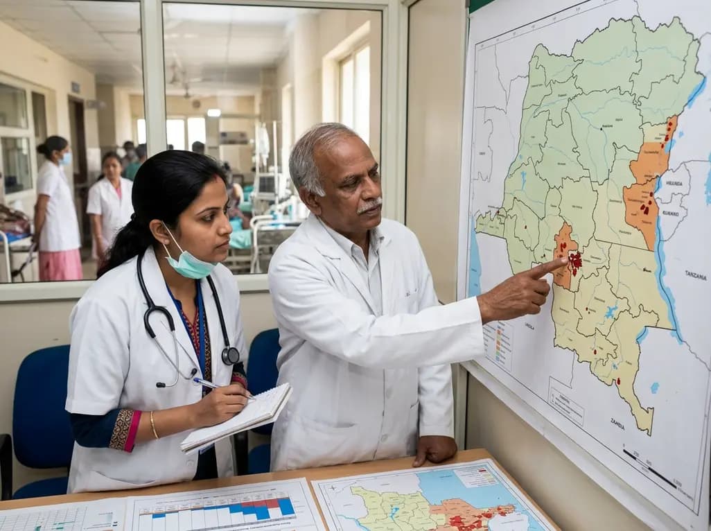
Epidemiologists and health leaders warn that active Ebola virus transmission in the Democratic Republic of Congo remains entrenched within communities. With fewer than ten percent of expected contacts monitored, public health officials urge rapid surveillance scale-up and reinforced healthcare precautions.
Today

A scoping review evaluates 116 studies on AI in women's reproductive health, finding that 83.6% carry high risk of bias. While ensemble models show promise, disparities and lack of validation hinder LMIC deployment.
Today

Differential oxygenation during peripheral TA-NRP can cause undetected coronary hypoxemia due to arch vessel clamping. A novel circuit with continuous aortic root monitoring detects desaturation early, facilitating left ventricular unloading and preventing allograft dysfunction and unnecessary heart discard.
Today

India faces significant opportunities to expand medical device exports to high-growth markets like Mexico, Brazil, and the UK. By addressing its trade deficit, advancing domestic component manufacturing, and aligning with global regulatory standards, the country aims to become an international healthcare hub.
Today

A post hoc analysis of the PARM-T2D study shows pemafibrate exerts baseline-dependent effects on LDL-C and particle size in type 2 diabetes and hypertriglyceridemia. It lowers triglycerides and remnants while favorably remodeling atherogenic LDL particles based on initial lipid control status.
Today
