.webp&w=384&q=75)

Loading, please wait...
Loading, please wait...

Controlled donation after circulatory death has transformed solid organ recovery across the globe. Thoracoabdominal normothermic regional perfusion delivers warm, oxygenated blood to in situ organs following circulatory arrest. However, clinicians increasingly report the risk of differential oxygenation in TA-NRP during peripheral cannulation. In protocols permitting ante-mortem femoral guidewire placement, teams establish peripheral circuits to restore organ perfusion rapidly after death determination. This approach shortens warm ischemic injury and avoids immediate manipulation of delicate cardiac anatomy. Furthermore, peripheral cannulation maintains simultaneous support to abdominal and thoracic organs without requiring an emergency sternotomy. Nevertheless, retrograde arterial inflow from the femoral cannula produces complex physiological competition against native cardiac ejection. Clinicians must appreciate that peripheral perfusion operates fundamentally differently from central aortic cannulation. Retrograde extracorporeal flow must advance upstream through the aorta to perfuse the coronary arteries effectively. Therefore, subtle hemodynamic disturbances can compromise cardiac reanimation and graft preservation. As transplant systems expand their donation protocols, surgical teams must recognize these circulatory dynamics to protect donor organs from occult ischemic damage.
A major challenge in peripheral extracorporeal life support is the occurrence of differential oxygenation, identical to Harlequin syndrome in venoarterial ECMO. This phenomenon develops when native left ventricular ejection collides with retrograde arterial flow from the femoral cannula. If pulmonary function remains impaired or donor ventilation is paused during initial cannulation, the recovering heart pumps deoxygenated blood into the ascending aorta. Concurrently, the extracorporeal circuit infuses fully oxygenated blood retrograde through the descending aorta. Consequently, a hemodynamic interface forms along the aorta depending on native cardiac output and pump flow rates. Moreover, donation after circulatory death protocols strictly mandate surgical ligation or clamping of all supra-aortic arch vessels. Transplant surgeons execute this step to prevent cerebral reperfusion, thereby respecting the dead donor rule. Because arch vessels remain clamped, standard right radial arterial lines and pulse oximeters cannot monitor ascending aortic blood. Thus, clinicians remain completely unaware of coronary arterial blood gas tensions. When the left ventricle pumps hypoxic blood, the coronaries suffer profound hypoxemia while lower body organs receive hyperoxygenated perfusion. Without direct surveillance, teams cannot detect this dangerous mismatch.
Unrecognized coronary desaturation produces severe clinical consequences for donor heart viability and organ utilization rates. The newly restarted myocardium requires high oxygen delivery to regenerate depleted cellular energy stores. However, persistent coronary hypoxemia quickly induces acute ventricular dysfunction, subendocardial ischemia, and metabolic acidosis. When recovery teams observe sluggish cardiac contractility or failing hemodynamics, they often misinterpret these reversible findings as permanent warm ischemic injury. Consequently, procurement teams frequently discard potentially viable organs, denying life-saving grafts to patients on waiting lists. Alternatively, teams may transplant an unsuspected, hypoxemically injured heart, resulting in severe primary graft dysfunction after transplantation. Primary graft dysfunction represents the leading cause of early recipient mortality and prolonged intensive care hospitalization. Furthermore, rescuing a failing graft requires expensive inotropic support or temporary mechanical circulatory devices. Therefore, eliminating undetected coronary desaturation preserves valuable donor organs and enhances recipient survival. Clinical teams must apply objective diagnostic tools to separate true organ failure from reversible perfusion artifacts. Ensuring proper oxygen delivery avoids premature graft discard and optimizes cardiac transplant outcomes.
To overcome this diagnostic blind spot, transplant researchers introduced a practical circuit modification using continuous aortic root oxygenation monitoring. Investigators placed an optical saturation probe directly into the aortic root to track coronary oxygen tensions in real time. Continuous monitoring proved indispensable across two critical clinical scenarios during organ recovery. First, the sensor immediately alerted clinicians to severe differential oxygenation during full peripheral support, allowing instant corrective action. Teams restored coronary oxygenation rapidly by modifying circuit dynamics and donor ventilation. Second, the device detected severe aortic root desaturation following discontinuation of the perfusion circuit. In this delicate weaning phase, conventional upper-body arterial lines remained nonfunctional because arch vessels were already ligated. Consequently, clinicians would have missed critical myocardial hypoxemia without direct root monitoring. Integrating a continuous optical sensor provides immediate, objective visualization of oxygen delivery throughout resuscitation, functional evaluation, and weaning. Furthermore, this inexpensive circuit addition eliminates dangerous diagnostic assumptions during procurement. Adopting real-time root monitoring significantly elevates the safety, precision, and consistency of donor heart assessment.
When continuous monitoring detects differential oxygenation, transplant teams must implement targeted physiological interventions without delay. Left ventricular unloading represents the most effective strategy to restore coronary oxygenation. Elevated afterload from retrograde femoral flow often distends the left ventricle and increases myocardial oxygen demand. Furthermore, the struggling ventricle continues ejecting desaturated blood into the aortic sinuses. By venting the left ventricle or decompressing the pulmonary artery, surgeons reduce chamber distension and suppress native forward ejection. Consequently, oxygenated blood from the extracorporeal circuit reaches the coronary ostia retrogradely. Additionally, anesthesiologists must resume lung ventilation using high inspired oxygen concentrations to oxygenate any residual ventricular output. These coordinated interventions systematically relieve myocardial ischemia and restore normal contractility. These physiological principles carry immense relevance for expanding transplant programs in India and worldwide. While donation after brain death remains standard practice in India, controlled donation after circulatory death protocols are rapidly developing. Indian surgical and critical care societies must establish comprehensive guidelines that incorporate continuous root monitoring during peripheral perfusion. Standardizing these protective measures will expand donor utilization safely and maximize long-term graft survival.
Differential oxygenation in TA-NRP occurs when deoxygenated blood ejected from the recovering heart collides with retrograde oxygenated flow from femoral arterial cannulation. Because surgeons clamp the supra-aortic vessels to prevent cerebral reperfusion, clinicians cannot assess upper-body oxygenation via standard radial arterial lines. Consequently, the left ventricle can pump desaturated blood directly into the aortic root and coronary arteries, depriving myocardial tissue of essential oxygen without detection.
Continuous aortic root oxygenation monitoring places an optical saturation sensor directly near the coronary ostia. Consequently, perfusionists immediately identify when desaturated blood perfuses the myocardial circulation. When clinicians identify hypoxemic coronary perfusion early, they can actively intervene by unloading the left ventricle, adjusting pump flow rates, or optimizing lung ventilation. Therefore, this real-time monitoring prevents ischemic myocardial injury, averts primary graft dysfunction, and avoids the unnecessary discard of donor hearts.
Transplant teams routinely ligate or clamp the brachiocephalic trunk, left carotid artery, and left subclavian artery during thoracoabdominal procurement protocols. This vascular occlusion strictly isolates the cerebral vasculature to respect the dead donor rule. As a direct result, standard right radial artery lines and upper-limb pulse oximeters lose physiological connection with central aortic flow. Hence, monitoring peripheral upper-body vessels fails completely to reflect true ascending aortic blood gases.
Disclaimer: This content is for informational and educational purposes only, and does not substitute professional medical advice, diagnosis, or treatment. It is intended solely for healthcare professionals. Readers should independently verify clinical data and drug dosages. The publisher and authors disclaim liability for any adverse outcomes resulting from the application of this information. Refer to the latest local and national guidelines for clinical practice.
References

Read summarized clinical updates, watch expert medical content, and earn CME certifications right from your smartphone.
Open your smartphone camera to scan and install.





Differential oxygenation during peripheral TA-NRP can cause undetected coronary hypoxemia due to arch vessel clamping. A novel circuit with continuous aortic root monitoring detects desaturation early, facilitating left ventricular unloading and preventing allograft dysfunction and unnecessary heart discard.
Today
A landmark cluster randomized trial demonstrates that community-based mass dog vaccination achieves superior coverage and sustains protective herd immunity against rabies far better than conventional campaign models, offering vital lessons for global and Indian elimination targets.
Today
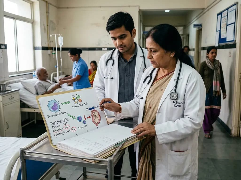
Explore the crucial link between dietary protein and immunocompetence. This review examines how low-protein nutrition shifts metabolic resources away from pathogen defense toward early reproduction, compromising host survival and immunity against severe bacterial infections.
Today

A longitudinal study reveals that patients undergoing staged bilateral hip arthroscopy experience progressive contralateral labral deterioration during surgical intervals. However, structural degradation does not compromise 2-year postoperative clinical outcomes or patient-reported satisfaction scores.
Today

A post hoc analysis of the PARM-T2D study shows pemafibrate exerts baseline-dependent effects on LDL-C and particle size in type 2 diabetes and hypertriglyceridemia. It lowers triglycerides and remnants while favorably remodeling atherogenic LDL particles based on initial lipid control status.
Today
