

Loading, please wait...
Loading, please wait...

Titanium and its alloys serve as the foundation of modern reconstructive procedures in joint arthroplasty and dental rehabilitation. These metallic substrates provide outstanding mechanical strength, chemical stability, and structural endurance under substantial physiological loads. However, untreated titanium remains inherently bioinert, which occasionally causes delayed biological anchoring or fibrous encapsulation in poor bone environments. To overcome this limitation, surface modification techniques seek to convert the passive metal into a dynamic osteogenic surface. Recent material science advances highlight strontium zinc phosphate coatings as an effective chemical conversion strategy. By incorporating therapeutic metal ions into crystalline phosphate lattices, researchers can actively stimulate surrounding skeletal stem cells. Furthermore, tuning the morphological pattern of these layers directly governs cell attachment and local mineral deposition.
Phosphate chemical conversion represents a cost-effective and scalable technique for depositing uniform functional layers onto complex implant geometries. Through controlled chemical bath reactions, surgeons and engineers can fabricate synthetic strontium zinc phosphate coatings with distinct crystalline morphologies. Recent characterization trials demonstrate that adjusting processing parameters alters surface topography without compromising core crystallographic phases. Specifically, energy-dispersive spectroscopy and X-ray diffraction verify identical chemical stoichiometry across varying coating architectures. Consequently, researchers can isolate the exact biological effects of surface topography without chemical confounding. When investigators compare lamellar flake-like grains against dense block crystals, the microstructural divergence produces noticeable physiological differences. The lamellar pattern provides higher specific surface area and finer submicron niches, which directly assist protein adsorption. Therefore, optimizing interfacial architecture through controlled chemical conversion provides a practical pathway toward fabricating next-generation bioactive surgical implants.
Surface wettability and chemical dissolution govern initial biological responses when an implant contacts blood and interstitial fluid. Interestingly, the lamellar microstructure delivers significantly superior hydrophilicity compared to conventional block crystal geometries. High surface wettability lowers contact angles, thereby promoting instantaneous capillary drawing of fluid and rapid fibronectin adsorption. In addition, the open architecture of lamellar grains accelerates the steady release of bioactive strontium and zinc ions into the peri-implant microenvironment. Strontium effectively replicates calcium signaling pathways, which stimulates osteoblast proliferation while simultaneously suppressing osteoclastic resorption. Meanwhile, sustained zinc ion release activates cellular alkaline phosphatase, enhances matrix mineralization, and delivers natural antimicrobial defense against colonizing pathogens. Consequently, the combination of enhanced surface energy and balanced ionic dissolution establishes a favorable regenerative milieu during initial post-surgical healing phases.
Bone marrow mesenchymal stem cells provide the primary cellular reservoir for de novo bone formation around metallic hardware. In vitro biological assays confirm that chemically converted surfaces display excellent cytocompatibility without inducing cellular toxicity. Furthermore, stem cells seeded onto modified substrates exhibit accelerated pseudopodial extension, robust focal contact development, and elevated cytoskeletal organization compared with smooth titanium. While all coated groups perform better than bare metal, the lamellar topography stimulates significantly greater cellular expansion over prolonged culture intervals. The nanoscale edges and interconnected pores of the lamellar network provide optimal mechanical anchorage for migrating progenitor cells. In contrast, smooth titanium surfaces fail to supply adequate spatial cues, leading to delayed cellular recruitment. Therefore, lamellar surfaces establish faster cellular confluence, securing the critical cellular mass necessary for downstream osteogenic differentiation.
The transition from stem cell proliferation to mature matrix mineralization dictates definitive structural stability in clinical implantology. Gene expression analyses reveal that lamellar architectures upregulate essential osteogenic markers, including runt-related transcription factor 2, osteocalcin, and collagen type I. As a result, developing cells deposit extensive calcium-phosphate nodules directly along the modified metallic interface. Moreover, the controlled release of strontium ions activates canonical Wnt signaling pathways, which rapidly channels undifferentiated stem cells toward an osteogenic fate. Early mineralized matrix deposition minimizes micro-motion at the bone-implant interface, which directly prevents fibrous tissue interposition. Because early implant loosening remains a prominent cause of early prosthetic failure, this accelerated osteogenesis offers profound clinical benefits. Thus, adjusting surface grains to a lamellar morphology maximizes bone-to-implant contact during early recuperation.
Orthopedic reconstructive surgeons and implant dentists constantly manage patients with compromised skeletal density, including elderly individuals and patients with severe osteoporosis. In these vulnerable populations, standard mechanical press-fit techniques often fail to achieve sufficient secondary biological stability. By implementing bioactive conversion coatings, clinicians can potentially shorten non-weight-bearing periods and accelerate dental prosthetic loading protocols. In spinal arthrodesis and total joint reconstruction, rapid bone bridging reduces revision burdens and enhances long-term construct survival. Similarly, in dental implantology, augmenting primary stability with accelerated osteoinduction prevents peri-implant crestal bone loss. Because chemical conversion processes operate reliably on complex porous geometries, manufacturers can easily apply this method to 3D-printed orthopedic cages and threaded dental fixtures. Consequently, translating these microstructural discoveries into clinical implant manufacturing could significantly improve patient outcomes across reconstructive surgical disciplines.
Strontium zinc phosphate coatings transform bioinert titanium into a biologically active surface. They improve hydrophilicity, facilitate rapid protein adsorption, and release therapeutic strontium and zinc ions into peri-implant tissues. This dual-ion activity stimulates osteoblast growth, suppresses bone resorption, and accelerates secure osseointegration, thereby lowering the risk of early implant loosening in orthopedic and dental procedures.
The lamellar microstructure provides a superior surface area, increased microscopic roughness, and better wettability compared to dense block crystals. These physical attributes accelerate favorable fluid infiltration and foster earlier bone marrow stem cell attachment. Furthermore, the lamellar configuration optimizes sustained ion release kinetics, promoting faster osteogenic differentiation and rapid early bone matrix deposition.
Strontium mimics calcium signaling, stimulating osteoblast maturation while dampening osteoclastic bone breakdown. Concurrently, zinc serves as an essential enzymatic cofactor that upregulates alkaline phosphatase activity, drives protein synthesis, and provides local antibacterial defense. Together, these complementary actions create a protected, highly osteogenic biological niche that accelerates mineralization and solid structural integration.
Disclaimer: This content is for informational and educational purposes only. It is not intended to be a substitute for professional medical advice, diagnosis, or treatment. Always seek the advice of your physician or other qualified health provider with any questions you may have regarding a medical condition. Never disregard professional medical advice or delay in seeking it because of something you have read on this website. The views expressed in this article are those of the author and do not necessarily reflect the official policy or position of any medical board or organization. Refer to the latest local and national guidelines for clinical practice.
References
1. Wang Q et al. Relative assessment of strontium-zinc-phosphate chemical conversion coatings on titanium for augmenting bioactivity: Effect of microstructure. J Biomater Appl. 2026 Nov 11. doi: 10.1177/08853282261507882. PMID: 43013816.
2. Gong ZY, Xiao GY, Lu YP, Zuo KQ. Microstructural evolution of strontium-zinc-phosphate coating on titanium via changing Zn2+ concentration in phosphate solution for enhanced osteogenic activity. Surf Coat Tech. 2022;433:128108.
3. Wang Q, Zhou P, Liu S, Attarilar S, Ma RLW, Zhong Y, et al. Multi-scale surface treatments of titanium implants for rapid osseointegration: a review. Nanomaterials. 2020;10(6):1244.

Read summarized clinical updates, watch expert medical content, and earn CME certifications right from your smartphone.
Open your smartphone camera to scan and install.





Discover how microstructural optimization of strontium zinc phosphate coatings enhances titanium implant bioactivity, accelerating stem cell adhesion and early osseointegration in orthopedic and dental applications.
Today

Targeting bacterial signaling pathways marks a major shift in antimicrobial strategies. By disrupting quorum sensing and biofilm formation, anti-virulence tools suppress foodborne pathogens without driving antimicrobial resistance, protecting vulnerable patients and the broader food ecosystem.
Today
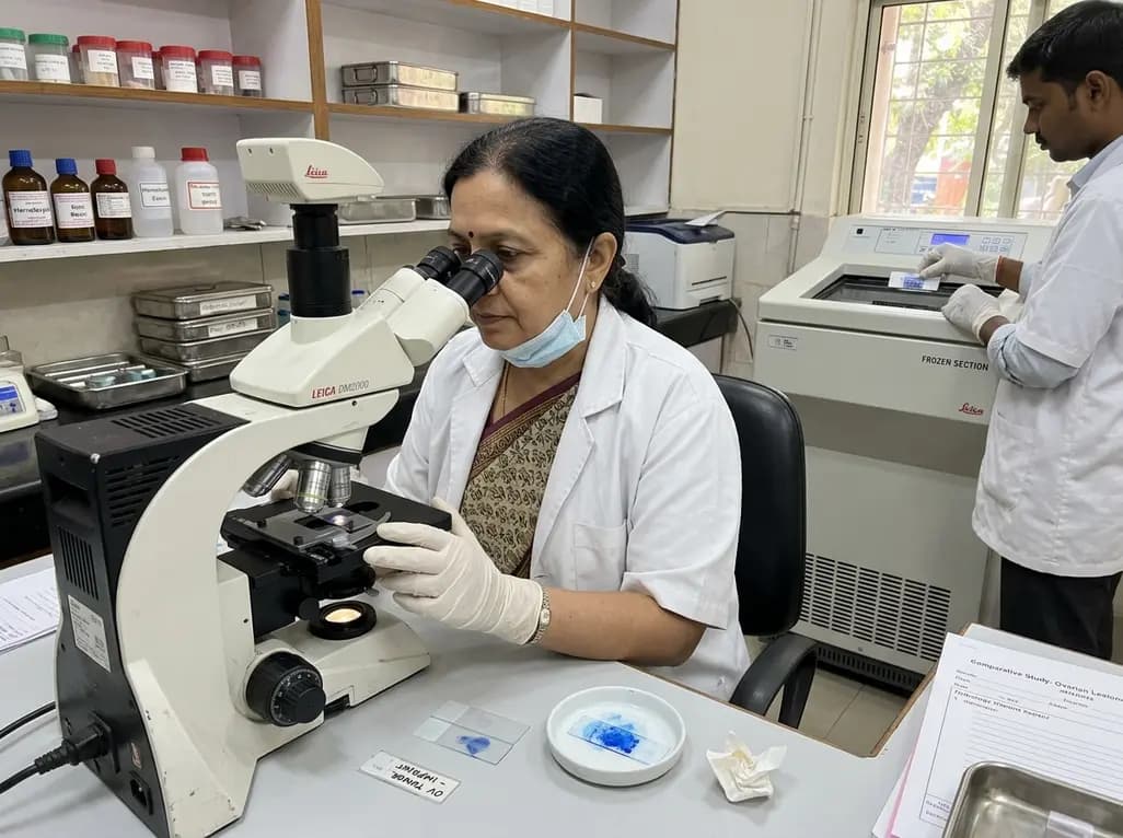
A new comparative study demonstrates that intraoperative imprint cytology achieves 96.6% diagnostic accuracy in ovarian tumours, outperforming frozen section in sensitivity for borderline lesions and offering an accessible, rapid alternative for resource-constrained surgical centres.
Today

Thoracic aortic aneurysm lacks etiologic medical therapy. A new study reveals that targeting specific αV integrins with Cilengitide attenuates TGF-β and mechanotransduction pathways, effectively limiting pro-fibrotic remodeling and aortic bulb dilation in both syndromic and sporadic disease models.
Today

A real-world retrospective study demonstrates that combining intravitreal ranibizumab with focal/grid macular laser significantly improves visual acuity and central macular thickness compared to laser monotherapy in patients with center-involved diabetic macular edema.
Today
