

Loading, please wait...
Loading, please wait...

Cranial radiation serves as an indispensable therapy for adult patients battling high-grade and low-grade gliomas. However, oncologists increasingly recognize that cranial radiation stroke risk represents a severe complication in long-term brain tumor survivors. Radiation therapy effectively arrests neoplastic cellular proliferation, but ionizing energy simultaneously induces extensive microvascular endothelial disruption. Consequently, patients who undergo brain radiotherapy experience an accelerated trajectory toward occlusive cerebrovascular events.
Moreover, underlying cardiovascular comorbidities dramatically amplify this baseline risk profile. In particular, pre-existing hyperlipidemia compounds radiation-induced arterial damage by driving accelerated atherosclerosis, vascular stiffness, and luminal stenosis. Clinical neuro-oncologists frequently encounter challenges when balancing oncologic survival against survivorship toxicity. While modern stereotactic modalities spare uninvolved brain parenchyma, major vascular trunks frequently receive significant radiation doses. Therefore, clinicians must assess how traditional vascular risk factors interact with radiation toxicity over time. Recent cohort analyses indicate that stroke incidence after cranial irradiation is substantially higher than historical general population averages. Recognizing these vulnerable patients enables multidisciplinary teams to implement targeted risk reduction protocols early during survivorship care.
A rigorous retrospective cohort study evaluated 297 adult glioma patients with hyperlipidemia who received cranial radiation therapy between 2005 and 2021. Within this specialized cohort, 65 patients (21.9%) developed incident strokes during post-treatment follow-up. Using sophisticated Cox proportional hazards modeling with death treated as a competing event, researchers established crucial predictive factors for ischemic outcomes. Notably, increasing age at diagnosis independently raised stroke likelihood, reflecting the natural vulnerability of aging cerebral vasculature.
Conversely, statin exposure demonstrated a profoundly protective association against subsequent cerebrovascular events. Specifically, patients who took statin medications experienced an impressive 64% reduction in incident stroke risk compared to non-users. This robust hazard ratio of 0.36 underscores the transformative therapeutic potential of lipid-lowering agents in neuro-oncology cohorts. Furthermore, these findings highlight that metabolic optimization confers substantial survival advantages even in complex cancer populations. Physicians often hesitate to prescribe preventive statins to glioma patients due to concerns regarding polypharmacy or poor prognosis. However, this study proves that timely statin therapy provides significant clinical benefits by safeguarding patients from disabling focal neurological deficits.
To understand these protective clinical observations, one must examine the biological cascade triggered by therapeutic ionizing radiation. Cranial irradiation generates abundant reactive oxygen species that damage endothelial cells lining medium and large intracranial vessels. Consequently, irradiated endothelial cells downregulate endothelial nitric oxide synthase, impairing vasodilation and disrupting regional vascular autoregulation. Furthermore, radiation triggers chronic expression of pro-inflammatory cytokines, including interleukin-6 and transforming growth factor-beta.
This sustained inflammatory milieu promotes monocyte adhesion, intimal proliferation, and medial fibrosis, generating accelerated atheroma formation. When systemic hyperlipidemia coexists, circulating low-density lipoprotein particles infiltrate these vulnerable, permeable endothelial layers at rapid rates. As a result, dense fibrofatty plaques develop within intracranial vessels, predisposing patients to sudden arterial thrombosis or downstream thromboembolism. Statins directly counteract these detrimental pathways through multiple pleiotropic mechanisms. Beyond lowering serum cholesterol levels, statins attenuate endothelial inflammation, stabilize fragile fibrous caps, reduce oxidative stress, and restore nitric oxide bioavailability. Therefore, statin therapy functions as both a lipid modifier and a potent endothelial stabilizer in irradiated cerebral vascular territories.
Modern neuro-oncology care requires proactive vascular risk surveillance alongside active antineoplastic therapies. Because primary brain tumor patients frequently experience extended survival with updated chemoradiation protocols, preventing vascular morbidity is paramount. Historically, oncologists prioritized immediate tumor control while cardiovascular comorbidities received less aggressive management. Nevertheless, clinicians now recognize that unmanaged dyslipidemia and hypertension significantly jeopardize functional independence. Therefore, multidisciplinary teams comprising neuro-oncologists, radiation oncologists, cardiologists, and neurologists should collaborate systematically to optimize metabolic health.
Additionally, managing cranial radiation stroke risk requires vigilant long-term neuroimaging surveillance. Clinicians must distinguish between radiation-induced ischemic stroke, delayed radiation necrosis, and progressive tumor recurrence on surveillance neuroimaging. Advanced magnetic resonance sequences, such as diffusion-weighted imaging and perfusion studies, help delineate acute cytotoxic edema from treatment-related necrosis. Furthermore, implementing comprehensive cardiovascular risk reduction protocols does not compromise primary antineoplastic regimens. In fact, initiating statin therapy in dyslipidemic patients mitigates vessel vulnerability without interfering with alkylating chemotherapies. Thus, comprehensive survivorship programs should integrate regular lipid panels, blood pressure monitoring, and glycemic surveillance into standard post-radiation follow-up visits.
Implementing timely lipid-lowering intervention requires structured clinical workflows throughout the oncology care continuum. Baseline cardiovascular assessments should ideally occur prior to radiation planning or immediately following radiotherapy completion. Physicians should obtain fasting lipid profiles, assess baseline blood pressure, and calculate aggregate cardiovascular risk scores. Furthermore, clinicians must evaluate specific radiotherapy details, including total radiation dose, fractionation schemes, and proximity of target fields to the circle of Willis. High-dose exposure to the internal carotid arteries or middle cerebral vessels warrants particularly aggressive preventative oversight.
When clinicians identify dyslipidemia, they should initiate statin therapy promptly while evaluating drug-drug interactions. For example, neuro-oncologists must carefully check whether chosen statins interact with antiepileptic medications commonly prescribed to glioma patients, such as carbamazepine or phenytoin. Moderate-intensity or high-intensity statins with minimal hepatic cytochrome P450 interactions often provide the optimal balance of safety and efficacy. In addition, healthcare teams must educate patients and caregivers regarding subtle stroke symptoms, emphasizing rapid emergency presentation. Ultimately, incorporating evidence-based statin pharmacotherapy safeguards neurological function, decreases acute hospitalizations, and substantially improves survivorship quality.
Cranial radiation damages vascular endothelial cells, triggering chronic inflammation, intimal thickening, and accelerated atherosclerosis in cerebral vessels. Consequently, irradiated arteries become stiff and stenotic over time. When combined with traditional vascular risk factors like hyperlipidemia, these structural vascular alterations significantly elevate the lifetime incidence of ischemic stroke.
Statins reduce stroke risk by lowering atherogenic low-density lipoproteins and providing critical pleiotropic vascular benefits. Specifically, they attenuate endothelial inflammation, enhance nitric oxide production, decrease oxidative stress, and stabilize vulnerable vascular plaques. These combined biological actions protect radiation-damaged intracranial arteries from progressive occlusion and acute thrombotic events.
Clinicians should screen lipid profiles and cardiovascular risk factors before or immediately after cranial radiation therapy. If hyperlipidemia is diagnosed, initiating a statin promptly is prudent. However, physicians must evaluate potential drug interactions with antiepileptic agents or chemotherapies, selecting statin regimens that safely minimize hepatic cytochrome P450 interference.
Disclaimer: This content is for informational and educational purposes only... Refer to the latest local and national guidelines for clinical practice.
References

Read summarized clinical updates, watch expert medical content, and earn CME certifications right from your smartphone.
Open your smartphone camera to scan and install.





A retrospective cohort study demonstrates that statin use is independently associated with a 64% reduction in stroke risk among glioma patients with hyperlipidemia who received cranial radiation therapy, highlighting the critical role of proactive lipid management and vascular risk mitigation in neuro-oncology care.
Today

Novel glucose-responsive mPEG-PSA nanocarriers deliver insulin aspart on demand, achieving dual postprandial and basal glycemic control without hypoglycemia in diabetic models. This biomaterial breakthrough offers promising clinical implications for physiologic diabetes management.
Today
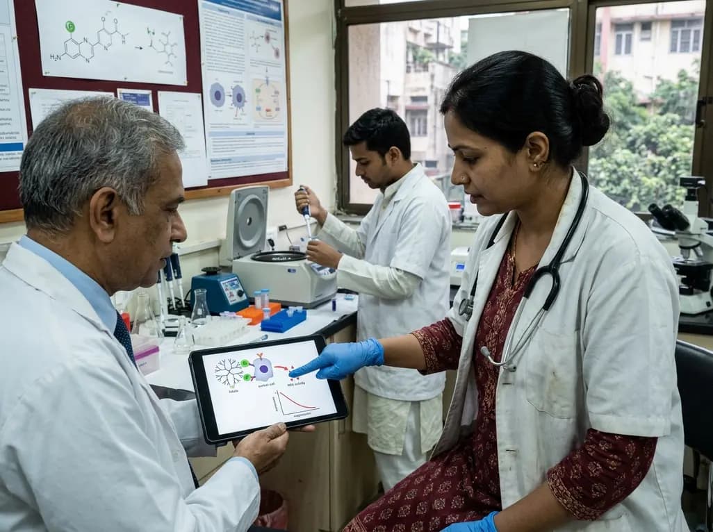
A novel folate-conjugated tecto-dendrimer nanoplatform successfully delivers siRNA to achieve targeted HK2 silencing, disrupting ovarian cancer glycolysis and suppressing lactate output.
Today

A comparative non-contact study reveals that high-power radiofrequency delivery with the QDOT MICRO catheter induces an artifactual contact force rise. Electrophysiologists must understand this power-dependent thermal and electrical phenomenon to avoid inadvertent catheter withdrawal and ineffective ablation.
Today

A novel proof-of-concept study presents a six-degree-of-freedom articulated manikin neck equipped with linear potentiometers. The system provides real-time kinematic motion feedback during cervical immobilisation, delivering sub-degree tracking accuracy to improve pre-hospital trauma training and patient safety.
Today

A retrospective comparative cohort study evaluated genitourinary safety in HFpEF between dapagliflozin and empagliflozin over six months. Rates of symptomatic UTI and asymptomatic bacteriuria showed no statistically significant differences, supporting comparable safety profiles across both SGLT2 inhibitors.
Today
