.webp&w=384&q=75)

Loading, please wait...
Loading, please wait...
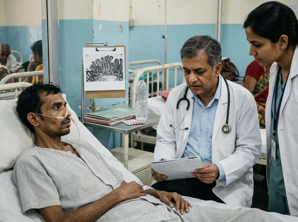
Malnutrition remains a formidable global health challenge, particularly in regions where environmental enteric dysfunction is already prevalent. Chronic starvation leads to a specialized condition known as starvation-induced enteropathy, which involves profound structural and functional changes in the gastrointestinal tract. A recent cross-sectional study conducted in Zambia, led by Besa et al., has shed light on how starvation specifically exacerbates mucosal damage in adult patients. By examining individuals with benign esophageal strictures, researchers could isolate the effects of starvation from other inflammatory factors. This research is critical for healthcare providers in India and similar settings, where malnutrition often complicates the management of surgical and medical conditions. The study highlights that the reduction in epithelial surface area is not merely a consequence of the environment but is significantly driven by the lack of nutrient intake. Consequently, understanding these mechanisms is essential for developing targeted nutritional rehabilitation protocols. Starvation-induced enteropathy represents a severe manifestation of nutrient deprivation that requires a multidisciplinary approach to clinical care.
The hallmark of starvation-induced enteropathy is the physical degradation of the duodenal lining. In the Zambian study, researchers utilized duodenal biopsies and morphometry to quantify these changes. They discovered that patients suffering from starvation had a median epithelial surface area of only 323 μm per 100 μm muscularis mucosae. This was significantly lower than the 448 μm observed in community controls. This reduction in surface area corresponds to villous blunting and a loss of the overall absorptive capacity of the small intestine. Importantly, these structural alterations persisted even after the researchers adjusted for confounding variables like Body Mass Index (BMI), HIV status, and C-reactive protein (CRP) levels. Therefore, the findings suggest that the gut undergoes a form of "autophagy" or atrophy when deprived of intraluminal nutrients. For clinicians, this implies that a severely malnourished patient has a compromised physical barrier and a reduced ability to absorb even if nutrition is reintroduced. Recognizing this structural baseline is vital when planning refeeding strategies to avoid complications such as malabsorption or refeeding syndrome.
Starvation does not only affect the physical structure of the gut; it also creates a state of systemic hormonal chaos. The study revealed significant shifts in pancreatic hormones among cases compared to controls. Specifically, patients with starvation-induced enteropathy exhibited lower median concentrations of C-peptide and insulin. Insulin levels were markedly reduced (639 pg/mL in cases versus 2037 pg/mL in controls), reflecting the body's attempt to conserve energy and utilize fat stores. Conversely, glucagon concentrations were notably higher in the starvation group. This elevated glucagon level facilitates gluconeogenesis and glycogenolysis, ensuring a baseline blood glucose level despite the absence of dietary intake. However, this shift toward a catabolic state can complicate the metabolic response when nutritional support begins. The relative insulin deficiency means that the body is not primed for an immediate influx of glucose. For medical educators and practitioners, these data underscore the importance of monitoring glycemic control and electrolyte balance during the initial stages of nutritional intervention in patients with chronic esophageal obstructions.
One of the most striking findings of the research was the significant reduction in Glucagon-Like Peptide-2 (GLP-2) levels. GLP-2 is a potent trophic hormone responsible for maintaining the integrity of the intestinal mucosa and promoting epithelial cell growth. In the starvation cases, GLP-2 concentrations were essentially undetectable (0 ng/mL), whereas the controls had a median of 2.3 ng/mL. This deficiency likely explains the severe villous blunting and reduced surface area noted in the morphometric analysis. Without the stimulatory effect of GLP-2, the intestinal lining cannot replace cells at a normal rate. Interestingly, the study also found elevated levels of secretin in starved patients. Typically, secretin is released in response to acid in the duodenum to stimulate bicarbonate secretion. Its elevation in a fasting state suggests a complex dysregulation of the enteroendocrine system. These hormonal markers provide a window into the biological mechanisms that sustain starvation-induced enteropathy and suggest that the absence of food-derived signals disrupts the entire gut-brain-endocrine axis, further compromising the patient's physiological resilience.
The findings from this study have direct relevance for clinical practice in India, where esophageal strictures due to caustic ingestion or tuberculosis are not uncommon. When a patient presents with a long-standing esophageal obstruction, the physician must assume that starvation-induced enteropathy is present. This means the patient is not just "thin," but their digestive system is structurally and hormonally altered. Traditional refeeding might fail if the mucosal surface area is too low to handle the nutrient load. Therefore, the use of specialized formulas or a gradual increase in caloric intake may be necessary. Furthermore, the role of gut-specific trophic factors might be an area for future therapeutic intervention. While GLP-2 analogs are currently expensive and used for short-bowel syndrome, understanding their role in starvation highlights the potential for future cost-effective biological therapies. Practitioners should focus on early identification of these hormonal and structural changes to improve the outcomes of esophageal dilation or surgical bypass procedures, ensuring that the patient is metabolically prepared for the recovery phase.
It is important to differentiate starvation-induced enteropathy from environmental enteropathy, which is ubiquitous in many low-income countries. Environmental enteropathy is a chronic inflammatory state of the gut caused by constant exposure to fecal pathogens, leading to similar structural changes like villous blunting. However, the Zambian study demonstrated that starvation imposes an additional layer of damage. The cases in the study already lived in an environment where environmental enteropathy was common, yet their mucosal surface area was significantly worse than the local controls. This proves that the lack of enteral nutrition is a potent, independent driver of gut atrophy. While environmental enteropathy is characterized by inflammation and increased permeability, starvation-induced enteropathy is more closely linked to a lack of trophic signaling and metabolic adaptation. For researchers, this distinction is vital for developing biomarkers that can separate these two conditions. For clinicians, it serves as a reminder that correcting the environmental factors is only half the battle; restoring the internal nutritional and hormonal environment is equally essential for total gut recovery.
Starvation-induced enteropathy refers specifically to the structural and functional changes in the small intestine, such as villous blunting and reduced surface area, caused by a lack of food intake. While general malnutrition affects the whole body, this enteropathy emphasizes the gut's inability to maintain its own lining. This condition makes it harder for the patient to recover because their digestive system cannot effectively process nutrients even when they are eventually provided.
Glucagon-Like Peptide-2 (GLP-2) is a critical hormone that stimulates the growth and repair of the intestinal lining. In patients with starvation, the lack of food in the gut prevents the release of GLP-2. This absence leads to severe mucosal atrophy, as the body can no longer maintain the healthy villi needed for absorption. Restoring GLP-2 signaling is a key focus for researchers looking to improve gut health in severely malnourished individuals.
The primary risks include severe insulin deficiency and high glucagon levels, which shift the body into a catabolic state. When such patients are suddenly refed, they are at high risk for refeeding syndrome, characterized by dangerous shifts in electrolytes and glucose. Because the gut and pancreas are not prepared for a sudden influx of calories, clinicians must reintroduce nutrition very slowly and monitor the patient's metabolic markers closely to prevent complications.
Disclaimer: This content is for informational and educational purposes only and does not constitute medical advice. It is intended for healthcare professionals. Always consult a qualified medical professional for diagnosis and treatment. Refer to the latest local and national guidelines for clinical practice.
References
Besa E et al. Structural and Hormonal Changes Associated With Starvation in Zambian Adult Patients With Esophageal Strictures: A Cross-Sectional Study. Health Sci Rep. 2026 Jul undefined. doi: 10.1002/hsr2.72772. PMID: 42437273.
Kelly P et al. Environmental enteropathy: a review of the literature. Journal of Gastroenterology and Hepatology. 2016; 31(1): 45-53.
Gartner L et al. Severe Acute Malnutrition in Childhood: Hormonal and Metabolic Status at Presentation. PMC. 2021; 12(4): 102-115.

Read summarized clinical updates, watch expert medical content, and earn CME certifications right from your smartphone.
Open your smartphone camera to scan and install.





A cross-sectional study in Zambia reveals that starvation due to esophageal strictures causes significant duodenal mucosal atrophy and hormonal dysregulation, including reduced GLP-2 and insulin levels, beyond the effects of environmental enteropathy alone.
3 months ago
Following Nigeria's first recorded Ebola case linked to regional travel, the Africa CDC has convened an emergency ministerial summit. This alert underscores critical clinical priorities for healthcare professionals, including differentiating early viral infection from malaria, implementing barrier nursing, and triage.
Today

Leucine Rich Bio has introduced South Asia's first comprehensive vaginal microbiome test, BugSpeaks. By profiling microbial dysbiosis, pathogens, and antimicrobial resistance, this next-generation diagnostic platform provides clinicians with deeper insights into recurrent infections, fertility, and reproductive health.
Today

Review how tailored exercise programs benefit frail older patients with heart failure. Explore pathophysiological mechanisms, individualized training prescriptions, and strategies to overcome adherence barriers in clinical practice.
Today

This comparative study evaluates posterior labral tears with and without glenohumeral instability, highlighting distinct demographic patterns, MRI underestimation in stable shoulders, and successful arthroscopic repair outcomes.
Today

A clinical study demonstrates that early intervention with tocilizumab, either as monotherapy or bridged with corticosteroids, achieves significant proptosis reduction and resolves active inflammation in treatment-naive thyroid eye disease.
Today
