

Loading, please wait...
Loading, please wait...

Acute abdominal vascular emergencies frequently present with misleading clinical features that redirect medical attention toward cardiopulmonary systems. Specifically, acute portal vein thrombosis represents a dangerous condition that can mimic catastrophic thoracic disorders. Patients may initially describe sudden, severe chest pain alongside diaphoresis and brief syncopal episodes. Because these dramatic symptoms suggest acute coronary syndrome or aortic dissection, emergency teams immediately prioritize cardiopulmonary investigations. However, electrocardiograms and serial cardiac troponin measurements remain reassuringly normal. In these challenging scenarios, an elevated D-dimer level often misleads physicians into suspecting acute pulmonary embolism. Consequently, clinicians promptly request computed tomography aortography to assess central intrathoracic vessels while visceral vascular occlusion evolves unnoticed. Over subsequent hours, splanchnic venous congestion progressively causes abdominal discomfort, ileus, or bloody diarrhea. Therefore, emergency clinicians must remain alert when unexplained chest pain precedes progressive gastrointestinal distress. Splanchnic ischemic pain can radiate cephalad into lower thoracic dermatomes, creating diagnostic confusion. Early identification preserves bowel viability and averts disaster. When primary cardiopulmonary testing reveals normal findings despite markedly elevated thrombotic biomarkers, clinicians should deliberately expand their diagnostic focus to consider visceral venous occlusions.
Selecting an appropriate computed tomography protocol dictates diagnostic accuracy in acute vascular illness. For example, clinicians routinely order arterial-phase CT aortography or dedicated pulmonary angiography during initial evaluations for chest pain. Although an arterial-phase examination clearly delineates the aorta, it fails to display the portal venous architecture. During this rapid arterial transit, contrast medium fills systemic arteries while unopacified splanchnic blood dilutes venous return. As a result, early scans generate false-negative evaluations of portal patency or produce confusing mixing artifacts. To visualize mesenteric and portal structures reliably, radiologists must acquire scans in the portal venous phase at sixty to seventy seconds post-injection. This specific timing provides dense intraluminal enhancement across the main portal, superior mesenteric, and splenic veins. Furthermore, venous-phase imaging allows accurate detection of secondary bowel wall ischemia, including mural hypoenhancement and mesenteric edema. Direct communication between clinicians and radiologists expedites this critical distinction. Therefore, when unexplained abdominal symptoms arise after a negative aortogram, physicians must promptly request dedicated portal venous imaging. Clinicians must remember that a normal arterial angiogram does not exclude extensive splanchnic thrombosis.
The progression of portal and mesenteric vein occlusion incites profound hemodynamic disturbances. When acute intraluminal thrombi obstruct the portal trunk and superior mesenteric vein, splanchnic outflow ceases abruptly. Consequently, capillary hydrostatic pressure rises sharply within the mucosal beds of the small intestine and colon. This unrelenting hydrostatic congestion forces fluid extravasation into the intestinal wall and peritoneal cavity. In addition, elevated venous resistance reduces the effective arteriovenous perfusion pressure gradient across the bowel wall. Although arterial delivery continues normally, severe microvascular congestion hampers oxygen diffusion, producing focal tissue ischemia and mucosal injury. As mucosal integrity deteriorates, patients experience bloody diarrhea or hematochezia. Furthermore, sudden splanchnic venous pooling and intense visceral pain can stimulate vagal hyperactivity, causing transient syncope and diaphoresis. Rapid restitution of portal perfusion mitigates these severe ischemic changes. Because the splanchnic reservoir accommodates a significant fraction of total blood volume, abrupt portal obstruction can temporarily compromise systemic venous return. Thus, recognizing this pathophysiological sequence helps clinicians comprehend why extensive abdominal venous occlusion can initially present as sudden circulatory instability before evolving into overt gastrointestinal distress.
Investigating the underlying predisposing factors represents an essential step in managing splanchnic venous thrombosis. In patients without liver cirrhosis, clinicians must identify underlying inherited or acquired hypercoagulable disorders. Notably, metabolic dysfunction-associated steatotic liver disease has gained growing attention as a potential prothrombotic contributor. Chronic hepatic steatosis causes low-grade vascular inflammation, altered platelet activity, and prothrombotic changes in endothelial cell signaling. However, physicians must also evaluate for occult hematological malignancies. For example, myeloproliferative neoplasms frequently drive spontaneous splanchnic thrombosis, requiring testing for the JAK2 V617F somatic mutation. Additionally, clinicians should screen for inherited thrombophilias, including Factor V Leiden, prothrombin gene G20210A mutations, and deficiencies of natural anticoagulants like protein C, protein S, and antithrombin. Furthermore, workups must exclude antiphospholipid syndrome and paroxysmal nocturnal hemoglobinuria. Local inflammatory pathology, such as subclinical pancreatitis or localized intra-abdominal infections, can also trigger localized venous clot formation. Such comprehensive evaluations reveal treatable disorders in many affected individuals. Therefore, clinicians must pursue a systematic diagnostic strategy. Identifying transient versus permanent prothrombotic risks directly dictates the safety and recommended duration of long-term anticoagulation.
Early therapeutic anticoagulation serves as the definitive treatment for acute non-cirrhotic splanchnic thrombosis. When clinicians identify extensive venous clots without active hemorrhage, they should promptly start therapeutic low-molecular-weight heparin or unfractionated heparin. Subsequently, physicians typically transition stable patients to direct oral anticoagulants or vitamin K antagonists. Anticoagulant therapy prevents clot extension into smaller mesenteric arcades, thereby averting intestinal infarction and avoiding emergent bowel resection. Moreover, rapid medical intervention substantially increases the probability of spontaneous clot dissolution and vascular recanalization. Evidence shows that initiating anticoagulants within the first week achieves the highest rates of complete vascular reopening. Conversely, therapeutic delay allows fibrous organization of the thrombus, resulting in permanent portal cavernoma and chronic portal hypertension. Clinicians should schedule follow-up contrast-enhanced imaging at three to six months to evaluate luminal recovery. Structured surveillance protects vulnerable patients against silent recrudescence. If repeat imaging demonstrates complete recanalization and comprehensive thrombophilia screens remain unremarkable, clinicians may safely discontinue therapy. However, patients harboring persistent thrombophilic defects or extensive unprovoked occlusions require lifelong anticoagulation to prevent recurrent splanchnic thrombosis.
An arterial-phase CT captures vascular structures within twenty to thirty seconds following contrast injection. At this early point, contrast agent fills the aorta and major arteries while unopacified splanchnic blood still circulates through the portal system. Consequently, the portal vein lacks adequate contrast enhancement. Clinicians require dedicated portal venous-phase scans acquired at sixty to seventy seconds to achieve dense luminal opacification and distinguish true intraluminal thrombi from flow artifacts.
Mesenteric ischemia typically manifests as severe abdominal pain disproportionate to physical examination findings, accompanied by anorexia, nausea, and abdominal distension. As severe venous congestion worsens, intestinal mucosal sloughing produces hematochezia or bloody diarrhea. Furthermore, physicians should monitor for systemic warning signs, including persistent tachycardia, metabolic acidosis, elevated serum lactate, and localized peritonitis. These clinical red flags signify impending transmural bowel necrosis requiring immediate surgical evaluation and aggressive intervention.
Current clinical guidelines recommend initiating therapeutic anticoagulation immediately upon diagnosis for a minimum duration of three to six months. Physicians routinely perform repeat cross-sectional imaging at three months to document complete vessel recanalization. If vascular patency is restored and comprehensive thrombophilia workup remains negative, therapy may conclude safely. However, patients with persistent prothrombotic mutations, chronic antiphospholipid antibodies, or unprovoked recurring thrombosis require indefinite, long-term anticoagulation to avoid recurrent life-threatening venous occlusions.
Disclaimer: This content is for informational and educational purposes only... Refer to the latest local and national guidelines for clinical practice.
References

Read summarized clinical updates, watch expert medical content, and earn CME certifications right from your smartphone.
Open your smartphone camera to scan and install.





Acute portal vein thrombosis can present deceptively with chest pain, collapse, and elevated D-dimer. Discover why venous-phase CT imaging and early anticoagulation are vital to prevent mesenteric ischemia and achieve complete recanalization.
Today
A 21-year-old postpartum female presenting with right heart failure was initially suspected of peripartum cardiomyopathy, but comprehensive imaging and serology confirmed MCTD-associated pulmonary hypertension. Early diuretic and immunosuppressive therapy led to successful clinical stabilization.
Today
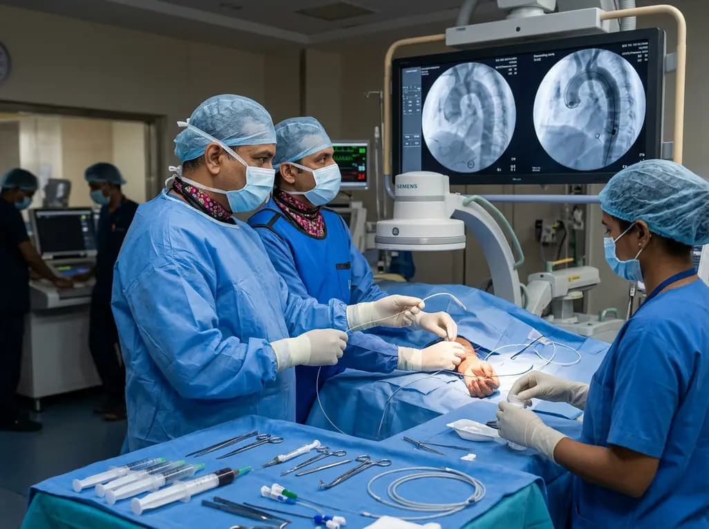
The novel CAST technique utilizes the indwelling Safari XS guidewire as a tracking rail to reposition a radial pigtail catheter into the descending aorta during minimalist TAVI, overcoming challenging vascular anatomy without requiring secondary femoral access or specialized equipment.
Today
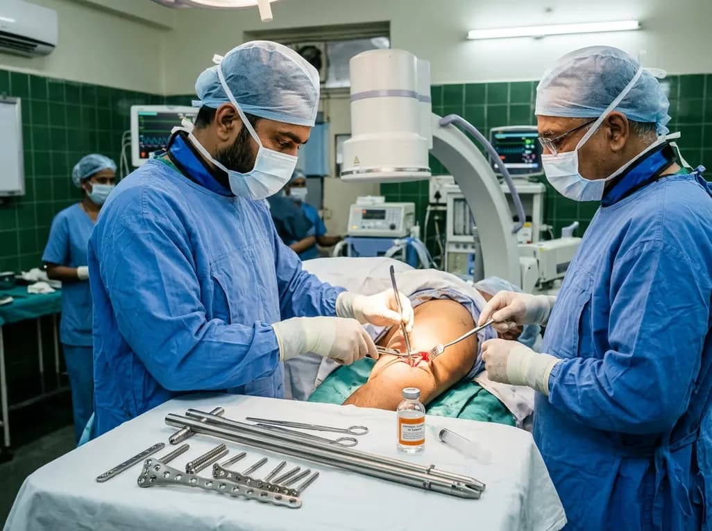
Septic humeral shaft nonunion in patients with obesity presents extreme mechanical and biological challenges. Discover how hybrid nail-plate osteosynthesis paired with targeted antibiotic therapy provides rigid stability, eradicates infection, and restores maximal upper extremity function.
Today

A case report details how reusing single-use insulin cartridges caused device failure and diabetic ketoacidosis in a type 1 diabetes patient using an advanced hybrid closed-loop system. The report highlights technological blindspots, mechanical degradation, and safety protocols during supply shortages.
Today
