

Loading, please wait...
Loading, please wait...
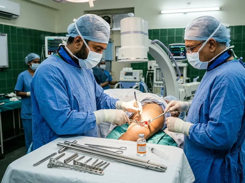
Managing a septic humeral shaft nonunion presents one of the most formidable clinical dilemmas in modern orthopedics. When chronic infection coexists with unstable mechanical failure, conventional single-modality implants frequently succumb to mechanical fatigue. In patients with elevated body mass index, excessive mechanical loading and prolonged lever arms dramatically heighten the risk of construct breakdown. Consequently, orthopedic surgeons require robust salvage strategies that achieve rigid mechanical stability while simultaneously eradicating persistent bone infection.
Patients presenting with a high body mass index experience immense mechanical demands across the upper extremity. Although the humerus does not bear body weight during quiet standing, it endures substantial torsional and bending moments during daily activities. For example, pushing up from a seated position, transfers, and ambulatory assistance impart repetitive axial and rotational forces. Furthermore, excessive adipose tissue increases the effective lever arm acting on the fracture site. Consequently, these amplified mechanical vectors create adverse biomechanical conditions that continuously jeopardize bone healing.
In addition to physical stresses, chronic low-grade bacterial infection compromises the local biological environment. Pathogens produce biofilms that coat retained implants and necrotic bone fragments. As a result, osteoclast activity increases while vascular supply diminishes, precipitating profound bone resorption. When mechanical instability coincides with septic osteolysis, standard osteosynthesis techniques often fail. Therefore, surgeons must evaluate both the host biology and construct biomechanics when planning revision procedures. Addressing only the pathogen or only the mechanical instability invariably leads to recurrent failure. Thus, successful reconstructive salvage requires a comprehensive strategy that harmonizes biological revitalization with unmatched rigid mechanical fixation.
Historically, clinicians managed humeral diaphyseal nonunions using either locked intramedullary nails or open compression plates. However, both isolated modalities demonstrate distinct biomechanical vulnerabilities under excessive load. Intramedullary nails provide excellent axial alignment and resist bending forces through load-sharing properties. Nevertheless, standard locked nails frequently permit subtle toggling and torsional micromotion within the wide humeral medullary canal. This persistent rotational instability inhibits osteoblast bridge formation across the nonunion gap.
Conversely, dynamic compression plating offers rigid direct compression and superior rotational control. Unfortunately, isolated compression plates function as load-bearing beams that bear immense repetitive bending moments. In obese individuals, these bending moments accelerate cyclic fatigue within the plate metal. Consequently, the implant eventually fractures before successful bony bridging can occur. Moreover, previous failed surgical attempts leave compromised cortical bone with scarce screw purchase. Therefore, single-implant revisions repeatedly fail in high-demand patients. Orthopedic surgeons must recognize that solitary plating or solitary nailing cannot provide sufficient stability in such difficult revision settings. Instead, combining both modalities creates a synergistic load-sharing construct that shields implants from early fatigue failure.
Hybrid fixation merges an intramedullary nail with an extramedullary dynamic compression plate to maximize structural rigidity. First, the surgical team positions the patient prone or in a lateral decubitus orientation. Surgeons perform an extensive posterior triceps-splitting or triceps-reflecting approach to visualize the humeral diaphysis directly. During deep exposure, the team carefully identifies, mobilizes, and neurolyses the radial nerve to safeguard neurological integrity. Next, the surgeon aggressively debrides all nonviable soft tissue, fibrous nonunion pseudarthrosis, and necrotic bone fragments.
Subsequently, the operator introduces an intramedullary nail to reconstitute longitudinal axial alignment and neutralize bending forces. Concurrently, the surgeon applies a narrow 4.5-millimeter dynamic compression plate over the nonunion site. By carefully inserting bicortical and unicortical screws around the nail, the plate locks the fracture fragments tightly together. Consequently, the plate completely abolishes rotational and translational shear stresses. In addition, the surgeon packs autologous or allogenic cancellous bone chips into the prepared gap to stimulate osteoinduction. Furthermore, this hybrid architecture distributes mechanical forces between the intramedullary axis and the external cortical plate. As a result, the construct delivers unmatched structural resilience capable of withstanding early functional rehabilitation.
Eradicating microbial colonization represents an indispensable requirement for achieving definitive osseous union. During the revision procedure, clinicians must harvest multiple deep tissue and bone specimens before administering perioperative antibiotics. Common indolent pathogens, such as Staphylococcus epidermidis, readily generate resilient extracellular biofilms that resist host immune clearance. Therefore, meticulous mechanical debridement remains the cornerstone of infection control. The surgeon must excise all contaminated hardware, devitalized bone, and pseudocapsular lining until healthy bleeding margins appear.
Following comprehensive surgical debridement, microbiological cultures direct pathogen-specific antimicrobial therapy. For sensitive Staphylococcus epidermidis strains, clinicians frequently select oral clindamycin due to its remarkable bioavailability and superior bone penetration. Additionally, clindamycin provides potent anti-staphylococcal activity and inhibits bacterial toxin production effectively. Patients generally complete a tailored four- to six-week course of targeted therapy while physicians monitor serum inflammatory markers regularly. Furthermore, the combination of rigid mechanical stability and potent antimicrobial coverage suppresses biofilm propagation. Consequently, the bone graft safely incorporates within a sterilised biological environment, fostering rapid vascularization and robust bone remodeling.
Posterior humeral exposure presents inherent risks of traction or compression injuries to the radial nerve. Even with meticulous neurolysis, transient postoperative radial neuropraxia can occur following extensive revision surgery. However, conservative treatment typically yields outstanding neurological recovery when the nerve continuity remains intact. Clinicians actively manage neuropathic discomfort and paresthesia using targeted pharmacotherapy, such as pregabalin. Furthermore, early dynamic splinting maintains functional wrist extension, preventing fixed contractures and preserving tendon excursion.
Simultaneously, a structured physical rehabilitation program commences immediately after surgical intervention. Because the hybrid construct provides superior multiplanar stability, patients can initiate early active-assisted shoulder and elbow range of motion. Moreover, rigid fixation accelerates bone trabecular bridging, allowing patients to regain robust functional independence. In validated assessments, patients achieve remarkable Upper Extremity Functional Scale scores, often regaining over 85 percent of maximal functional capacity. Therefore, combining rigid biomechanical fixation with vigilant postoperative neuro-rehabilitation successfully salvages limbs from catastrophic nonunion. Ultimately, this comprehensive management model restores full daily living independence while preventing permanent disability.
Obesity substantially elevates mechanical demands on the upper extremity. Although the humerus is not a classic weight-bearing bone, obese individuals routinely apply heavy forces during bed transfers, pushing from chairs, and ambulatory stabilization. Furthermore, increased limb girth lengthens the effective mechanical lever arm. These factors create massive cyclic bending and torsional moments across the nonunion site. Consequently, isolated plates or nails experience accelerated fatigue and hardware fracture before bony union can occur.
Hybrid nail-plate fixation combines the complementary biomechanical advantages of two distinct osteosynthesis devices. Specifically, the intramedullary nail resists axial collapse and bending forces through effective load sharing within the canal. Concurrently, the narrow dynamic compression plate provides rigid compression and neutralizes dangerous torsional and shear forces. By distributing stresses between the intramedullary nail and the extramedullary plate, the construct prevents hardware fatigue and maintains perfect fracture reduction until union finishes.
When intraoperative radial nerve continuity is confirmed, surgeons manage postoperative neuropraxia conservatively. First, clinicians prescribe neuropathic agents such as pregabalin or gabapentin to control paresthesias and burning discomfort. Additionally, therapists provide a dynamic wrist extension splint to prevent flexion contractures while preserving hand function. Physical therapy maintains passive finger and wrist mobility. Most transient traction neuropraxias resolve spontaneously within three to six months as nerve regeneration and remyelination proceed.
Disclaimer: This content is for informational and educational purposes only. It is not intended to provide medical advice, diagnosis, or treatment. Clinicians must always exercise their own clinical judgment for any individual patient. Refer to the latest local and national guidelines for clinical practice.
References
Chiquini-Ramírez P et al. Hybrid Osteosynthesis for Septic Humeral Shaft Nonunion in a Patient with Obesity: A Case Report. Cureus. 2026 Nov undefined. doi: 10.7759/cureus.119283. PMID: 42999717.
Riopelle D, Chung BC, Driscoll C, Azad A, Nicholson L. Humerus Plate-Nail Construct for Salvage of Humeral Nonunion with Failed Free Fibula Flap: Technique and A Report of 2 Cases. JBJS Case Connect. 2025;15(4):e25.00250.
Marti RK, Verheyen CC, Besselaar PP. Treatment of humeral shaft nonunions: nailing versus plating. Arch Orthop Trauma Surg. 2004;124(1):50-54.

Read summarized clinical updates, watch expert medical content, and earn CME certifications right from your smartphone.
Open your smartphone camera to scan and install.





Septic humeral shaft nonunion in patients with obesity presents extreme mechanical and biological challenges. Discover how hybrid nail-plate osteosynthesis paired with targeted antibiotic therapy provides rigid stability, eradicates infection, and restores maximal upper extremity function.
Today

Acute portal vein thrombosis can present deceptively with chest pain, collapse, and elevated D-dimer. Discover why venous-phase CT imaging and early anticoagulation are vital to prevent mesenteric ischemia and achieve complete recanalization.
Today
A 21-year-old postpartum female presenting with right heart failure was initially suspected of peripartum cardiomyopathy, but comprehensive imaging and serology confirmed MCTD-associated pulmonary hypertension. Early diuretic and immunosuppressive therapy led to successful clinical stabilization.
Today
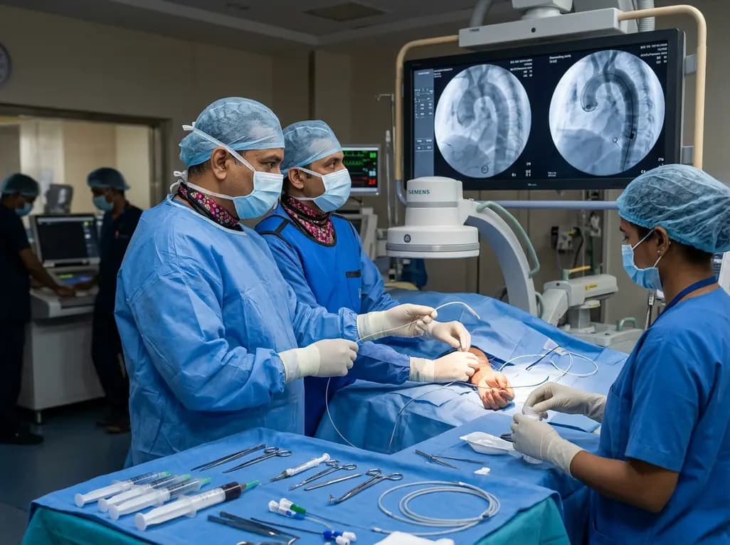
The novel CAST technique utilizes the indwelling Safari XS guidewire as a tracking rail to reposition a radial pigtail catheter into the descending aorta during minimalist TAVI, overcoming challenging vascular anatomy without requiring secondary femoral access or specialized equipment.
Today
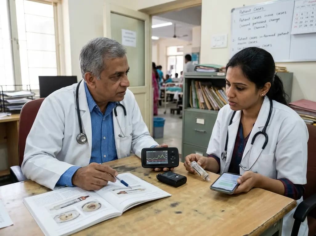
A case report details how reusing single-use insulin cartridges caused device failure and diabetic ketoacidosis in a type 1 diabetes patient using an advanced hybrid closed-loop system. The report highlights technological blindspots, mechanical degradation, and safety protocols during supply shortages.
Today
