

Loading, please wait...
Loading, please wait...

Postpartum dyspnea and volume overload present an urgent diagnostic dilemma for treating physicians. Mixed connective tissue disease is an autoimmune condition featuring overlapping manifestations of systemic lupus erythematosus, systemic sclerosis, and polymyositis. Clinicians recognize that pulmonary arterial involvement is the leading cause of mortality in these patients. However, encountering newly decompensated MCTD pulmonary hypertension during the puerperium remains exceedingly uncommon and dangerous. Patients typically present with progressive fatigue, bilateral leg swelling, and orthopnea several weeks or months after delivery. In this clinical scenario, a 21-year-old female presented three months following an uneventful vaginal delivery. She experienced worsening shortness of breath, a persistent cough, and bilateral lower limb edema. Physical examination revealed marked hypotension, elevated jugular venous pressure, a loud pulmonic second heart sound, and a tricuspid regurgitation murmur. In addition, bilateral fine basal crepitations were noted on lung auscultation. Because the initial clinical signs mimicked common postpartum complications, clinicians initially evaluated her for peripartum cardiomyopathy. Consequently, this case underscores how insidious vascular remodeling can evade detection until acute postpartum hemodynamic shifts unmask overt right ventricular failure.
Differentiating primary pulmonary vascular disorders from postpartum left ventricular failure is vital for patient survival. When a young postpartum woman develops anasarca and respiratory distress, physicians routinely prioritize peripartum cardiomyopathy on their differential list. Nevertheless, true peripartum cardiomyopathy characteristically demonstrates left ventricular systolic dysfunction with a reduced ejection fraction. In stark contrast, isolated pulmonary arterial hypertension primarily impairs the right ventricle due to excessive pulmonary vascular resistance. Therefore, bedside transthoracic echocardiography serves as an indispensable primary screening tool. In this patient, echocardiographic evaluation revealed moderate to severe pulmonary arterial hypertension alongside significant tricuspid regurgitation. Importantly, the left ventricular chamber and ejection fraction showed preserved function without intrinsic myocardial failure. Clinicians must recognize that misclassifying right heart failure as left ventricular failure delays specific life-saving interventions. Furthermore, aggressive afterload reduction intended for left ventricular disease can precipitate hemodynamic collapse in patients with severe right ventricular strain. Thus, meticulous physical examination combined with urgent echocardiography prevents dangerous therapeutic delays. Recognizing dominant right heart pathology promptly shifts the clinical investigation toward underlying pulmonary vascular and autoimmune etiologies.
Once clinicians establish right ventricular pressure overload, determining the underlying etiology requires comprehensive multimodal investigation. First, clinicians must evaluate pulmonary parenchymal architecture and exclude chronic thromboembolic disease. High-resolution computed tomography of the chest in this patient demonstrated bilateral moderate pleural effusions. Additionally, imaging revealed subtle honeycombing with partial atelectasis of the dependent lung zones and cardiomegaly. Computed tomography pulmonary angiography subsequently excluded acute pulmonary thromboembolism while confirming vascular changes consistent with pulmonary hypertension. Simultaneously, physicians conducted an extensive rheumatologic serological evaluation. Autoimmune antibody profiling revealed positive antinuclear antibodies, high-titer anti-U1-RNP antibodies, and concomitant positivity for SS-A, SS-B, and Jo-1 antibodies. The presence of high-titer anti-U1-RNP antibodies serves as the serological hallmark defining mixed connective tissue disease. Moreover, the coexistence of Sjögren and myositis-related autoantibodies highlighted the complex autoimmune overlap phenotype. As a result, these definitive immunological findings bridged the clinical gap, confirming connective tissue disease-associated pulmonary hypertension rather than an idiopathic cardiac pathology. Clinicians in tertiary centers should maintain a low threshold for early autoantibody testing in unexplained postpartum heart failure.
Managing severe right-sided heart failure secondary to autoimmune pulmonary vascular disease requires an aggressive, multi-targeted approach. Initial medical therapy must prioritize fluid decongestion without inducing systemic hypotension. In this clinical scenario, medical teams initiated intravenous furosemide at forty milligrams daily and spironolactone at twenty-five milligrams daily. Because the patient presented with borderline hypotension, physicians carefully monitored her fluid balance and venous filling pressures. Furthermore, clinicians added lisinopril at ten milligrams daily and therapeutic heparin anticoagulation to mitigate vascular stasis and thromboembolic risks. Concurrently, controlling the underlying immunological inflammation remains paramount in mixed connective tissue disease flares. Therefore, physicians commenced systemic immunosuppressive therapy with oral prednisolone at twenty milligrams daily. Over subsequent hospital days, the medical team monitored her response and gradually tapered the corticosteroid dose. Consequently, the patient demonstrated significant clinical improvement, with resolution of anasarca, reduction in jugular venous distension, and substantial relief of dyspnea. Early initiation of combined diuretic and immunosuppressive therapy successfully interrupted the vicious cycle of right ventricular failure. This clinical success demonstrates the necessity of prompt hemodynamic optimization alongside targeted immunosuppression.
Following initial hemodynamic stabilization, long-term ambulatory management requires coordinated multidisciplinary surveillance involving rheumatology, cardiology, and pulmonology. Mixed connective tissue disease-associated pulmonary arterial hypertension carries a guarded prognosis if pulmonary vascular remodeling continues unchecked. Therefore, outpatient management must focus on formal hemodynamic staging via right heart catheterization. Although initial corticosteroid treatment effectively suppresses inflammatory vasculitis, most patients eventually require targeted pulmonary arterial hypertension medications. Specifically, therapies such as endothelin receptor antagonists, phosphodiesterase-5 inhibitors, or prostacyclin analogues prevent progressive right heart deterioration. In addition, clinicians must emphasize contraception and reproductive counseling, because future pregnancies carry immense maternal morbidity and mortality risks. The profound physiological changes of pregnancy, such as increased plasma volume and cardiac output, impose severe stress on a compromised right ventricle. Hence, physicians should educate young female patients about these risks immediately before hospital discharge. Regular clinical reassessment with serial echocardiograms, six-minute walk tests, and biomarker tracking ensures sustained disease control. Ultimately, prompt recognition during the index presentation offers patients the greatest opportunity for long-term functional recovery.
Both clinical conditions present during the puerperium with overlapping symptoms, including severe dyspnea, fatigue, orthopnea, and peripheral edema. Because peripartum cardiomyopathy is more widely recognized among obstetric clinicians, physicians often assume left ventricular failure first. However, echocardiography readily differentiates them. Peripartum cardiomyopathy demonstrates reduced left ventricular ejection fraction, whereas connective tissue disease-associated pulmonary hypertension primarily causes isolated right ventricular enlargement, elevated pulmonary pressures, and severe tricuspid regurgitation with preserved left ventricular function.
Pregnancy produces profound physiological adaptations, including a fifty percent expansion in blood volume and increased cardiac output. Following placental expulsion, sudden autotransfusion and relief of inferior vena cava compression rapidly augment venous return. In patients with preexisting subclinical vascular injury, this dramatic fluid shift overwhelms the noncompliant pulmonary vascular bed. Consequently, right ventricular afterload rises precipitously, precipitating acute right-sided chamber dilation, functional tricuspid valve regurgitation, and decompensated heart failure within weeks or months after delivery.
Unlike pure idiopathic pulmonary arterial hypertension, connective tissue disease-associated cases often feature active perivascular inflammation and microvascular injury. In conditions like mixed connective tissue disease or lupus, systemic corticosteroids effectively dampen pulmonary vascular endothelial inflammation and arrest progressive tissue injury. While vasodilators specifically decrease pulmonary artery pressures, corticosteroids and steroid-sparing immunosuppressants target underlying autoimmune activity. Therefore, early immunosuppressive treatment improves right ventricular hemodynamics, accelerates symptomatic recovery, and prevents irreversible plexiform vascular remodeling in selected responsive phenotypes.
Disclaimer: This content is for informational and educational purposes only. It is not intended to be a substitute for professional medical advice, diagnosis, or treatment. Always seek the advice of your physician or other qualified health provider with any questions you may have regarding a medical condition. Refer to the latest local and national guidelines for clinical practice.
References

Read summarized clinical updates, watch expert medical content, and earn CME certifications right from your smartphone.
Open your smartphone camera to scan and install.





A 21-year-old postpartum female presenting with right heart failure was initially suspected of peripartum cardiomyopathy, but comprehensive imaging and serology confirmed MCTD-associated pulmonary hypertension. Early diuretic and immunosuppressive therapy led to successful clinical stabilization.
Today
Acute portal vein thrombosis can present deceptively with chest pain, collapse, and elevated D-dimer. Discover why venous-phase CT imaging and early anticoagulation are vital to prevent mesenteric ischemia and achieve complete recanalization.
Today
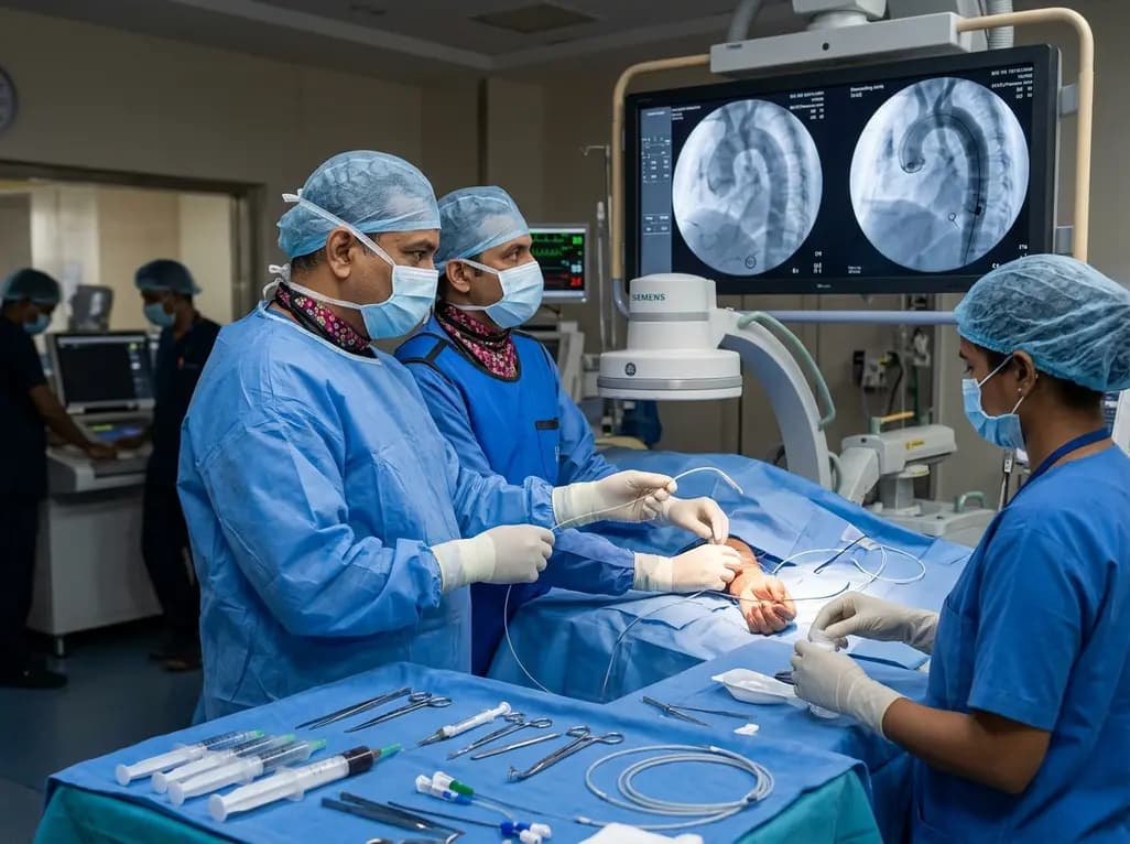
The novel CAST technique utilizes the indwelling Safari XS guidewire as a tracking rail to reposition a radial pigtail catheter into the descending aorta during minimalist TAVI, overcoming challenging vascular anatomy without requiring secondary femoral access or specialized equipment.
Today
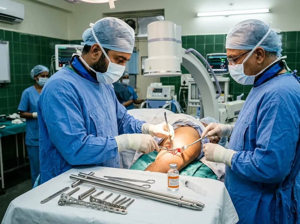
Septic humeral shaft nonunion in patients with obesity presents extreme mechanical and biological challenges. Discover how hybrid nail-plate osteosynthesis paired with targeted antibiotic therapy provides rigid stability, eradicates infection, and restores maximal upper extremity function.
Today
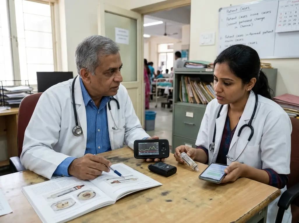
A case report details how reusing single-use insulin cartridges caused device failure and diabetic ketoacidosis in a type 1 diabetes patient using an advanced hybrid closed-loop system. The report highlights technological blindspots, mechanical degradation, and safety protocols during supply shortages.
Today
