

Loading, please wait...
Loading, please wait...

Intradural extramedullary lesions of the spinal canal represent diagnostic dilemmas for spine surgeons worldwide. Most intradural tumors in the lumbar spine consist of schwannomas, meningiomas, or myxopapillary ependymomas. In contrast, primary intradural vascular neoplasms occur exceptionally rarely within the thecal sac. Clinicians recently identified a spinal epithelioid hemangioma in the cauda equina region of a sixty-three-year-old male. Historically, pathologists categorize epithelioid hemangioma as a benign vascular tumor affecting skin, soft tissue, and tubular bone. However, this unique presentation represents the first documented intradural manifestation across the entire neuroaxis. The patient initially presented with three months of progressive radiculopathy radiating down the left L5 distribution. Because the patient had previous cutaneous melanoma, oncologists initially suspected metastatic spread. Consequently, differentiating between an unusual primary neoplasm and metastatic disease represented an urgent clinical priority. Magnetic resonance imaging demonstrated an enhancing mass occupying the lumbar thecal sac. Furthermore, neurological examination confirmed marked dermatomal sensory deficits and radiating pain. This extraordinary presentation emphasizes why clinicians must expand their differential diagnosis for intraspinal lesions. Early identification ensures timely operative intervention before permanent neurological decline develops.
Diagnostic neuroimaging plays an indispensable role during the initial assessment of lumbosacral pain syndromes. Specifically, high-resolution contrast-enhanced magnetic resonance imaging provides unmatched anatomical clarity within the cauda equina. In this patient, imaging scans revealed a well-circumscribed mass measuring one by two centimeters at L4-L5. Furthermore, the scan revealed concurrent left L5-S1 foraminal stenosis contributing to radicular compression. Vascular intradural lesions typically show intense, uniform gadolinium enhancement on T1-weighted sequences. Moreover, they often demonstrate heterogeneous hyperintensity on T2-weighted acquisitions due to variable blood flow. However, standard imaging cannot reliably separate benign vascular growths from nerve sheath tumors or solitary metastases. Cavernous and capillary hemangiomas comprise the vast majority of spinal vascular anomalies reported in surgical literature. In contrast, epithelioid vascular entities remain remarkably scarce in intradural spaces. Therefore, neurosurgeons cannot establish a definitive histological diagnosis based solely on pre-operative radiological features. Additionally, radiologists must scrutinize adjacent osseous structures for erosion or direct extradural infiltration. Preoperative identification of hypervascularity alerts the surgical team to potential hemorrhagic complications during exposure. Consequently, precise preoperative imaging protocols optimize operative planning and minimize unpredictable intraoperative events.
Surgical excision remains the gold standard for symptomatic intradural extramedullary lesions of the lumbar canal. Operating within the cauda equina demands extreme technical precision because clustered nerve roots surround the mass. Therefore, the neurosurgical team utilized real-time intraoperative ultrasound alongside continuous multimodal neurophysiological monitoring. Ultrasound localization confirmed the exact craniocaudal borders of the tumor before the surgeons opened the thecal sac. Subsequently, surgeons performed a targeted L4-L5 laminectomy to access the spinal subarachnoid space cleanly. Electrophysiological monitoring tracked transcranial motor evoked potentials and somatosensory evoked potentials continuously throughout the procedure. In addition, triggered electromyography helped differentiate nonfunctional neoplastic adhesions from viable, functional lumbar nerve roots. As a result, the surgical team achieved a successful en bloc gross-total resection without disrupting critical neural bundles. Surgeons meticulously coagulated feeding microvessels to eliminate the risk of uncontrollable subarachnoid hemorrhage. Furthermore, this controlled microdissection preserved neurological integrity throughout the entire cauda equina arborization. Postoperative recovery proceeded smoothly without the emergence of new sensorimotor deficits or sphincter disturbances. Ultimately, modern intraoperative adjuncts maximize patient safety during complex spinal intradural resections.
Definitive diagnosis of epithelioid vascular lesions depends entirely on rigorous microscopic examination and comprehensive immunohistochemical testing. Gross specimen evaluation in this case revealed a firm, well-delineated red-brown vascular nodule measuring two centimeters. Microscopically, hematoxylin and eosin staining demonstrated abundant well-formed blood vessels lined by prominent, plump endothelial cells. Notably, these epithelioid endothelial cells projected into vascular lumina, displaying characteristic intracytoplasmic vacuoles without cytological atypia. Pathologists observed no atypical mitotic figures, marked nuclear pleomorphism, or necrosis, which ruled out high-grade malignant angiosarcoma. Furthermore, distinguishing this lesion from metastatic melanoma required an exhaustive immunohistochemistry panel due to previous malignancy. Immunohistochemical analysis demonstrated intense diffuse positivity for endothelial markers, specifically ERG, CD31, and CD34. Conversely, the tumor showed total negativity for melanocytic markers, including SOX10, Melan-A, and HMB-45. In addition, negative staining for human herpesvirus-8 excluded Kaposi sarcoma, while negative inhibin staining excluded hemangioblastoma. Similarly, negative CAM5.2 and cytokeratin markers effectively ruled out epithelioid hemangioendothelioma and metastatic carcinoma. Consequently, this detailed profiling firmly confirmed the diagnosis of benign epithelioid hemangioma within the thecal canal. Pathologists must maintain high suspicion when evaluating atypical vascular architectures in uncommon anatomic locations.
Postoperative management of uncommon spinal vascular neoplasms centers on symptom resolution and regular radiological surveillance. Seven weeks following surgical intervention, postoperative magnetic resonance imaging verified complete gross-total excision without residual mass effect. Furthermore, the patient experienced dramatic relief from debilitating left lower extremity pain and resumed normal daily activities. In bone and soft tissue, epithelioid hemangiomas occasionally demonstrate locally aggressive behavior or recurrent growth patterns. However, complete surgical resection typically yields an excellent long-term prognosis with minimal risk of recurrence. Therefore, surgeons consider complete en bloc removal curative when margins remain free of pathological tissue. Clinicians nonetheless recommend serial contrast-enhanced spinal imaging at scheduled intervals to monitor the resection cavity meticulously. In addition, oncologists must continue systemic surveillance because patients with concurrent or past malignancies require vigilance. For Indian neurosurgical centers, this landmark case underscores the importance of multidisciplinary collaboration among neurosurgeons, radiologists, and pathologists. Advanced intraoperative neuro-monitoring and precision microsurgery allow safe execution of total tumor resection even in crowded neural spaces. Ultimately, documenting unusual entities in the cauda equina expands current neuro-oncological literature and refines future spinal clinical protocols.
Epithelioid hemangioma displays lobular capillary formations lined by plump, histiocytoid endothelial cells. In contrast, cavernous hemangiomas contain large sinusoidal blood spaces without epithelioid morphology. Furthermore, epithelioid hemangioma demonstrates strong ERG and CD31 immunoreactivity without nuclear atypia or atypical mitoses. These distinctive markers separate it from malignant angiosarcoma and intermediate hemangioendothelioma. Consequently, comprehensive histological evaluation provides reliable diagnostic differentiation from other vascular spinal anomalies.
Yes, spinal epithelioid hemangioma can closely mimic metastatic malignancies on pre-operative radiological evaluations. Because these tumors enhance intensely on magnetic resonance imaging, clinicians frequently mistake them for metastatic deposits, schwannomas, or ependymomas. Furthermore, when patients possess a prior cancer history, such as melanoma, diagnostic confusion increases significantly. Therefore, pathologists must deploy specialized immunohistochemical markers, including SOX10 and HMB-45. Consequently, this targeted profiling successfully excludes metastatic disease and confirms primary vascular pathology.
Complete en bloc gross-total surgical resection represents the definitive curative treatment for spinal epithelioid hemangioma. Surgeons achieve optimal outcomes by utilizing intraoperative ultrasound guidance and continuous neurophysiological monitoring. Following successful surgery, patients generally experience rapid neurological recovery. However, clinicians recommend baseline contrast-enhanced magnetic resonance imaging at seven weeks post-surgery. In addition, neurosurgeons advise periodic radiological surveillance over subsequent years to ensure long-term stability and identify potential local recurrence promptly.
Disclaimer: This content is for informational and educational purposes only... Refer to the latest local and national guidelines for clinical practice.
References

Read summarized clinical updates, watch expert medical content, and earn CME certifications right from your smartphone.
Open your smartphone camera to scan and install.





Surgeons have reported the first intradural extramedullary epithelioid hemangioma of the cauda equina. The 63-year-old patient underwent successful en bloc gross-total resection under neurophysiological monitoring, confirmed by immunohistochemistry showing ERG and CD31 positivity.
Today
Standardizing continuous physiological waveforms in EHRs has long challenged clinical teams. The novel TIDE framework leverages HL7 FHIR R4 to decouple queryable clinical metadata from massive raw signal repositories, unlocking scalable telemetry exchange for bedside care and predictive research.
Today

A secondary analysis of the MOMS clinical trial reveals that infant sex does not influence neonatal opioid withdrawal syndrome severity in buprenorphine-exposed newborns. Maternal co-exposures and clinical factors drive outcomes, highlighting the need for objective assessment and supportive breastfeeding.
Today
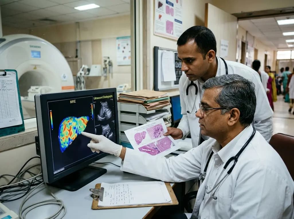
A comparative study reveals magnetic resonance elastography outperforms ultrasound elastography in assessing liver fibrosis in Fontan-associated liver disease, demonstrating stronger histological agreement and superior discrimination.
Today
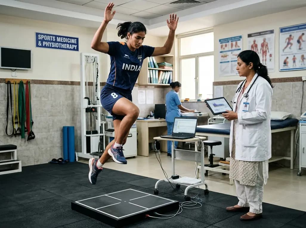
A systematic review and meta-analysis highlights the substantial impact of plyometric jump training on jump performance, reactive strength, and landing kinematics in female team-sport athletes, providing evidence-based programming guidelines for sports medicine and orthopedics.
Today

A comprehensive review of the role of growth hormone in diabetic kidney disease and podocytopathy. Learn how elevated GH activates ZEB2, Notch, and TGF-beta1, driving podocyte loss and glomerulosclerosis, and explore novel targeted therapeutic approaches.
Today
