

Loading, please wait...
Loading, please wait...
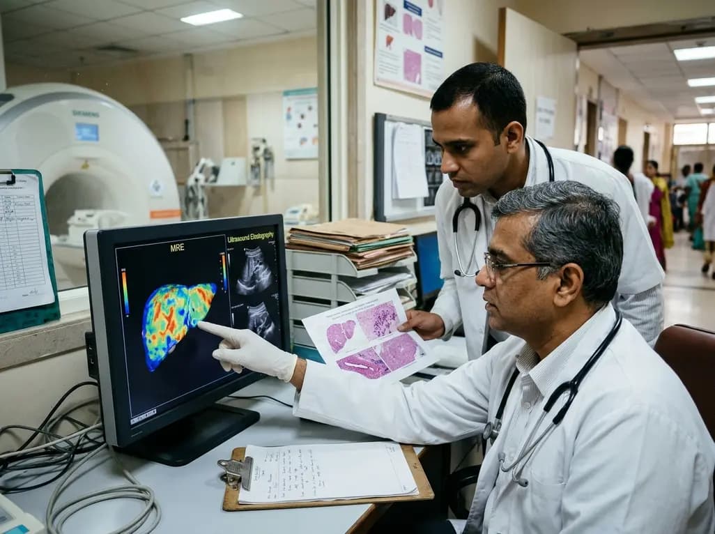
Fontan-associated liver disease has emerged as a significant universal sequela in patients surviving single-ventricle palliation. Consequently, clinicians require reliable noninvasive modalities to monitor hepatic architecture over time. Furthermore, chronic passive venous congestion and diminished cardiac output trigger progressive sinusoidal fibrosis. This pathological process can ultimately culminate in cirrhosis and hepatocellular carcinoma. However, accurate noninvasive staging remains notoriously difficult because passive congestion directly increases parenchyma stiffness independently of true fibrosis. Recently, an important cross-sectional study evaluated magnetic resonance elastography alongside ultrasound elastography against histological scoring. Therefore, these comparative findings offer critical clinical guidance for post-Fontan surveillance.
The Fontan circuit fundamentally alters systemic and splanchnic hemodynamics. Because this surgical configuration lacks a subpulmonary pump, central venous pressure remains persistently elevated. Consequently, chronic retrograde hydrostatic pressure transmits directly through the hepatic veins into the sinusoids. This persistent hypertension induces severe sinusoidal dilation and perivenular ischemia. In response to continuous mechanical stretch and hypoxia, hepatic stellate cells activate and synthesize collagen fibers. Therefore, liver remodeling occurs predominantly within the centrilobular zones rather than the periportal regions typically affected by viral hepatitis. In addition, low cardiac output impairs hepatic arterial buffer responses, which accelerates cellular injury and architectural remodeling. As a result, patients inevitably develop progressive congestive hepatic fibrosis over several decades. Unfortunately, noninvasive assessment tools often stumble because venous engorgement elevates tissue stiffness even before dense fibrotic bands appear. Clinicians must therefore understand that elevated stiffness reflects both fluid congestion and architectural scarring.
To resolve diagnostic uncertainties, investigators conducted a rigorous cross-sectional study evaluating thirty-four Fontan patients. The cohort had a mean age of twenty years, and most individuals had received an extracardiac conduit Fontan. Furthermore, pathologists staged biopsy specimens using the validated congestive hepatic fibrosis score system. This specialized histological framework specifically evaluates sinusoidal dilation, centrilobular collagen deposition, and portal-central bridging. Investigators categorized participants into non-significant fibrosis (score 2A or less) and advanced fibrosis (score 2B or greater) cohorts. Notably, twenty-three patients, representing nearly sixty-eight percent of the cohort, displayed advanced fibrosis on tissue analysis. Meanwhile, invasive hemodynamic parameters remained remarkably similar between the two fibrosis categories. Specifically, systemic venous pressures and cardiac index values showed no significant statistical disparity across cohorts. This critical observation indicates that hemodynamic values alone cannot accurately differentiate early fibrosis from advanced parenchymal remodeling. Consequently, clinicians must rely on objective tissue characterization rather than resting pressure metrics during routine surveillance.
The investigation directly compared quantitative stiffness measurements from magnetic resonance elastography and ultrasound elastography against biopsy findings. Magnetic resonance elastography measurements were significantly higher in the advanced fibrosis cohort compared to the mild fibrosis group. Specifically, patients with advanced fibrosis recorded mean values of 5.0 kilopascals, whereas patients with non-significant fibrosis averaged 4.1 kilopascals. In contrast, ultrasound elastography failed to show any statistically significant difference between the two histological groups. Furthermore, magnetic resonance elastography demonstrated fair agreement with liver histology, yielding a quadratic-weighted kappa of 0.468. Conversely, ultrasound elastography displayed an absolute lack of meaningful agreement, producing a negative kappa value of minus 0.057. Receiver operating characteristic analyses further reinforced these stark differences. Magnetic resonance elastography demonstrated acceptable discriminative performance, reaching an area under the curve of 0.77. However, ultrasound elastography showed poor discrimination with an area under the curve of only 0.44. The DeLong test confirmed that magnetic resonance elastography provided statistically superior diagnostic accuracy.
Clinicians frequently question why ultrasound elastography performs reliably in chronic viral hepatitis yet fails in Fontan patients. First, acoustic shear wave elastography interrogates only a localized acoustic window within the right hepatic lobe. Consequently, it cannot capture the patchy, heterogeneous distribution typical of congestive fibrosis. Second, ultrasound shear wave speed depends heavily on instantaneous vascular congestion and intrathoracic respiratory pressures. Because post-Fontan hemodynamics fluctuate with systemic volume status, ultrasound measurements capture vascular engorgement rather than collagen density. In addition, deep acoustic attenuation, thick subcutaneous tissue, and surgical chest wall scars frequently degrade acoustic wave propagation. Therefore, ultrasound elastography exhibits wide measurement variability and poor reproducibility in this specific clinical population. In contrast, magnetic resonance elastography evaluates almost the entire hepatic volume through cross-sectional mechanical wave propagation. Furthermore, magnetic resonance techniques effectively avoid acoustic shadowing and anatomical sampling errors. Thus, magnetic resonance elastography provides a far more stable assessment of structural remodeling, even when background venous pressure remains chronically elevated.
Although magnetic resonance elastography outperformed ultrasound elastography, its discriminative capacity remained lower than values typically reported in viral hepatitis. In non-congestive liver diseases, magnetic resonance elastography routinely achieves areas under the curve exceeding 0.90. This discrepancy underscores that chronic passive congestion inevitably elevates baseline liver stiffness across all Fontan patients. Therefore, clinicians cannot extrapolate standard stiffness thresholds validated in non-congestive etiologies to post-Fontan individuals. Instead, the medical community must establish disease-specific cut-off values linked directly to congestive histology systems. Moreover, clinicians should integrate elastography findings into a comprehensive multimodality framework. Such surveillance protocols must combine serial magnetic resonance imaging, biochemical markers, cardiopulmonary exercise performance, and periodic cardiac catheterization. When magnetic resonance elastography indicates escalating stiffness values over time, clinicians should promptly investigate potential cardiac hemodynamic deterioration. In particular, treating physicians must rule out subclinical conduit stenosis, elevated pulmonary vascular resistance, or systemic ventricular dysfunction before presuming isolated liver failure.
Optimizing long-term outcomes in congenital heart disease requires close collaboration between pediatric cardiologists, adult congenital heart disease specialists, and hepatologists. Surveillance should ideally commence within five to ten years after the Fontan operation. Furthermore, medical centers should establish standardized surveillance pathways that incorporate annual laboratory testing and noninvasive imaging. Although ultrasound elastography remains accessible and inexpensive, clinicians should avoid using it as a stand-alone tool for fibrosis staging. Instead, centers equipped with magnetic resonance elastography should consider incorporating baseline elastography during routine cardiac magnetic resonance examinations. Consequently, simultaneous assessment of conduit patency, ventricular function, and hepatic stiffness becomes feasible in a single clinical visit. When elastography values trend upward, clinicians should consider transjugular liver biopsy paired with right heart catheterization. This combined approach safely measures resting Fontan pressures while securing histological confirmation. Ultimately, proactive multimodal surveillance enables early intervention, preventing decompensated cirrhosis, refractory portal hypertension, and liver failure.
Liver biopsy remains the definitive diagnostic standard because noninvasive elastography values are confounded by chronic venous congestion. Histological examination uniquely differentiates sinusoidal engorgement from genuine collagen deposition and architectural distortion. Furthermore, biopsy specimens permit accurate staging using the congestive hepatic fibrosis score, which identifies bridging fibrosis and nodular architecture. Consequently, biopsy provides indispensable anatomical confirmation whenever noninvasive surveillance yields ambiguous findings or when clinicians consider heart transplantation.
Chronic venous hypertension increases physical tissue tension by engorging the hepatic vascular bed with blood. Because ultrasound elastography measures acoustic shear wave propagation speed, heightened fluid pressure artificially elevates stiffness readings independently of collagen accumulation. Moreover, ultrasound samples only a narrow acoustic window, making it vulnerable to localized vascular fluctuations and respiratory artifacts. Consequently, ultrasound elastography frequently misclassifies simple hemodynamic congestion as severe structural fibrosis, producing unreliable results in post-Fontan surveillance.
Current consensus guidelines recommend initiating hepatocellular carcinoma surveillance approximately ten years following Fontan completion, or earlier if advanced fibrosis appears. Hepatologists advise performing cross-sectional contrast imaging with magnetic resonance or computed tomography combined with serial serum alpha-fetoprotein measurements every six to twelve months. Because hypervascular benign regenerative nodules occur frequently in congestive hepatopathy, multidisciplinary imaging review remains critical. Therefore, expert consensus ensures accurate differentiation between benign regenerative lesions and early malignant transformation.
Disclaimer: This content is for informational and educational purposes only... Refer to the latest local and national guidelines for clinical practice.
References

Read summarized clinical updates, watch expert medical content, and earn CME certifications right from your smartphone.
Open your smartphone camera to scan and install.





A comparative study reveals magnetic resonance elastography outperforms ultrasound elastography in assessing liver fibrosis in Fontan-associated liver disease, demonstrating stronger histological agreement and superior discrimination.
Today

Overexpression of glycoprotein nonmetastatic melanoma protein B (GPNMB) mediates resistance to CDK4/6 inhibitors like abemaciclib and palbociclib in ER+ breast cancer through integrin-driven AKT/ERK signaling. Targeting GPNMB with antibody-drug conjugates like glembatumumab vedotin may overcome this resistance.
Today
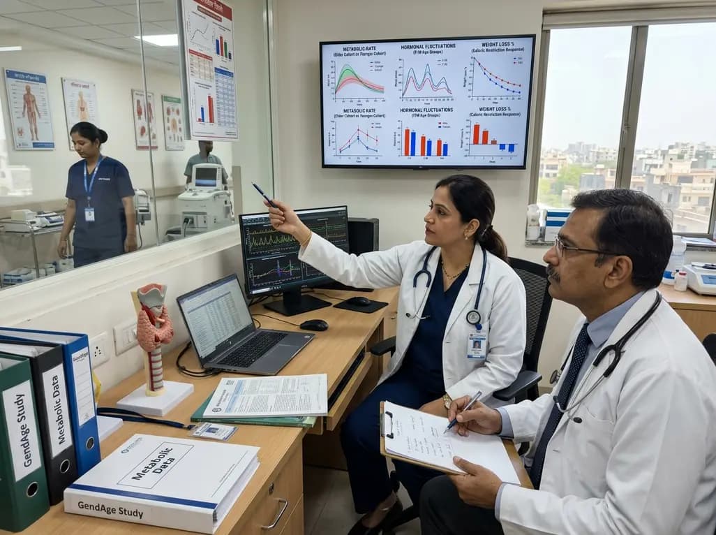
The GendAge weight loss study investigates how sex hormones, age, and menopausal status modulate caloric restriction response. By comparing metabolic adaptations across younger and older cohorts, this controlled feeding trial aims to refine precision dietary guidance for effective obesity management.
Today
A systematic review and meta-analysis highlights the substantial impact of plyometric jump training on jump performance, reactive strength, and landing kinematics in female team-sport athletes, providing evidence-based programming guidelines for sports medicine and orthopedics.
Today

A comprehensive review of the role of growth hormone in diabetic kidney disease and podocytopathy. Learn how elevated GH activates ZEB2, Notch, and TGF-beta1, driving podocyte loss and glomerulosclerosis, and explore novel targeted therapeutic approaches.
Today
