.webp&w=384&q=75)

Loading, please wait...
Loading, please wait...
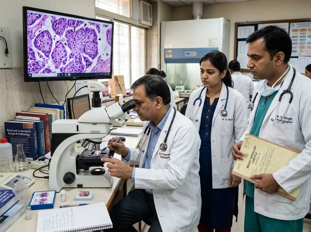
Solid papillary carcinoma breast neoplasms represent a distinct, rare malignancy that primarily affects elderly postmenopausal women. Historically, clinical literature characterizes this entity as an indolent breast tumor with favorable patient outcomes. Pathologists typically recognize these lesions by their circumscribed expansile nodules supported by delicate fibrovascular cores. However, recent clinical reports highlight unusual presentations that challenge this traditional concept of uniform indolence. Clinicians increasingly encounter cases displaying prominent high-grade nuclear atypia, marked cellular pleomorphism, and elevated proliferation indices. Consequently, managing these atypical cases requires a re-evaluation of established diagnostic frameworks. While standard solid papillary lesions demonstrate monotonous cellular features and low mitotic activity, emerging evidence demonstrates significant biological heterogeneity. Practitioners must look beyond standard architectural patterns to identify underlying aggressive markers. Early recognition ensures that patients receive tailored risk stratification rather than routine conservative management. Multidisciplinary collaboration remains crucial to properly diagnose these complex tumors.
Patients presenting with solid papillary carcinoma frequently exhibit non-specific clinical symptoms that closely mimic other breast conditions. Most elderly women report a painless, slow-growing retroareolar or central breast mass. Spontaneous or bloody nipple discharge occurs in nearly half of affected individuals due to ductal origin. Preoperative imaging plays an essential supportive role in identifying suspicious breast lesions. On mammography, clinicians typically observe well-circumscribed, round, or lobulated masses. Consequently, ultrasonography serves as an invaluable diagnostic tool, revealing frond-like solid components or complex cystic structures within dilated ducts. On ultrasound, these suspicious lesions routinely receive a BI-RADS 4 or BI-RADS 5 classification, prompting further tissue sampling. Magnetic resonance imaging further enhances diagnostic accuracy by displaying circumscribed tumor margins and rapid contrast enhancement. However, radiologic findings alone cannot reliably distinguish between low-grade indolent variants and aggressive high-grade cytological forms. Therefore, image-guided biopsy remains mandatory to obtain representative histological material for definitive evaluation.
Definitive histological evaluation provides the foundation for diagnosing solid papillary breast lesions. Under low-power magnification, pathologists observe characteristic solid expansile cellular sheets punctuated by thin fibrovascular stalks. A defining diagnostic hallmark is the partial or complete loss of the peripheral myoepithelial cell layer along tumor borders. Furthermore, standard tumors exhibit monotonous, low-grade cuboidal cells with neuroendocrine or mucinous differentiation. However, recent atypical cases reveal striking cytological discordance within these classic architectural frameworks. Microscopic examination of high-grade variants demonstrates pronounced nuclear enlargement, hyperchromasia, prominent nucleoli, and marked pleomorphism. Pathologists also identify frequent atypical mitotic figures and a significantly elevated Ki-67 proliferation index. Surprisingly, these aggressive cytological changes can develop even in the absence of conventional infiltrative ductal growth patterns. This stark contrast creates a diagnostic challenge for pathology departments. Consequently, thorough sectioning of surgical excision specimens is essential to accurately evaluate the entire tumor volume.
The identification of high-grade cytological features in solid papillary tumors highlights the underlying biological heterogeneity of this rare neoplasm. Historically, indolent behavior justified conservative management strategies, as low-grade cases rarely metastasize or recur. However, tumors exhibiting increased mitotic activity and strong Ki-67 staining possess a significantly higher potential for local recurrence and systemic spread. Consequently, clinicians cannot assume an indolent course based solely on circumscribed solid papillary architecture. Biomarker profiling further elucidates the biological characteristics of these aggressive variants. Immunohistochemical studies demonstrate strong estrogen and progesterone receptor expression, alongside variable human epidermal growth factor receptor 2 expression. Additionally, neuroendocrine markers such as synaptophysin frequently show positive reactivity. Despite these reassuring hormonal profiles, high proliferation markers indicate aggressive cellular turnover. Therefore, risk stratification must incorporate cytological grade alongside traditional TNM staging criteria. Understanding biological heterogeneity allows multidisciplinary teams to identify high-risk patients who require intensive surveillance.
Pathologists face multiple diagnostic pitfalls when evaluating solid papillary breast lesions due to overlapping morphological features. On initial examination, low-power microscopic views can closely resemble benign conditions, including florid ductal hyperplasia and intraductal papilloma. However, benign lesions preserve an intact myoepithelial cell layer and lack high-grade cellular atypia. Solid papillary tumors must also be carefully differentiated from lobular carcinoma in situ and invasive ductal carcinoma. Immunohistochemical staining plays a crucial role in resolving these challenging differential diagnoses. For example, positive E-cadherin expression rules out lobular lesions, which typically demonstrate total loss of cell adhesion proteins. Similarly, myoepithelial markers such as p63 and calponin help confirm the loss of basal support cells. When high-grade cytology is present, pathologists must rule out high-grade ductal carcinoma in situ. Core needle biopsies may fail to capture small foci of microinvasion. Consequently, clinical teams should perform comprehensive diagnostic panel testing.
Surgical excision remains the primary therapeutic intervention for solid papillary breast neoplasms. Breast-conserving surgery with clear margins provides excellent local disease control for localized in situ lesions. However, larger masses or central retroareolar involvement may necessitate total mastectomy depending on patient anatomy and oncologic feasibility. In cases where high-grade cytological features or invasive components exist, surgeons frequently perform sentinel lymph node biopsy to assess regional nodal status. Postoperative management depends heavily on comprehensive histopathological and immunohistochemical findings. Hormone receptor-positive tumors typically warrant adjuvant endocrine therapy, such as tamoxifen or aromatase inhibitors, to reduce recurrence risks. Patients presenting with marked nuclear atypia, high Ki-67 levels, or lymph node involvement may benefit from adjuvant systemic chemotherapy or radiation therapy. Long-term follow-up protocols should include periodic clinical examinations and annual mammographic surveillance. Collaborative multidisciplinary care ensures that surgical approach and systemic therapy align with individual tumor biology, optimizing patient outcomes.
Solid papillary carcinoma of the breast is a rare, distinct breast neoplasm primarily affecting postmenopausal women. Architecturally, it features solid cellular sheets supported by delicate fibrovascular cores with loss of peripheral myoepithelial cells. Although historically regarded as a low-grade, indolent malignancy, recent evidence demonstrates that some cases harbor aggressive high-grade cytological features, marked nuclear atypia, and high proliferation rates requiring tailored clinical management.
Diagnosis begins with preoperative imaging, including mammography and ultrasonography, which typically show BI-RADS 4 or BI-RADS 5 suspicious lesions. Core needle biopsy provides initial tissue sampling, but definitive diagnosis requires surgical excision and histopathological examination. Pathologists utilize immunohistochemical markers such as p63, calponin, and Ki-67 to confirm myoepithelial loss, assess cellular proliferation, and evaluate high-grade nuclear atypia alongside hormone receptor status.
Primary surgical treatment involves breast-conserving lumpectomy or total mastectomy depending on tumor size and patient preference. Sentinel lymph node biopsy is recommended when high-grade cytological features or invasive components are identified. Adjuvant care includes endocrine medications like tamoxifen or aromatase inhibitors for hormone receptor-positive tumors. Furthermore, high-grade cases exhibiting elevated Ki-67 proliferation markers may require systemic chemotherapy or postoperative radiation therapy.
Disclaimer: This content is for informational and educational purposes only. It is not intended to serve as professional medical advice, diagnosis, or treatment. Refer to the latest local and national guidelines for clinical practice.
References

Read summarized clinical updates, watch expert medical content, and earn CME certifications right from your smartphone.
Open your smartphone camera to scan and install.





Solid papillary carcinoma of the breast is traditionally considered an indolent, low-grade neoplasm. However, recent findings highlight cases with high-grade cytological atypia, elevated Ki-67, and increased mitotic activity. Pathologists and clinicians must recognize these aggressive features for accurate risk stratification.
last month

This review outlines multidimensional intervention strategies to enhance adult vaccination coverage, emphasizing behavioral motivation, clinical workflow optimization, community engagement, and digital health tools to protect high-risk populations across their life course.
Today

A school-based intervention using the Health Action Process Approach (HAPA) significantly enhanced adolescents' perceived benefits of consulting healthcare providers and promoted proactive menstrual health behaviors, offering key clinical insights for managing adolescent dysmenorrhea and endometriosis.
Today

A global dose-response meta-analysis of 8.8 million adults reveals that consuming an extra 100 grams of processed food daily raises cardiovascular event risk by 14%. Explore how industrial additives, systemic inflammation, and metabolic disruption heighten long-term multimorbidity and cardiometabolic risks.
Today

A psychometric validation study confirms that the Turkish Copenhagen Hip and Groin Outcome Score (HAGOS-T) offers excellent reliability, construct validity, and interpretability for patients with hip osteoarthritis, providing clinicians with a robust tool for functional assessment.
Today

A landmark dose-response meta-analysis of 8.8 million adults reveals that high ultra-processed food intake significantly elevates risks for cardiovascular disease, cancer, diabetes, depression, and all-cause mortality, highlighting urgent needs for clinical lifestyle interventions.
Today
