.webp&w=384&q=75)

Loading, please wait...
Loading, please wait...

Tooth extraction triggers inevitable alveolar bone resorption that complicates subsequent implant placement. Consequently, maintaining structural ridge volume remains a top priority in contemporary dentistry. Clinicians frequently perform socket preservation to arrest bone loss and secure an optimal foundation for restorations. Combining bioactive scaffolds with autologous concentrates offers an exciting pathway to optimize regenerative outcomes.
Following exodontia, the alveolar bone undergoes rapid osteoclastic remodeling. As a result, the ridge experiences severe vertical and horizontal resorption within the first six months. The thin buccal bone plate resorbs most rapidly because it lacks adequate vascular supply. This progressive dimensional collapse frequently complicates ideal three-dimensional implant placement. In addition, severe ridge defects often require invasive secondary bone augmentation, increasing overall treatment time, financial expenditure, and patient morbidity.
Therefore, dental clinicians prioritize proactive ridge management during tooth removal. Atraumatic extraction techniques preserve fragile alveolar walls and protect vital osteogenic cells from mechanical trauma. Furthermore, immediately filling the fresh extraction defect with an osteoconductive matrix prevents soft tissue invagination into the healing space. Although traditional grafting materials provide structural volume, they often resorb slowly and exhibit limited osteoinductive potential. Consequently, modern implantology actively explores autologous, biologically active alternatives that accelerate natural bone repair while effectively preserving anatomical ridge contours.
Modern socket preservation strategies increasingly utilize autologous platelet concentrates to enhance tissue regeneration. While standard platelet concentrates use glass tubes, titanium-prepared platelet-rich fibrin utilizes medical-grade titanium tubes. Notably, titanium initiates thorough platelet activation without shedding microscopic silica particles into the coagulum. This distinct preparation creates an organized fibrin network with thicker, highly resilient fibers. Moreover, the dense matrix sustains continuous growth factor delivery throughout the critical early healing phase.
Concurrently, investigators incorporate chitosan nanoparticles to boost cellular osteogenesis within the site. Chitosan is a natural biopolymer that displays excellent biocompatibility, biodegradability, and intrinsic antibacterial properties. When formulated into nanoparticles, its elevated surface-area-to-volume ratio strongly promotes osteoblast adhesion and proliferation. Additionally, chitosan nanoparticles stimulate alkaline phosphatase activity, facilitating active calcium phosphate deposition. Combining chitosan nanoparticles with titanium-prepared platelet-rich fibrin creates a synergistic composite scaffold. This bioactive combination provides essential structural stability while delivering osteoinductive signals directly to healing alveolar tissues.
Rigorous clinical protocols require standardized, multi-interval evaluations to validate regenerative outcomes. In this single-arm study design, researchers assess systemically healthy patients requiring permanent tooth extraction. Clinicians measure horizontal ridge width and vertical socket height immediately after tooth removal to establish accurate baseline values. Subsequently, scheduled follow-up visits at three and six months monitor soft tissue epithelialization and keratinized mucosal dimensions. Preserving thick keratinized tissue around future implant sites prevents plaque accumulation and future peri-implant diseases.
Simultaneously, volumetric cone-beam computed tomography provides precise radiographic records at identical time points. Dental specialists evaluate horizontal and vertical bone alterations across standardized reference planes. Furthermore, high-resolution scans allow researchers to measure bone density changes within the healing extraction socket. This radiographic assessment quantifies mineral apposition over time, confirming progressive socket mineralization. In addition, repeated-measures statistical analysis evaluates within-subject dimensional stability across the entire observation window, demonstrating reproducible clinical efficacy.
While radiographic imaging reveals macroscopic dimensions, histomorphometric examination provides critical insights into cellular bone quality. During implant surgery at six months, clinicians harvest standardized bone core biopsies using trephine drills. Pathologists prepare and stain these core specimens to assess tissue composition under light microscopy. Specifically, digital histomorphometry quantifies vital bone percentage, residual graft particles, and connective tissue volume. High proportions of newly formed vital bone indicate superior osteogenic remodeling and ensure primary implant stability.
Furthermore, histological evaluation examines trabecular architecture and microvascular angiogenesis within the treated socket. Osteoblasts deposit new bone matrix along degrading chitosan nanoparticles, confirming high cytocompatibility. Meanwhile, new microcapillaries sprout throughout the titanium-prepared fibrin network, supplying oxygen, nutrients, and osteoprogenitor cells. Consequently, robust neovascularization accelerates mineralized osteoid formation without eliciting foreign-body reactions or chronic inflammation. These cellular findings validate the biological efficacy of this combined regenerative protocol prior to functional loading.
Translating novel biological protocols into daily dental practice requires efficient, accessible workflows. Clinicians can easily obtain titanium tubes and standard centrifuges to prepare autologous titanium-prepared platelet-rich fibrin clots. Similarly, incorporating chitosan nanoparticles requires straightforward chairside mixing during exodontia. This protocol eliminates reliance on expensive allografts or xenografts, reducing financial hurdles for dental patients across India.
Moreover, preserving alveolar ridge dimensions substantially simplifies secondary dental implant placement. Adequate bone volume prevents the need for complex lateral guided bone regeneration or sinus augmentation procedures. Consequently, surgeons can position implants with optimal prosthetic trajectories, ensuring favorable load distribution and cleansable emergence profiles. In addition, preserving intact alveolar architecture maintains natural papilla contours and soft tissue aesthetics. Ultimately, combining chitosan nanoparticles with titanium-prepared platelet-rich fibrin offers an evidence-based pathway to achieve predictable, long-lasting implant restorations.
Titanium-prepared platelet-rich fibrin utilizes medical-grade titanium tubes rather than silica-coated glass tubes during blood centrifugation. Titanium possesses superior hemocompatibility, which activates platelets thoroughly without introducing silica contaminants from tube walls. Consequently, this method produces a more organized, dense fibrin architecture with thicker fibrils. Furthermore, this durable matrix resists premature enzymatic breakdown and steadily releases vital growth factors like vascular endothelial growth factor for extended healing durations.
Chitosan nanoparticles provide significant osteoconductive and biological benefits during early alveolar healing. Specifically, their microscopic size and elevated surface area promote rapid osteoblast adhesion, proliferation, and alkaline phosphatase secretion. In addition, chitosan exhibits intrinsic antibacterial properties that suppress localized infection within fresh extraction sockets. When clinicians mix these nanoparticles with platelet-rich fibrin, the scaffold facilitates osteogenic differentiation, stabilizes blood clots, and accelerates mineralization across newly developing alveolar trabecular bone.
Clinicians typically place dental implants approximately six months after performing this socket preservation protocol. At this six-month milestone, the combination of chitosan nanoparticles and titanium-prepared platelet-rich fibrin has facilitated adequate mature vital bone formation. Radiographic cone-beam scans confirm stable ridge dimensions, while histomorphometric core biopsies reveal dense trabecular mineralization. Consequently, dental surgeons achieve excellent primary implant stability and predictable osseointegration without requiring secondary grafting procedures.
Disclaimer: This content is for informational and educational purposes only and should not be taken as professional medical advice. Always consult a healthcare provider for medical concerns. Refer to the latest local and national guidelines for clinical practice.
References

Read summarized clinical updates, watch expert medical content, and earn CME certifications right from your smartphone.
Open your smartphone camera to scan and install.





Alveolar ridge resorption following extraction complicates implant placement. A protocol evaluates chitosan nanoparticles with titanium-prepared platelet-rich fibrin (T-PRF) for socket preservation, analyzing clinical, radiographic, and histomorphometric bone healing outcomes.
Today
A rapid evidence review highlights that digital health fragmentation scatters clinical data across disparate systems, undermining antimicrobial prescribing and stewardship. Addressing these interoperability gaps is vital to mitigate patient safety risks and combat rising antimicrobial resistance.
Today

A cross-sectional study of over 33 million US live births from 2016 through 2024 reveals a 74% increase in hypertensive disorders of pregnancy. This clinical overview explores demographic drivers, maternal-fetal outcomes, and essential preventive strategies for obstetric practice.
Today
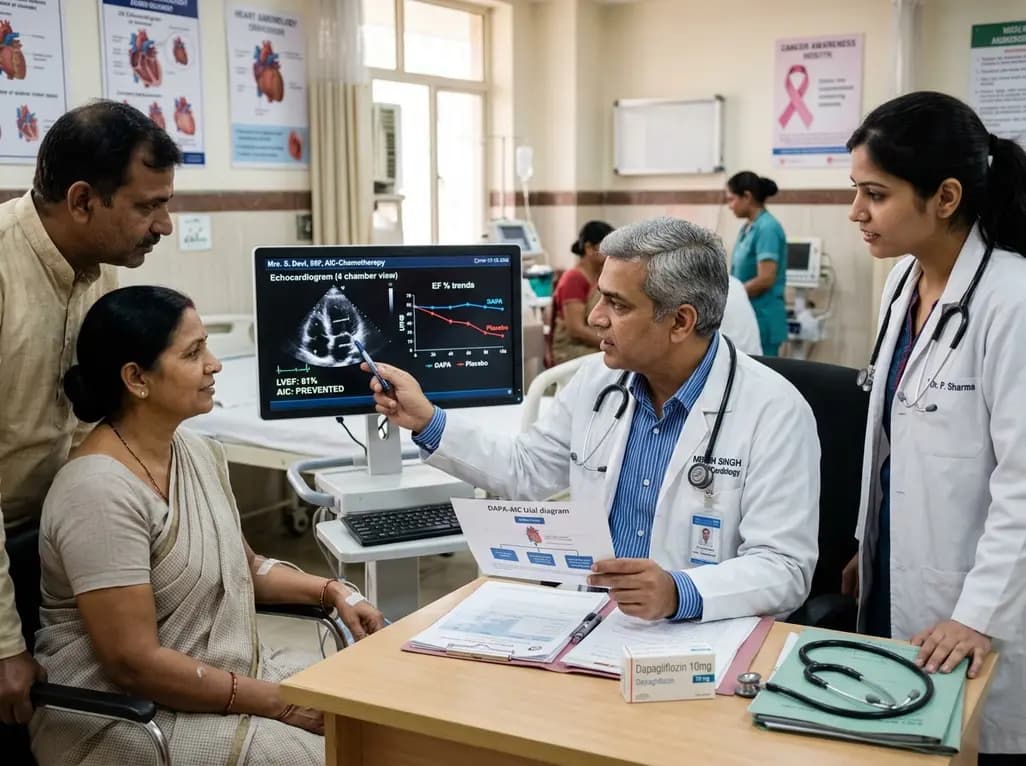
The phase II DAPA-AIC trial demonstrates that dapagliflozin 10 mg daily significantly preserves left ventricular ejection fraction compared to placebo over four months in adults undergoing anthracycline chemotherapy, highlighting a potential preventive strategy in cardio-oncology.
Today

A scoping review reveals severe gaps in long-term assistive technology provision following humanitarian emergencies. Addressing post-disaster rehabilitation requires systematic follow-up, workforce training, and integration into national health systems to sustain mobility and recovery.
Today
A randomized controlled trial investigated whether virtual reality thyroid biopsy reduces procedural pain, anxiety, and fear. While adjusted analyses showed modest pain reduction, unadjusted differences remained statistically insignificant, highlighting the need for larger confirmatory trials.
Today
