.webp&w=384&q=75)

Loading, please wait...
Loading, please wait...

Vascular pathology frequently connects systemic microvascular dysfunction across distant organ systems. Consequently, clinicians increasingly recognize the intricate cross-talk between the brain and kidneys, commonly termed the kidney-brain axis. Both organs possess low-resistance capillary beds that expose delicate microvessels to continuous high-volume blood flow. As a result, hemodynamic stress, chronic hypertension, and systemic endothelial injury often cause simultaneous microvascular damage in both organs. Recent large-scale neuroimaging studies highlight that cumulative small vessel disease burden in the brain closely reflects progressive microvascular deterioration in the renal parenchyma. Among various neuroimaging signatures, enlarged perivascular spaces serve as key markers of impaired glymphatic clearance and structural microvascular degeneration. Specifically, enlarged perivascular spaces within the basal ganglia signal severe hypertensive arteriolosclerosis. Conversely, spaces within the centrum semiovale correlate with cerebral amyloid angiopathy. Evaluating how reduced renal function and glomerular hyperfiltration relate to distinct neuroimaging phenotypes provides crucial mechanistic insights. Therefore, understanding this systemic connection allows clinicians to identify patients at high risk for concomitant cerebrovascular and renal complications early in the disease process.
Enlarged perivascular spaces represent fluid-filled compartments surrounding small cerebral penetrating arteries, arterioles, capillaries, and venules. Under normal physiological conditions, these interstitial pathways participate actively in waste clearance through the cerebral glymphatic network. However, chronic vascular inflammation, blood-brain barrier disruption, and vessel wall stiffening impair fluid movement, causing interstitial fluid retention and visible space enlargement on magnetic resonance scans. Clinicians routinely assess these neuroimaging features across two primary anatomical regions: the basal ganglia and centrum semiovale. Basal ganglia involvement predominantly reflects deep perforating vessel damage caused by systemic arterial hypertension and arteriolosclerosis. In contrast, centrum semiovale changes reflect cortical vessel pathology linked to amyloid deposition. Furthermore, total cerebral small vessel disease burden integrates these fluid spaces along with white matter hyperintensities, lacunes, and cerebral microbleeds into a single comprehensive score. Consequently, analyzing these neuroimaging markers allows physicians to stratify cerebrovascular disease risk effectively. Recent research emphasizes that assessing both regional spatial distributions and global vascular scores provides essential clues regarding underlying microvascular pathology and renal filtration abnormalities.
To examine how renal parameters associate with specific neuroimaging markers, investigators analyzed pooled individual patient data from the Microbleeds International Collaborative Network. This multi-center international registry includes a large cohort of patients presenting with acute ischemic stroke or transient ischemic attack. Researchers meticulously evaluated baseline estimated glomerular filtration rates alongside brain magnetic resonance imaging scans. They categorized renal function into distinct clinical strata, including impaired glomerular filtration (rates between thirty and sixty or under thirty mL/min/1.73 m²) and glomerular hyperfiltration (filtration rates exceeding the age-adjusted and sex-adjusted ninety-fifth percentile). Neuroimaging specialists rated enlarged perivascular space severity in both the basal ganglia and centrum semiovale using validated five-point ordinal visual scales. Simultaneously, they calculated total cerebral small vessel disease burden by assigning cumulative points for severe white matter hyperintensities, cerebral microbleeds, lacunes, and enlarged perivascular spaces. Through rigorous multivariable ordinal regression modeling, investigators adjusted for key demographic and cardiovascular confounders, including age, sex, vascular risk factors, and prior antithrombotic therapy.
The research study revealed striking differences in anatomical relationships between renal function markers and perivascular space distributions. Decreased estimated glomerular filtration rate demonstrated a clear, independent association with increased perivascular space severity specifically in the basal ganglia. Specifically, patients with marked renal impairment exhibited significantly higher odds of severe basal ganglia involvement compared to individuals with normal renal function. Conversely, reduced renal filtration showed no significant association with enlarged perivascular spaces in the centrum semiovale. This remarkable contrast highlights fundamental mechanistic differences between deep arteriolosclerotic pathology and superficial amyloid-related vascular disease. Furthermore, the presence of severe basal ganglia changes strongly mirrored overall small vessel disease burden across the brain. Statistical models confirmed that renal function impairment correlates robustly with higher global neuroimaging lesion scores. Thus, structural damage within deep subcortical penetrating arterioles closely parallels microvascular rarefaction and endothelial breakdown in the renal glomeruli. These findings confirm that basal ganglia markers act as reliable surrogates for systemic hypertensive microangiopathy.
Interestingly, the investigators also evaluated glomerular hyperfiltration, a state characterized by pathologically elevated glomerular filtration rates often seen in early stage diabetes or early vascular remodeling. Glomerular hyperfiltration represents intraglomerular hypertension and compensatory hyperperfusion prior to overt nephron loss. In this stroke cohort, hyperfiltration did not show a statistically significant association with perivascular space expansion in either anatomical region after adjusting for key risk factors. In contrast, progressive decline in estimated glomerular filtration rate consistently predicted higher subcortical microvascular lesion burdens. Therefore, structural cerebrovascular remodeling aligns more closely with established renal tissue damage and reduced filtration capacity than with early hemodynamically driven hyperfiltration states. Moreover, combining traditional microvascular markers like white matter hyperintensities and microbleeds with perivascular space ratings significantly enhanced the strength of the association between kidney dysfunction and total cerebral small vessel disease burden. These results suggest that advanced renal impairment serves as a reliable marker of widespread, end-organ microvascular degeneration throughout both the brain and kidneys.
These crucial findings carry profound clinical implications for multidisciplinary management across neurology, nephrology, and primary care. Recognizing that basal ganglia perivascular space enlargement reflects impaired renal function empowers clinicians to implement comprehensive vascular risk factor management strategies promptly. Consequently, when neurologists identify extensive basal ganglia changes or high global vessel disease scores on brain magnetic resonance imaging, they should routinely screen patients for chronic kidney disease through estimated glomerular filtration rate testing and urine albumin-to-creatinine ratio assessments. Conversely, nephrologists managing chronic kidney disease patients must maintain heightened clinical vigilance for subclinical cerebrovascular disease, cognitive decline, and increased stroke risk. Early aggressive control of blood pressure, optimized glycemic control, lifestyle modification, and careful selection of nephroprotective and neuroprotective pharmacotherapies remain essential steps. Ultimately, adopting an integrated, cross-specialty approach helps mitigate progressive target organ damage, reduces future cerebrovascular events, and improves overall long-term patient outcomes across clinical settings.
Enlarged perivascular spaces are fluid-filled compartments surrounding small cerebral vessels. They signal impaired fluid drainage and structural breakdown in the brain. Anatomically, enlarged spaces in the basal ganglia indicate hypertensive small vessel disease, whereas spaces in the centrum semiovale reflect cerebral amyloid angiopathy, serving as important diagnostic biomarkers.
Reduced estimated glomerular filtration rate directly correlates with increased small vessel disease burden, particularly within the basal ganglia. Both organs share vulnerable low-resistance vascular beds. Consequently, systemic hypertension and microvascular injury damage renal glomeruli and deep cerebral penetrating arteries simultaneously, accelerating parallel end-organ damage in both systems.
Yes, clinicians should regularly screen stroke and small vessel disease patients for kidney dysfunction. Detecting elevated basal ganglia perivascular space severity on magnetic resonance imaging warrants prompt evaluation of estimated glomerular filtration rate and urinary albumin excretion, allowing early initiation of multidisciplinary cardioprotective and nephroprotective therapies.
Disclaimer: This content is for informational and educational purposes only and does not constitute medical advice, diagnosis, or treatment. Always seek the advice of a qualified healthcare provider with any questions regarding a medical condition. Refer to the latest local and national guidelines for clinical practice.
References

Read summarized clinical updates, watch expert medical content, and earn CME certifications right from your smartphone.
Open your smartphone camera to scan and install.





A pooled cohort study from the MICON network demonstrates that reduced eGFR is independently associated with basal ganglia enlarged perivascular spaces and overall cerebral small vessel disease burden, underlining shared microvascular pathology in the brain-kidney axis.
last month
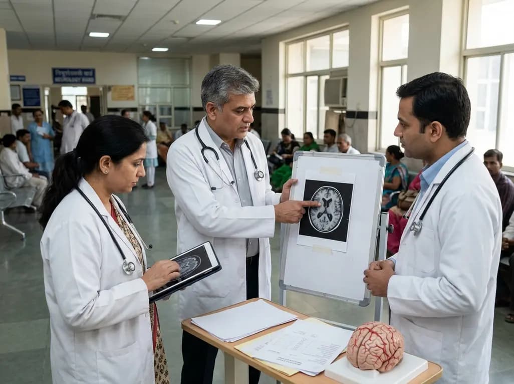
A new study reveals that lipid-related metabolic dysregulation, marked by elevated TG/HDL-C ratio and glymphatic changes, independently impacts survival in idiopathic normal pressure hydrocephalus.
Today

A premature neonate developed upper limb compartment syndrome after uterine rupture extruded the arm through a scar defect. Conservative management with continuous monitoring yielded complete functional recovery and normal limb growth at 10-year follow-up, highlighting non-operative safety in selected cases.
Today
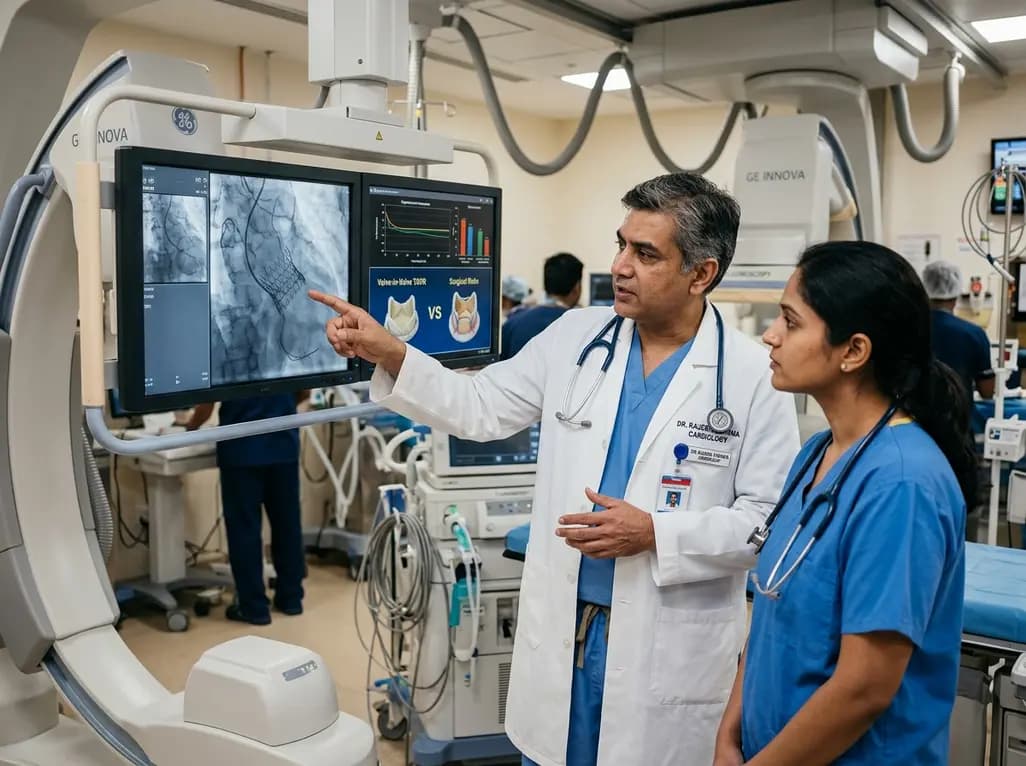
A meta-analysis of 13 propensity score-matched studies shows ViV-TAVR delivers lower early mortality and reduced bleeding compared to redo-SAVR for degenerated bioprosthetic aortic valves, though long-term hemodynamics warrant careful anatomical and patient-centered evaluation.
Today
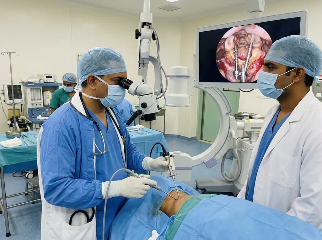
Endoscopic posterior cervical fusion combines minimally invasive decompression, joint preparation, and rigid screw-rod fixation for atlantoaxial pathologies. Early clinical findings demonstrate solid bony union, excellent symptom relief, and minimal soft-tissue morbidity without significant vascular compromise.
Yesterday

The All-India Food Processors' Association has approached the Supreme Court to oppose FSSAI's proposed per-100g benchmark for front-of-pack warning labels, advocating instead for a per-serving threshold. We explore the regulatory showdown, nutritional evidence, and implications for clinical lifestyle counseling.
Today
