

Loading, please wait...
Loading, please wait...

Modern cranial surgery demands extreme technical accuracy, particularly when navigating narrow anatomical corridors surrounded by critical neural and vascular networks. The deployment of skull base intraoperative technology has redefined surgical boundaries, allowing clinicians to optimize tumor resection while safeguarding patient function. Recently, the Society of British Neurological Surgeons MedTech Committee conducted a comprehensive cross-sectional national survey evaluating current operative practices across the United Kingdom and Ireland. By assessing three benchmark procedures—transsphenoidal pituitary adenoma resection, retrosigmoid vestibular schwannoma excision, and pterional aneurysm clipping—the study established a crucial descriptive baseline. Notably, the investigators demonstrated that technology utilization is heavily procedure-specific rather than surgeon-dependent. Consequently, established modalities cluster tightly around recurring anatomical challenges, whereas emerging platforms face slower, more selective implementation. In the past, surgical departments lacked standardized, procedure-level data detailing how frequently specialists employ these tools during everyday interventions. This survey bridges that gap across twenty-one major neurosurgical units, revealing that surgical anatomy fundamentally dictates equipment selection. As healthcare systems increasingly demand cost-effectiveness and objective patient safety data, understanding these established patterns enables surgical leaders to modernize operative suites efficiently.
Transsphenoidal approaches for pituitary tumors have undergone an extraordinary technological shift over the past two decades. The national survey revealed that endoscopic visualization has now almost completely replaced the operating microscope in transsphenoidal adenoma surgery. Specifically, all responding centers reported consistent use of rigid endoscopes, reflecting widespread appreciation for panoramic visualization and superior illumination within deep sellar corridors. Furthermore, neuronavigation systems proved indispensable during these procedures, with two-thirds of participating units utilizing optical tracking routinely. Because stereotactic navigation clearly delineates the midline and identifies the sphenoid sinus architecture, it drastically lowers the risk of catastrophic carotid artery injury. In addition, surgical teams consistently deployed micro-drills and powered micro-debriders to navigate bony barriers and soft tissue interfaces safely. Interestingly, while optical guidance remains routine in transnasal approaches, its reported use dropped markedly in open vascular or posterior fossa surgeries. This divergence occurs because transsphenoidal corridors experience negligible brain shift, allowing registration accuracy to persist throughout the operation. Therefore, transsphenoidal surgery exemplifies how targeted technology integration directly addresses the specific spatial constraints of rigid skull base corridors.
In contrast to sellar interventions, retrosigmoid approaches for vestibular schwannoma resection demand continuous bimanual dexterity and deep stereoscopic perception. Accordingly, survey results confirmed that the traditional operating microscope remains the undisputed visual standard in this anatomical region. Every responding neurosurgical center reported universal reliance on microscopy, which allows precise microscopic dissection along fragile cranial nerve interfaces. Furthermore, intraoperative neurophysiological monitoring achieved complete unanimity among respondents. Clinicians routinely deployed continuous facial nerve electromyography and brainstem auditory evoked potentials to prevent permanent neurological deficits during tumor mobilization. Ultrasonic surgical aspirators also served as a universal adjunct. They allow controlled, low-traction internal debulking without transmitting mechanical stress to the brainstem. Meanwhile, rigid endoscopes saw only secondary utilization, functioning primarily as angled inspection tools to verify complete clearance within the lateral internal auditory canal. Robotic arms and augmented reality overlays continue to develop. Nevertheless, surgeons remain committed to stereomicroscopy, ultrasonic cavitation, and electrophysiological monitoring. Consequently, vestibular schwannoma protocols prioritize mature technologies with proven records of neural preservation.
Pterional craniotomy for intracranial aneurysm clipping requires immediate intraoperative verification of both complete dome occlusion and distal branch patency. Within this high-stakes vascular domain, operating microscopes remain the standard visual platform, supported by dedicated vascular assessment tools. Specifically, surgical respondents reported routine reliance on indocyanine green videoangiography to verify real-time capillary and arterial perfusion under near-infrared fluorescence. Additionally, micro-Doppler ultrasonography proved equally essential, providing instantaneous acoustic feedback regarding blood flow velocities in parent and perforating vessels. By combining these modalities, operative teams promptly detect unintended branch stenosis or lingering aneurysm remnants before dural closure. However, the survey revealed notable discordance regarding intraoperative neurophysiological monitoring during aneurysm clipping. Participating centers were evenly divided between routine and selective monitoring. This division reflects differing opinions regarding evoked potential utility during temporary clipping. Moreover, five out of thirteen vascular specialists reported utilizing augmented reality systems to project angiographic reconstructions directly onto the surgical field. Nevertheless, indocyanine green and micro-Doppler remain the foundational intraoperative safeguards during open cerebrovascular interventions.
Although medical device manufacturers frequently promote novel hardware, the survey revealed that real-world integration of emerging technologies remains exceptionally limited. Specifically, exoscopes, intraoperative magnetic resonance imaging, and surgical robotics demonstrated minimal uptake across reporting centers. Multiple pragmatic hurdles explain this hesitant adoption. Foremost among these barriers are substantial capital procurement costs and specialized maintenance requirements, which place significant strain on institutional budgets. Additionally, digital exoscopes require operative teams to adjust to detached monitor viewing. Many microsurgeons hesitate to adopt them without demonstrated superiority over binocular optics. Crucially, the survey authors noted that non-use could not be differentiated from a lack of equipment access, suggesting institutional availability limits clinical utilization. Interestingly, statistical analysis using Kendall's tau-b revealed no correlation between surgical experience and technology usage rates. Both early-career and senior neurosurgeons demonstrated similar patterns of technology selection based strictly on operative indications. Consequently, future innovations will only gain widespread acceptance if they address unmet clinical needs and demonstrate undeniable improvements in patient outcomes.
Rigid endoscopy provides panoramic, wide-angle illumination that significantly enhances anatomical visualization within the sellar region. Unlike the linear view of traditional microscopes, endoscopic optics allow surgeons to inspect deep lateral corners, suprasellar extensions, and hidden recesses, which facilitates complete tumor resection while preventing inadvertent damage to internal carotid arteries.
Intraoperative neurophysiological monitoring provides real-time functional assessment of fragile cranial nerves during cerebellopontine angle dissection. Continuous electromyography and evoked potential tracking immediately alert the surgical team to mechanical stretching, thermal injury, or ischemia. Consequently, this continuous feedback enables surgeons to adjust their dissection trajectory and successfully preserve vital facial and cochlear functions.
Indocyanine green videoangiography provides rapid visual confirmation of complete aneurysm occlusion and capillary perfusion under near-infrared light. Concurrently, micro-Doppler ultrasonography delivers real-time acoustic verification of arterial flow velocities in adjacent parent and branching vessels. Used together, these tools immediately reveal residual neck filling or inadvertent vessel stenosis before dural closure.
Disclaimer: This content is for informational and educational purposes only and does not constitute medical advice, diagnosis, or treatment. Healthcare professionals should exercise independent clinical judgment and verify information with primary medical literature and authorized institutional guidelines. Refer to the latest local and national guidelines for clinical practice.
References

Read summarized clinical updates, watch expert medical content, and earn CME certifications right from your smartphone.
Open your smartphone camera to scan and install.





A UK national survey reveals how intraoperative technology use in skull base and vascular neurosurgery is strictly procedure-specific. While endoscopy, neuronavigation, neuromonitoring, and ICG dominate routine procedures, emerging tools like exoscopes and robotics see limited real-world uptake.
Today

The 2025 Australian meningococcal surveillance report shows a 13% decline in invasive meningococcal disease notifications to 118 cases. Genogroup B accounted for 82.2% of typed infections, predominantly impacting infants and young adults, highlighting critical vaccination and rapid diagnostic imperatives.
Today
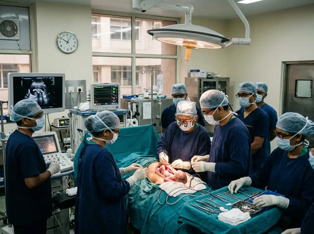
Doctors at AIIMS New Delhi have achieved a historic milestone by performing India's first open prenatal myelomeningocele repair on a 25-week unborn fetus. This landmark multidisciplinary procedure provides vital neuroprotective benefits, lowers hydrocephalus risks, and transforms maternal-fetal medicine nationwide.
Today
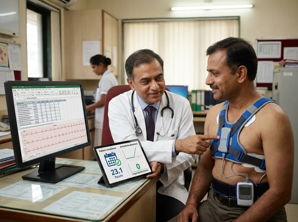
The ACE-PAS registry evaluated 21,612 patients fitted with the ASSURE wearable cardioverter defibrillator. The study demonstrated a 100% ventricular arrhythmia conversion rate, an inappropriate shock incidence of 0.0065 per patient-month, and high daily compliance of 23.1 hours, confirming real-world safety.
Today

A clinical overview of resveratrol supplementation in mitigating exercise-induced muscle damage. This narrative review explores physiological mechanisms, reductions in soreness markers like creatine kinase, dosage strategies, and bioavailability enhancements for sports medicine and nutrition practitioners.
Today

Childhood insulinoma is a rare cause of recurrent hyperinsulinemic hypoglycemia that often eludes early diagnosis. Advanced GLP-1 receptor-targeted PET/CT imaging using 68Ga-exendin-4 complements anatomic MRI to localize occult pancreatic lesions, facilitating focused, curative surgical enucleation.
Today
