.webp&w=384&q=75)

Loading, please wait...
Loading, please wait...
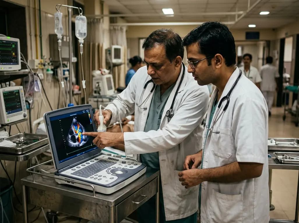
A sinus of Valsalva aneurysm represents an uncommon aortic root dilatation that develops between the aortic valve annulus and the sinotubular junction. Although congenital deficiency in the aortic media serves as the primary etiology, acquired factors such as infective endocarditis, atherosclerosis, or connective tissue disorders can also weaken the aortic wall. Consequently, the affected sinus gradually thins and expands under systemic intra-aortic pressure, creating a characteristic windsock deformity.
While many affected patients remain asymptomatic during early life, structural thinning predisposes the vessel wall to catastrophic perforation. Rupture of a sinus of Valsalva aneurysm abruptly transforms a quiescent vascular defect into an acute aorto-cardiac fistula. Consequently, high-pressure arterial blood rapidly decompresses into lower-pressure adjacent chambers, provoking massive left-to-right shunting. In younger individuals, particularly adolescents and young adults, this acute mechanical breakdown frequently triggers profound hemodynamic instability. Because the clinical evolution can be rapidly progressive, timely anatomical recognition and immediate clinical intervention are essential to preserve cardiac function and prevent sudden death.
The clinical manifestation of a ruptured aortic sinus varies significantly depending on the rate of perforation and the receiving chamber. Traditionally, acute onset dyspnea, sudden retrosternal chest pain, and severe fatigue dominate the classic presentation. However, atypical prodromal symptoms frequently mislead clinicians, creating substantial diagnostic delays in emergency departments.
Recent clinical and forensic reviews highlight that unexpected gastrointestinal disturbances, particularly isolated vomiting, nausea, and epigastric discomfort, can serve as the initial herald of acute circulatory failure. Therefore, emergency physicians must maintain heightened vigilance when young patients present with sudden gastrointestinal complaints coupled with unexplained tachycardia or diaphoresis. In addition, physical examination typically reveals signs of acute high-output volume overload. Clinicians frequently auscultate a classic continuous machinery-like murmur along the left sternal border. Furthermore, wide pulse pressure, bounding peripheral pulses, and elevated jugular venous pulsation often develop rapidly. When massive rupture occurs without prior detection, acute cardiogenic collapse and fatal arrhythmias may rapidly ensue before medical teams can establish definitive vascular access.
The anatomical origin of the aneurysm dictates both the trajectory of rupture and the subsequent hemodynamic disruption. Anatomically, the right coronary sinus represents the most frequent site of aneurysm formation, followed by the noncoronary sinus and rarely the left coronary sinus. When a noncoronary sinus aneurysm ruptures, the fistulous tract predominantly enters the right atrium. Conversely, right coronary sinus ruptures typically track directly into the right ventricle or right atrium.
As high-pressure aortic flow enters the thin-walled right atrium, right-sided chamber pressures escalate precipitously. Consequently, the massive left-to-right shunt severely increases pulmonary blood flow while compromising effective systemic cardiac output. Furthermore, acute right atrial volume overload compromises systemic venous return, culminating in rapid central venous congestion and acute hepatosplenomegaly. In addition, continuous stretching of the atrial myocardium and adjacent conduction tissue triggers malignant supraventricular or ventricular arrhythmias. When rupture extends into the pericardial space instead of a cardiac chamber, acute cardiac tamponade develops instantly. This catastrophic mechanical restriction arrests ventricular filling, leading to immediate electromechanical dissociation and sudden death.
Prompt identification of a sinus of Valsalva aneurysm demands multimodality cardiovascular imaging. Transthoracic echocardiography (TTE) functions as the indispensable first-line diagnostic modality in both emergency and intensive care settings. Specifically, color Doppler imaging visualizes the characteristic windsock projection and delineates the continuous high-velocity jet traversing into the recipient cardiac chamber.
However, transesophageal echocardiography (TEE) offers superior spatial resolution, particularly for noncoronary sinus lesions and posterior cardiac structures. TEE precisely defines the size, shape, and exact entry site of the fistulous tract while evaluating adjacent aortic valve competence. In addition, cardiac computed tomography (CT) and magnetic resonance imaging (MRI) provide comprehensive three-dimensional anatomical roadmaps. These advanced imaging techniques accurately assess aortic root geometry, evaluate coronary ostial displacement, and identify associated structural defects such as ventricular septal defects or bicuspid aortic valves. In stable patients requiring invasive assessment, cardiac catheterization and aortic root angiography quantify shunt fraction (Qp/Qs) and measure pulmonary vascular resistance. Therefore, integrating rapid echocardiographic screening with advanced cross-sectional imaging ensures precise anatomical localization and guides subsequent surgical planning.
Once a diagnosis of ruptured sinus of Valsalva aneurysm is established, immediate therapeutic intervention becomes imperative to prevent progressive circulatory collapse. Medical management plays an initial supportive role, focusing on acute stabilization. Clinicians administer intravenous loop diuretics, afterload-reducing vasodilators, and careful inotropic support to alleviate pulmonary vascular congestion and maintain systemic perfusion. Nevertheless, medical therapy serves only as a temporary bridge to definitive mechanical repair.
Surgical correction under cardiopulmonary bypass remains the gold standard treatment, offering exceptional long-term survival and low perioperative mortality. During open repair, surgeons utilize either a transaortic approach, a transcameral approach through the affected chamber, or a combined dual incision. The surgical team resects the aneurysmal sac and securely closes the communication using a synthetic patch or autologous pericardium, thereby avoiding leaflet distortion. Alternatively, percutaneous transcatheter closure has emerged as a safe and less invasive option for selected patients with favorable defect anatomy. Transcatheter device placement achieves rapid defect occlusion without sternotomy. However, strict anatomical evaluation is mandatory to prevent device embolization, coronary ostial impingement, or aortic valve regurgitation.
Comprehensive post-mortem investigations and histopathological evaluations have illuminated the structural mechanisms underlying sinus of Valsalva aneurysm rupture. Microscopic examination of the resected aortic wall or autopsy specimens typically demonstrates severe disruption and fragmentation of the medial elastic laminae. Furthermore, marked thinning of collagen fibers and loss of smooth muscle cells within the tunica media establish the structural substrate for progressive aneurysmal dilation.
Forensic series reveal that fatal cases frequently harbor secondary pathological abnormalities alongside the primary aortic defect. These accompanying features include marked cardiomegaly, myocyte hypertrophy, microscopic interstitial fibrosis, and focal inflammatory infiltration around the rupture site. In some adolescents and young adults, underlying congenital bicuspid aortic valve architecture or subclinical connective tissue dysplasia accelerates mechanical wall failure. Pathologists must differentiate acute rupture from infective endocarditis with mycotic pseudoaneurysm formation by performing detailed histological staining and tissue cultures. Therefore, thorough forensic autopsy, toxicological analysis, and cardiovascular histopathology remain indispensable for confirming the cause of death in sudden, unexplained adolescent fatalities.
A sinus of Valsalva aneurysm primarily arises from a congenital deficiency in the normal elastic tissue of the aortic media. Acquired factors like atherosclerosis, infections, or connective tissue disorders can also weaken the vessel wall. Over time, high intra-aortic pressure causes the weakened sinus wall to thin and dilate. Eventually, physical exertion, progressive hemodynamic stress, or localized tissue necrosis leads to sudden rupture into adjacent low-pressure cardiac chambers.
Patients experiencing acute rupture typically develop sudden-onset severe chest pain, profound dyspnea, palpitations, and rapid hemodynamic compromise. Interestingly, prodromal symptoms such as nausea and persistent vomiting can also occur initially. On physical examination, clinicians frequently detect a loud, continuous machinery-like murmur along the sternal border, elevated jugular venous pressure, wide arterial pulse pressure, and clinical signs of acute right heart failure or cardiogenic shock.
Definitive treatment requires urgent mechanical closure to eliminate the aorto-cardiac shunt and restore hemodynamic stability. Open surgical patch repair using cardiopulmonary bypass represents the established gold standard with excellent long-term outcomes. In selected patients with suitable anatomical margins and no associated complex cardiac defects, percutaneous transcatheter device closure provides an effective, minimally invasive alternative that avoids median sternotomy and shortens post-procedural hospital recovery.
Disclaimer: This content is for informational and educational purposes only. It is not intended to substitute professional medical advice, diagnosis, or treatment. Always seek the advice of a qualified healthcare provider with any questions you may have regarding a medical condition. Refer to the latest local and national guidelines for clinical practice.
References

Read summarized clinical updates, watch expert medical content, and earn CME certifications right from your smartphone.
Open your smartphone camera to scan and install.





Rupture of a sinus of Valsalva aneurysm into the right atrium is a rare and fatal cardiac anomaly. Learn about its prodromal signs, hemodynamic impact, diagnostic imaging modalities, and emergency surgical interventions to prevent sudden cardiac death.
last month

A secondary analysis of an HIV cure-related trial in South Africa highlights the acceptability and safety of serial excisional lymph node and gut biopsies among young women. Clear communication, empathetic care, and altruistic motives helped mitigate initial apprehension and sustain longitudinal trial engagement.
Today

Explore the crucial link between dietary protein and immunocompetence. This review examines how low-protein nutrition shifts metabolic resources away from pathogen defense toward early reproduction, compromising host survival and immunity against severe bacterial infections.
Today
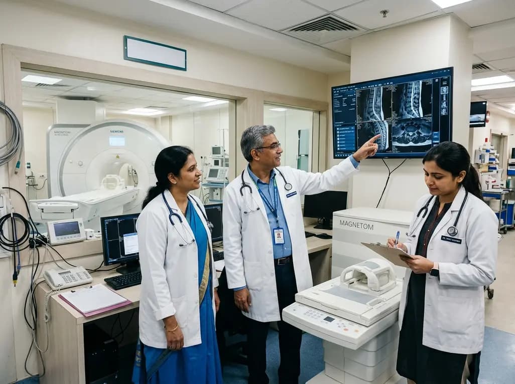
Under India's PLI initiative, domestic production of 57 critical medical devices has commenced, including MRI and CT scanners, cath labs, and heart valves. Global partnerships and local investments are expanding high-end technology, curbing import reliance, and strengthening clinical accessibility nationwide.
Today

A longitudinal study reveals that patients undergoing staged bilateral hip arthroscopy experience progressive contralateral labral deterioration during surgical intervals. However, structural degradation does not compromise 2-year postoperative clinical outcomes or patient-reported satisfaction scores.
Today

New immunological research reveals how PTPN22 forms a delayed inhibitory signaling complex to restrain T cell activation, explaining how the autoimmune-linked R620W variant disrupts feedback control and promotes persistent inflammation in conditions like rheumatoid arthritis and type 1 diabetes.
Today
