.webp&w=384&q=75)

Loading, please wait...
Loading, please wait...
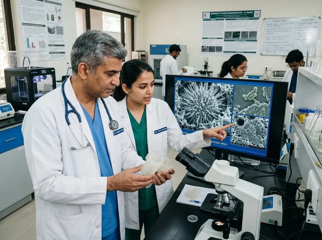
Reconstructing critical-sized skeletal defects remains a complex challenge in modern orthopedics and maxillofacial surgery. Clinicians frequently encounter severe structural loss caused by high-energy trauma, osteomyelitis, and aggressive tumor resections. In recent years, customized 3D-printed bone implants have emerged as an adaptable solution for anatomical reconstruction. By utilizing patient-specific imaging data, surgeons can manufacture personalized scaffolds made from biodegradable polymers such as polylactic acid. These polymer frameworks conform precisely to irregular defect margins, thereby providing immediate architectural stability. However, untreated polylactic acid surfaces possess significant biological drawbacks. The intrinsic hydrophobicity and bio-inert profile of the polymer impede cellular attachment, which often slows down natural osteogenesis. Consequently, delayed osseointegration increases the clinical vulnerability of the reconstruction site. Furthermore, prolonged exposure without cellular coverage invites opportunistic microbial colonization. Therefore, material scientists and orthopedic investigators have focused on developing bioactive surface modifications. Researchers at the Indian Institute of Technology Mandi have introduced a bio-inspired surface treatment that addresses these limitations simultaneously. By drawing inspiration from marine echinoderms, the engineering team has synthesized a biomimetic coating that transforms inert scaffolds into bioactive osteoconductive constructs.
Although additive manufacturing has revolutionized personalized reconstruction, biological integration remains an elusive objective when using synthetic polymers alone. Polylactic acid offers favorable mechanical properties and predictable degradation kinetics inside the body. Nevertheless, the lack of natural cell-adhesion ligands on untreated polymers hampers osteoblast recruitment. As a result, host osteoprogenitor cells fail to adhere securely to the underlying scaffold walls. This delayed interfacial bonding may eventually lead to micro-motion and aseptic loosening under cyclic physiological loads. In addition, bacterial pathogens frequently exploit this vulnerable window after implantation. Staphylococcal strains and other common hospital pathogens rapidly adhere to synthetic materials, forming resilient biofilms. Once established, these mature biofilms resist standard systemic antibiotic regimens and host immune surveillance. Consequently, deep-seated orthopedic infections often necessitate prolonged intravenous antimicrobial therapy or aggressive revision surgery. In extreme cases, complete hardware removal becomes unavoidable. Thus, modern orthopedic engineering demands multifunctional materials that stimulate osteoblast differentiation while actively resisting microbial colonization. The novel biomimetic surface design successfully bridges this gap by establishing an active defensive and regenerative interface.
To overcome surface inertness, the research team engineered a microstructured dual-layer coating that mimics the natural skeletal mineral of human bone. Hydroxyapatite constitutes the primary inorganic phase of native osseous tissue, making it an ideal candidate for osteoconductive coatings. However, standard hydroxyapatite coatings often present flat or unorganized topographies that lack antibacterial capabilities. In this breakthrough approach, investigators synthesized needle-like hydroxyapatite nanostructures arranged in radial clusters that closely resemble sea urchins. This urchin-like topography creates a dynamic three-dimensional nano-environment. Consequently, the high surface area enhances protein adsorption from surrounding interstitial fluids, which subsequently accelerates osteogenic signaling. Furthermore, the sharp, radially oriented hydroxyapatite micro-spicules exert localized mechanical pressure against approaching bacterial cell walls. Instead of releasing soluble pharmacological agents, the micro-architecture physically ruptures the bacterial membrane upon direct physical contact. Therefore, the physical topography achieves robust bactericidal activity purely through mechanobiological mechanisms. This mechanical defense strategy operates completely independent of conventional biochemical mechanisms, providing continuous protection across the material surface.
Applying bioceramic coatings to polymeric scaffolds usually presents significant chemical and thermal obstacles. Many conventional ceramic deposition methods require extreme sintering temperatures that easily melt or distort heat-sensitive polymers. To preserve scaffold geometry, the investigators formulated a gentle two-stage chemical synthesis protocol. First, the 3D-printed polylactic acid construct undergoes surface activation through a controlled alkaline bath. This initial pre-treatment introduces polar functional groups and localized mineral nucleation sites across the polymeric scaffold. Following surface activation, the scaffold undergoes hydrothermal mineralization in an aqueous solution at ninety degrees Celsius. Because this synthesis temperature remains well below the polymer melting point, the porous architecture retains its precise custom dimensions. During the hydrothermal reaction, hydroxyapatite crystals nucleate uniformly and assemble into dense clusters of microscopic mineral needles. Consequently, the entire porous scaffold gains a conformal ceramic coating without compromising structural integrity. This reliable, low-temperature method facilitates reproducible production, making it practical for clinical translation and large-scale industrial adoption.
The rise of multidrug-resistant pathogens represents a critical threat to successful surgical outcomes worldwide. Standard prophylactic strategies often rely heavily on local antibiotic-eluting cements and systemic drug administration. However, persistent exposure to localized sub-inhibitory antimicrobial concentrations promotes the emergence of resistant microbial strains. In contrast, the sea urchin-inspired micro-topography provides a robust physical defense against bacterial colonizers. When bacterial cells settle onto the scaffold, the sharp hydroxyapatite nanoneedles pierce the peptidoglycan cell wall and outer membrane. This severe physical disruption causes immediate loss of cellular turgor pressure, leading to rapid bacterial lysis. Because this bactericidal mechanism relies entirely on mechanical forces, bacteria cannot develop biochemical defenses or enzymatic resistance against the surface. Moreover, the coating eliminates the risk of systemic drug toxicity and localized cytotoxic reactions commonly associated with heavy metal nanoparticles. Therefore, mechanobactericidal surface designs represent a sustainable alternative for infection control in complex reconstructive procedures. Orthopedic surgeons can thus achieve reliable prophylactic protection without exacerbating global antimicrobial resistance challenges.
The clinical applications of this biomimetic nanotechnology extend across various surgical subspecialties. In trauma and oncologic orthopedics, surgeons can utilize these coated constructs to repair massive segmental bone voids with reduced complication rates. The accelerated osseointegration minimizes recovery times and supports earlier patient mobilization. In addition, craniofacial and maxillofacial surgeons can leverage these 3D-printed constructs to reconstruct complex aesthetic and structural facial deformities. Furthermore, the dental implantology field can directly apply this low-temperature coating to custom subperiosteal and endosseous devices. By facilitating rapid alveolar bone bonding and preventing peri-implantitis, the urchin-like ceramic layer improves long-term implant survival. In the future, researchers plan to evaluate the biomechanical performance and degradation rates of these coated constructs across large animal models. Standardizing the manufacturing process according to strict medical device regulatory standards will represent the next decisive milestone. Ultimately, merging additive manufacturing with bio-inspired surface engineering will deliver safer, patient-matched implants that optimize clinical recovery worldwide.
Q1: Why is polylactic acid modified before use in bone implants?
Polylactic acid offers excellent biocompatibility and customizable 3D printing capabilities for patient-specific implants. However, its hydrophobic and bio-inert nature limits natural bone cell adhesion, which delays structural osseointegration. Modifying the scaffold surface with bioactive minerals improves osteoconductivity and promotes rapid cellular bonding.
Q2: How does the sea urchin-inspired coating destroy bacteria without antibiotics?
The hydrothermal treatment generates dense, radial clusters of microscopic hydroxyapatite needles across the scaffold. When bacterial cells attempt to adhere, these sharp nano-spicules physically puncture and tear the microbial membrane. This mechanobactericidal action induces rapid bacterial lysis without causing biological toxicity or antimicrobial resistance.
Q3: What makes the low-temperature coating process clinically advantageous?
Many traditional ceramic coating techniques require high-temperature sintering, which melts or deforms biodegradable polymeric structures. The alkaline hydrothermal method operates at ninety degrees Celsius, preserving the exact geometry and mechanical integrity of the customized scaffold while generating a uniform mineralized coating.
Disclaimer: This content is for informational and educational purposes only. It does not constitute medical advice or replace professional judgment. Refer to the latest local and national guidelines for clinical practice.
References

Read summarized clinical updates, watch expert medical content, and earn CME certifications right from your smartphone.
Open your smartphone camera to scan and install.





IIT Mandi researchers have engineered a biomimetic sea urchin-inspired hydroxyapatite coating for 3D-printed scaffolds. This innovation mechanically destroys bacteria and accelerates bone healing without relying on conventional antibiotics.
last month

Pancreatic cancer cachexia affects over 80% of patients, driving severe muscle wasting, systemic inflammation, and treatment intolerance. This review highlights key pathophysiological drivers, exocrine insufficiency, objective CT imaging, and emerging therapies like GDF-15 inhibitors to improve clinical outcomes.
Today

A scoping review reveals how pregnancy affects phonation through laryngeal edema and reduced phonation time rather than major acoustic pitch alterations. Learn how to identify, evaluate, and manage vocal symptoms in expectant mothers.
Today

According to WHO data, average lifespan reached 73.3 years in 2023, recovering from pandemic lows. However, healthy life expectancy lags at 62.8 years due to surging noncommunicable conditions like cardiovascular disease and dementia, demanding proactive primary screening and sustained chronic care.
Today

Researchers have engineered an artificial sensory-pain integrated receptor using a multi-threshold organic synaptic transistor. The system distinguishes benign touch from noxious stimuli, enabling safer, more responsive prosthetic limbs with built-in memory and simplified hardware architecture.
Today

Medical robotics is transitioning beyond operative suites into diagnostics, cardiology, and endocrinology. Explore how innovations in tele-robotics, AI integration, and nanomedicine are overcoming geographical barriers and transforming precision healthcare delivery across India's evolving disease landscape.
Today
